 LEN
Why bother with ACL anatomy?
I treat ACL patients every single day. Most of us learned ACL anatomy in school, memorized that it runs from the back of the femur to the front of the tibia, and never really thought about it again.
But the more I read, the more I realize how much the anatomy explains what we see in the clinic. Why a 15 year old girl’s ACL isn’t the same as a 25 year old guy’s. Why surgeons talk about footprints and tunnel placement. Why the graft we put in doesn’t look or act like the ligament it replaced.
A 2019 systematic review in JBJS Reviews pulled together 92 studies on the size and shape of the ACL. It’s a great starting point, so let’s take a look.
What the review found
Cone and colleagues pulled every study they could find from 1900 to 2018 that measured the ACL. Here are the numbers in adults:
| Measure | What studies report |
|---|---|
| Length at full extension | About 27 to 38 mm |
| Cross-sectional area (midsubstance) | About 30 to 53 mm² |
| Volume | About 854 to 1,858 mm³ |
| Sagittal angle | About 45 to 65° |
| Femoral footprint | Usually 60 to 130 mm² |
| Tibial footprint | Usually 100 to 160 mm² |
A few things jumped out at me.
The ACL is a lot smaller in the middle than at the ends. The footprints on the bone are 2 to 3 times bigger than the ligament’s midsubstance. That’s why some describe it as an hourglass or a bow tie.
Women have a smaller ACL. Compared with males, the female ACL was about 4 to 12% shorter, 17 to 39% smaller in cross-sectional area, and 10 to 35% smaller in volume. Even after adjusting for body size, one study found the male ACL was still about 15% bigger.
Kids are not small adults. ACL length grew from about 25 mm at age 4 to about 39 mm at 18, then leveled off around 13 to 15 years old. Boys and girls were about the same until 10 to 12, then the boys pulled ahead by about 5 mm. The ACL also gets steeper with growth, about 20° from birth to maturity.
How you measure it matters. The numbers vary a lot between studies depending on MRI, CT, calipers or cadaver work. So take any single number with a grain of salt.
Where the ACL attaches
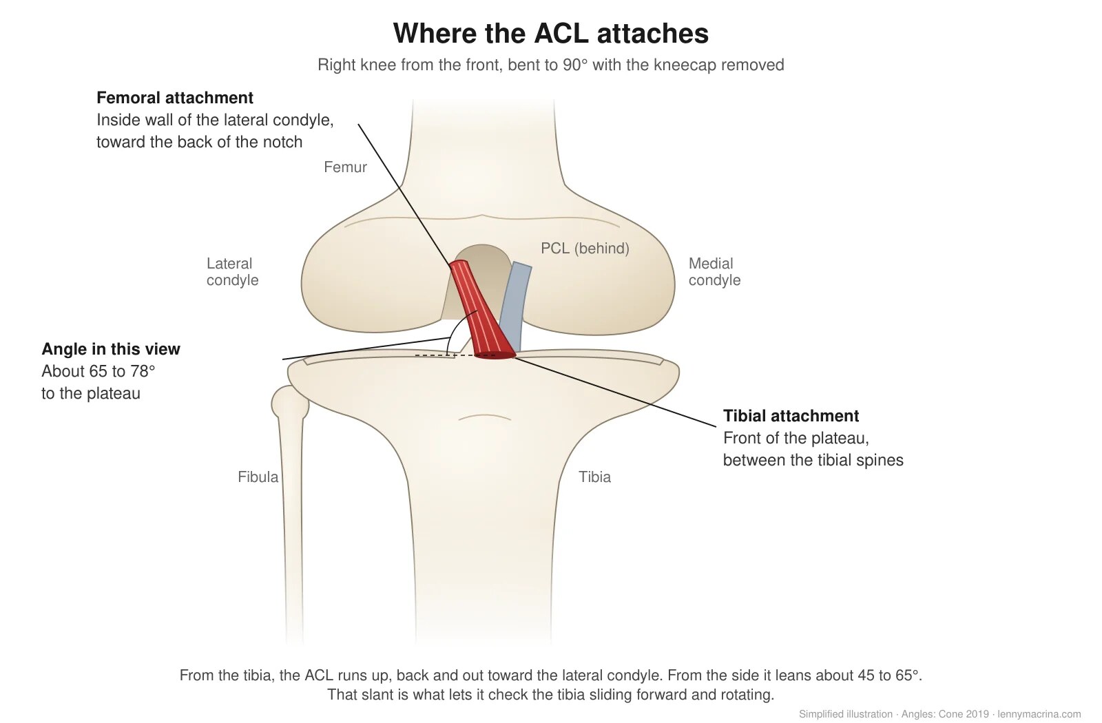
On the femur, the ACL attaches to the inside wall of the lateral femoral condyle, toward the back of the notch. Ferretti and colleagues found a bony ridge there, the lateral intercondylar ridge (the resident’s ridge), in every knee they studied. Most knees also had a smaller ridge splitting the AM and PL attachments. Surgeons use these ridges as landmarks for tunnel placement.
Iwahashi and colleagues looked at it under the microscope. The ligament’s direct attachment is a crescent about 17 mm long and 8 mm wide, sitting just behind that ridge.
On the tibia, Siebold and colleagues found the attachment is flat and C-shaped. It runs along the medial tibial spine and wraps around the front root of the lateral meniscus.
That slant, from the front of the tibia up and back to the outside wall of the notch, is what lets the ACL check both forward sliding of the tibia and rotation.
Two bundles, three bundles or a ribbon?
Most of us learned the ACL has two bundles, the anteromedial (AM) and the posterolateral (PL). That’s still the most common way to describe it, and Duthon and colleagues give a good overview in their 2006 review.
The two-bundle idea goes back to Girgis and colleagues in 1975. Ten years later, Odensten and Gillquist found no distinct bundles at all, and in 1991 Amis and Dawkins described three. So people have been arguing about this for 50 years.
Petersen and Zantop explain how the two bundles work in simple terms. As the knee bends, the femoral attachment turns more horizontal, so the AM bundle tightens and the PL bundle relaxes. The AM bundle is the main check on the tibia sliding forward. The PL bundle steadies the knee near full extension, especially against rotation.
- The PL bundle works hardest near full extension. In a study out of Pittsburgh, it carried the most load at 0° and its force dropped as the knee bent. It also matters more for rotational control.
- The AM bundle picks up more as the knee bends, peaking around 60° of flexion in that same study.
- They work together. A study out of Mass General found the two bundles share the load rather than taking turns.
Some researchers describe a third, intermediate bundle. News to me, for sure!
A 2026 cadaver study found three bundles in all 19 knees it looked at, and argued the ACL’s twist comes from where the bundles attach, not the fibers themselves twisting. It’s a small study, but it shows how much we’re still learning.
Then there’s the ribbon. Śmigielski and colleagues dissected 111 cadaver knees. About 2 to 3 mm from where it attaches to the femur, the ACL was a flat ribbon in every single knee, about 11 to 16 mm wide and only 2.5 to 3.4 mm thick. They saw no clear split between bundles.
Siebold and colleagues found the same flat shape in all 20 knees they dissected, about 10 mm wide and 4 mm thick.
So which is it? Probably both. The ACL is a flat ribbon that twists on itself. Its fibers rotate about 44 to 84° from the femur to the tibia, which makes it look rounder in the middle. That twist changes as the knee bends.
Nerves and blood supply
The ACL isn’t just a rope… it’s a sense organ with:
- Nerves. The ACL has mechanoreceptors, including Ruffini endings, Pacinian corpuscles and Golgi tendon organ like endings, plus free nerve endings. Those nerves come from branches of the tibial nerve. Johansson’s classic review showed these receptors talk to the muscle spindle system and help set muscle stiffness around the knee. If you disrupt the ACL then you lose part of that feedback loop. A 2026 review by Lee and colleagues points out that the problem doesn’t stop at the knee. After an ACL injury, the brain changes how it controls the leg too.
- Blood supply. The ACL gets most of its blood from the middle genicular artery through the synovial sheath that wraps around it. Arnoczky’s work showed the bony attachments add very little. That thin, wrapped blood supply is a big reason a torn ACL doesn’t heal on its own very well.
Why ACL anatomy matters in the clinic
Anatomy can feel complicated, but here’s how this stuff shows up in my rehab.
The new graft is not an ACL. We take a round tendon and put it where a flat, twisting ribbon used to be. That graft then goes through a long remodeling process called ligamentization. A systematic review by Claes and colleagues found grafts stay alive through that process and may end up looking like a normal ACL under the microscope, but differences in the collagen still hang around. Nobody agrees on the exact timeline and some studies have shown changes occur at 2+ years out of surgery. That’s one more reason I respect tissue biology and don’t rush the late stages of rehab. I wrote more about what changes in the knee after surgery here.
Rotation is a big deal. The PL bundle does a lot of its work near full extension and with rotational loads, which is exactly where athletes cut and land. A single round graft doesn’t fully copy that. It’s part of why more surgeons are adding an LET for higher risk athletes. I wrote about what that changes in rehab here.
Proprioception doesn’t come back on its own. When the ACL tears, those mechanoreceptors go with it. The graft won’t come with a fresh set of sensors. That’s why balance, landing work, reactive drills and dual task training aren’t extras. They’re a core part of getting the athlete back. I wrote more about neurocognitive training after ACL reconstruction here.
Kids and female athletes aren’t small adult men. The ACL is smaller in females and keeps changing shape until the mid teens. I’m not saying a smaller ACL causes tears, the research doesn’t say that. But it’s a good reminder to set expectations and testing targets by age and sex, not off one norm for everyone.
Look at how a number was measured. ACL sizes vary a lot depending on whether it was MRI, CT or a cadaver. Same thing applies to our own testing. Compare apples to apples.
Want to see how I put all of this together for the knee? Mike Reinold and I teach it step by step in our online course, Complete Knee Evaluation and Treatment.
Check out the course →Wrapping it up
The ACL is a small, flat, twisting band that’s thinner in the middle than at its ends. It has its own sensors and a pretty limited blood supply, and it looks different in a 12 year old girl than in a 25 year old guy.
When we reconstruct it, we’re putting something different in its place. Knowing ACL anatomy helps us explain to patients why rehab takes time, why we push neuromuscular training so hard, and why one set of numbers doesn’t fit everyone.
What part of ACL anatomy do you talk about most with your patients? DM me on Instagram @lenmacpt and let me know. And if you want more, all of my ACL rehab posts are in one place.
References
- Cone SG, Howe D, Fisher MB. Size and shape of the human anterior cruciate ligament and the impact of sex and skeletal growth: a systematic review. JBJS Rev. 2019;7(6):e8. doi.org/10.2106/JBJS.RVW.18.00145
- Śmigielski R, Zdanowicz U, Drwięga M, et al. Ribbon like appearance of the midsubstance fibres of the anterior cruciate ligament close to its femoral insertion site: a cadaveric study including 111 knees. Knee Surg Sports Traumatol Arthrosc. 2015;23(11):3143-3150. doi.org/10.1007/s00167-014-3146-7
- Gabriel MT, Wong EK, Woo SL, et al. Distribution of in situ forces in the anterior cruciate ligament in response to rotatory loads. J Orthop Res. 2004;22(1):85-89. doi.org/10.1016/S0736-0266(03)00133-5
- Wu JL, Seon JK, Gadikota HR, et al. In situ forces in the anteromedial and posterolateral bundles of the anterior cruciate ligament under simulated functional loading conditions. Am J Sports Med. 2010;38(3):558-563. doi.org/10.1177/0363546509350110
- Johansson H, Sjölander P, Sojka P. A sensory role for the cruciate ligaments. Clin Orthop Relat Res. 1991;(268):161-178. PubMed
- Arnoczky SP. Anatomy of the anterior cruciate ligament. Clin Orthop Relat Res. 1983;(172):19-25. PubMed
- Claes S, Verdonk P, Forsyth R, Bellemans J. The “ligamentization” process in anterior cruciate ligament reconstruction: what happens to the human graft? Am J Sports Med. 2011;39(11):2476-2483. doi.org/10.1177/0363546511402662
- Lee Z, Zhang Y, Wang P, et al. Disrupted sensorimotor control after ACL injury: from mechanoreceptor degeneration to neuroplasticity-oriented rehabilitation. Ann Med. 2026;58(1):2604403. doi.org/10.1080/07853890.2025.2604403
- Muhammed SB, Jasim AM. Anatomy of the anterior cruciate ligament: three-bundle concept with an apparent twist, a cadaveric study. J Arthrosc Surg Sports Med. 2026;7(1):63-69. doi.org/10.25259/JASSM_38_2025
- Girgis FG, Marshall JL, Monajem A. The cruciate ligaments of the knee joint: anatomical, functional and experimental analysis. Clin Orthop Relat Res. 1975;(106):216-231. doi.org/10.1097/00003086-197501000-00033
- Odensten M, Gillquist J. Functional anatomy of the anterior cruciate ligament and a rationale for reconstruction. J Bone Joint Surg Am. 1985;67(2):257-262. PubMed
- Amis AA, Dawkins GP. Functional anatomy of the anterior cruciate ligament: fibre bundle actions related to ligament replacements and injuries. J Bone Joint Surg Br. 1991;73(2):260-267. doi.org/10.1302/0301-620X.73B2.2005151
- Petersen W, Zantop T. Anatomy of the anterior cruciate ligament with regard to its two bundles. Clin Orthop Relat Res. 2007;454:35-47. doi.org/10.1097/BLO.0b013e31802b4a59
- Duthon VB, Barea C, Abrassart S, et al. Anatomy of the anterior cruciate ligament. Knee Surg Sports Traumatol Arthrosc. 2006;14(3):204-213. doi.org/10.1007/s00167-005-0679-9
- Ferretti M, Ekdahl M, Shen W, Fu FH. Osseous landmarks of the femoral attachment of the anterior cruciate ligament: an anatomic study. Arthroscopy. 2007;23(11):1218-1225. doi.org/10.1016/j.arthro.2007.09.008
- Iwahashi T, Shino K, Nakata K, et al. Direct anterior cruciate ligament insertion to the femur assessed by histology and 3-dimensional volume-rendered computed tomography. Arthroscopy. 2010;26(9 Suppl):S13-S20. doi.org/10.1016/j.arthro.2010.01.023
- Siebold R, Schuhmacher P, Fernandez F, et al. Flat midsubstance of the anterior cruciate ligament with tibial “C”-shaped insertion site. Knee Surg Sports Traumatol Arthrosc. 2015;23(11):3136-3142. doi.org/10.1007/s00167-014-3058-6
Want to go deeper on the knee? This is the online course I teach with Mike Reinold. ACL, patellofemoral pain, meniscus, progressive loading and return to sport.
Enroll Now →
Evidence based sports rehab content, course updates and clinical pearls. No spam.
ACL Reconstruction With an LET: What Changes in Rehab?
More ACL patients are getting a lateral extra-articular tenodesis (LET), even with patellar and quad tendon grafts. What the research says and what changes in rehab.
Read Post →
What Actually Changes After ACL Surgery
A study tracked 30 athletes at 3, 6, 9 and 12 months after ACL reconstruction. Here is what changed, and why symmetry alone does not mean ready.
Read Post →
Why Your Quad Tendon ACL Patient’s Quad Is Lagging
Quad tendon ACL grafts are on the rise. Here is why the quad lags after a QT graft and the rehab change most clinicians are missing.
Read Post →