Tag Archive for: rehabilitation

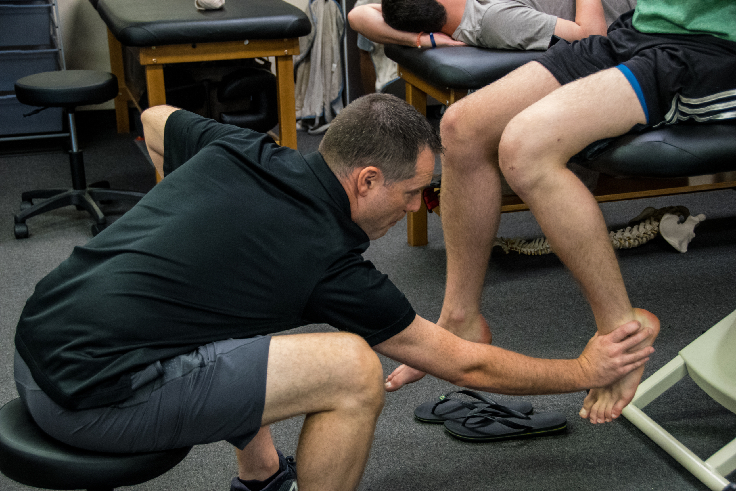
Is Early Physical Therapy Safe After a Rotator Cuff Repair?

Physical therapy is vital after a rotator cuff repair and continues to be common in an outpatient setting. Unfortunately, there’s no true consensus on when to actually begin PT.  Is early physical therapy safe after a rotator cuff repair or should we delay PT to protect the healing tendons?

Let’s dive into this and see what the research is saying…

Why I’m writing this post on rotator cuff rehabilitation

It seems as if we’re all over the place despite the research, which is pretty typical. Some docs prefer early passive range of motion (PROM) while others wait 6-8 weeks (and even up to 12+ weeks for a revision repair) before they allow any form of PT.

For the record, I’m going into this blog post as a firm believer of early PROM. It’s what we’d been doing for years in Birmingham at Champion Sports Medicine.

It’s what I only knew until I moved to Boston in 2014. Now I’ve seen a nearly 180-degree turn in rehab thoughts. Much more conservative!

I recently got in a Twitter discussion (debate) about this same topic with some very respected and prominent PT’s in the field.

It made me think about things so I decided to do a little research to see what the literature says. You can check out the discussion here.

Twitter can be confusing and tough to follow but just trust me, it goes on for a while!

Rotator Cuff Anatomy

The tendon most commonly torn is the supraspinatus tendon. Don’t get me wrong, you can tear the other rotator cuff tendons (infraspinatus, teres minor, subscapularis).

Keep in mind a medium, large or massive rotator cuff tear often will involve the infraspinatus tendon. If it does, then you need to consider modifying your progression appropriately.

But for the sake of this post, I’m going to stick to an isolated supraspinatus tendon for now.

Rotator Cuff Tendon Size and Location

Look at the size of the tendons as they insert on the greater tuberosity. You’ll see it differentiated by antero-posterior and medial-lateral directions.

According to my colleague and friend Jeff Dugas out of Birmingham, he showed in 2002 that the mean dimensions of the supraspinatus insertion were 1.27 cm in the medial-to-lateral direction. For the anterior-to-posterior direction, the supraspinatus dimension was 1.63 cm.

It helps to know this information because you may need to read an operative report and see the size of the tear. The docs will usually mention a 2 cm tear or something like that.

That means that the supraspinatus tendon and a small portion of the infraspinatus tendon were involved (and repaired.)

This is critical information to have when you’re trying to plot the post-op rehab progressions and determine the prognosis. The more tendons involved, then the higher the chance of repair failure.

There are many other factors that influence retear rates but tendon repair size is definitely one to consider.

Rotator Cuff Repair Surgery Types

I’m not going to bore you with the details of a repaired rotator cuff. There are numerous surgical techniques being used by orthopaedic surgeons.

Techniques such as a single row, double row, suture bridge or transosseous repairs are commonly performed. The picture below shows the difference between a single row and a double row repair, for example.

As you can see below, the double row tends to repair more of the tissue back to the humeral insertion point, which in theory has led to better tendon healing. This has been shown in numerous research studies and has become the best technique available.

So you had shoulder surgery…when to start physical therapy?

That seems to be the million dollar question! The research is all over the place. This means that doctors’ opinions are all over the place too, right?

Keep in mind that I’ve written about this in the past. Rotator cuff repair surgery is not always warranted and can be avoided!

Since I joined the group in Birmingham in 2002 (as a PT student), we had our post-op rotator cuff repair patients starting PT post-op day 1. They started PT early regardless of the tear size. This means a small tear of 1 cm in length started PT the same time a massive, 5 cm repair would start PT.

Some may disagree with this start time but it worked…it just worked. At least I think!

Why Early PT after a Rotator Cuff Repair

This is Key!!

There were several reasons why I think it worked:

  • They could chat with a professional.
  • Patients better understand their pain and get reassurance that what they were feeling was normal.
  • Someone could monitor their incisions and answer any and all questions.
  • Begin early, gentle ROM which often helps with pain control, too.

But, that was our ‘protocol’ and it continues to be that way many years later. Most other doctors that I have dealt with outside of Birmingham have taken a far more conservative approach to post-op rehab.

Agree to Disagree

Here in Boston, most docs wait at least 2-3 weeks and even up to 8-12 weeks to begin PT. Talk about eye-opening!

I don’t agree with this premise and wanted to dive a bit deeper into the literature to see if early physical therapy had a detrimental effect on short-term, mid-term and long-term outcomes.

Structure vs Function

The problem that continues to plague the research is the measurement of outcomes. Doctors care about the structural integrity of their rotator cuff repair. They see the research and are concerned with retear rates that hover in the 25-70%+ stratosphere. Of course I’d be concerned with retear rates that high!

Can you imagine if ACL re-tear rates were that high? Well, guess what they still hover in the 6-40% range even with our tremendous rehab skills and return to play testing.

But fortunately, we have other parameters to consider with our patients after a rotator cuff repair. We can look at the pain-free function!

Huh, what a novel idea. Regardless of the integrity of the repair, many patients can still live their lives to the fullest and in most cases without any pain.

Will Ferrell Yes GIF - Find & Share on GIPHY

Start PT Early after a Rotator Cuff Repair? What does the literature say…

I’m going to do my best and unbiased research to figure out if early PT after a rotator cuff repair is safe and effective compared to a delayed protocol. Let’s take a look…

There are a bunch of studies out there that you need to sift through. You can tell when the lead authors are MD’s or PT’s because the docs want to make sure their repair integrity is intact and the PT’s are concerned with restoring ROM, strength, and function.

With that, I’ve done my best to pull out some studies that have helped guide my practice and continue to influence me today.

Literature Review Findings

Age a BIG Factor!

Mind you, Cho et al showed that healing rates after a rotator cuff repair drastically change for older patients compared to younger patients.

The slide below was taken from my rotator cuff lecture that I’ve done in the past and helps to put things in perspective.

In no particular order…

Parsons et al JSES 2010 looked at 43 full thickness RTC repairs who were in a sling for 6 weeks. All were without PT for that time then evaluated for stiffness in PROM. They defined stiffness as 100° flexion/ 30° ER.

Overall, of the 43 surgeries, 23% (n=10) became stiff after that 1st evaluation session. The whole cohort displayed a 56% retear rate overall at 1 year, which to me seems crazy high!

To break it down further there was:

  • 30% retear in stiff group
  • 64% retear in non-stiff group
  • There was no significant difference in ROM or functional scores.

In my opinion, there were some pretty big limitations to the study that should be exposed, like:

  • Single row repair
  • No consideration for Diabetes or smoking
  • MRI without contrast to re-evaluate the repair status
  • What is “ER by the side???”- need to better define what degree of abduction.

So getting stiff may be a good thing but the repairs were done as single row repairs. We know these did not heal as well as they do with double row repairs.

More Literature Reviews

Moving on to a 2014 Level II systematic review and Meta-analysis, the authors said “the results contradicted our hypothesis that immobilization would increase tendon healing compared with an early-motion rehabilitation protocol, as structural outcomes were similar in the two groups 1 year after the arthroscopic repair of rotator cuff tears.

From the paper: “We speculate that rehabilitation is not the sole factor affecting tendon–bone recovery; the effects of other factors, such as older age, fatty degeneration, larger tears, and surgical technique, may outweigh those of the rehabilitation protocol.”

Kim et al AJSM 2012 looked at small to medium sized RTC repairs. They compared immediate PROM (0-120 degrees) to 4 weeks of absolute immobilization. They eventually showed no difference in ROM, pain or tendon healing. So seems like a smaller tear of less than 3 cm may be appropriate for immediate ROM, albeit it was limited to 120 degrees for some reason.

Not sure why they limited to 120 degrees because it seems as if the tendon would shorten as the humerus is placed in further flexion. Maybe they were concerned with subacromial impingement or something but the limitation is a bit confusing to me.

Healing Affected?

Another study by Lee et al AJSM 2012 wanted to compare ROM and healing rates between 2 different rehabilitation protocols after arthroscopic single-row repair (use caution) for full-thickness rotator cuff tear.

They showed pain, ROM, muscle strength, and function all significantly improved after arthroscopic rotator cuff repair, regardless of early postoperative rehabilitation protocols.

They also looked at the repair integrity with postoperative MRI scans, 7 of 30 cases (23.3%) in the immediate ROM group and 3 of 34 cases (8.8%) in the delayed group had re-tears, but the difference was not statistically significant (P = .106).

Well then, only a trend and all had similar functional outcomes regardless of when they started ROM…I’d say that helps the case to start early.

But again, these repairs were done via a single row repair and they allowed manual therapy 2 times per day and unlimited self-passive stretching exercise, which seems a bit aggressive anyway.

Do we even need a sling for 6 weeks?

No Functional Difference Between Three and Six Weeks of Immobilization After Arthroscopic Rotator Cuff Repair: A Prospective Randomized Controlled Non-Inferiority Trial Arthroscopy 2018

This study looked to compare clinical and radiologic results among patients with 3 versus 6 weeks of immobilization after arthroscopic rotator cuff repair in a prospective randomized controlled non-inferiority trial.

They concluded that “3 weeks of postoperative immobilization with sling use was non-inferior to the commonly used regimen involving 6 weeks of immobilization in a brace.” For the structurally concerned people out there, MRI indicated similar degrees of healing between the groups. 

Well then, that throws a wrench in things for the docs!

Ryan Gosling Giggle GIF - Find & Share on GIPHY

Does Early vs Delayed PT Affect Outcomes?

A systematic review by Gallagher et al 2015 looked to determine if there are differences between early and delayed rehabilitation after arthroscopic rotator cuff repair in terms of clinical outcomes and healing.  Six articles matched their criteria and reported significantly increased functional scores within the first 3-6 months with early rehabilitation compared to the delayed group.

To me, this is huge! Put yourself in their position. Imagine feeling better and being able to get back to work a little quicker. That’s my major argument for starting rehab sooner. Earlier pain relief, improved function and a feeling of being normal again.

Furthermore, none of the included studies reported any significant difference in rates of rotator cuff re-tear.

Medium-Large Tears use Caution

However, two studies noted a trend towards increased re-tear with early rehabilitation that did not reach significance. This was more pronounced in studies including medium-large tears. A similar trend that I’ve seen in the literature.

Here’s a nice table from that Gallagher study that summarized their findings for each research paper they included:

I want my patients to feel good as quick as possible and get back to some semblance of a normal life. But of course I want the long-term integrity of the repair to remain intact. And it seems as if this study helps that argument.

Then you look at a group of PT’s from Turkey (Duzgun et al Acta Orthop Traumatol Turc. 2011) that looked to compare the effects of the slow and accelerated protocols on pain and functional activity level after arthroscopic rotator cuff repair. Patients were randomized in two groups: the accelerated protocol group (n=13) and slow protocol group (n=16).

There was no significant difference between the slow and accelerated protocols with regard to pain at rest.

The accelerated protocol was superior to the slow protocol in terms of functional activity level, as determined by DASH at weeks 8, 12, and 16 after surgery.

I’ll take that!

Function over Structure (at times!)

Told you that the PT groups tend to favor function over structure, haha!

Early passive Motion ok- The French Version

This next study out of France that included French Society for Shoulder & Elbow ( 2012) looked to compare the clinical results after two types of postoperative management: immediate passive motion versus immobilization. Patients were randomized to receive postoperative management of immediate passive motion or strict immobilization for 6 weeks.

They concluded that their results suggested that early passive motion should be authorized: the functional results were better with no significant difference in healing. Functional results were statistically better after immediate passive motion and a lower rate of adhesive capsulitis and complex regional pain syndrome.

Seems as if there may be a trend but certainly not an overwhelming conclusion that early ROM is guaranteed to lead to inferior structural results. But, it seems pretty conclusive that ROM, pain, and function are improved after early ROM.

Let’s continue to look at the research

This group from China (Shen et al Arch Orthop Trauma Surg. 2014 Sep) performed a systematic review and meta-analysis to determine whether immobilization after arthroscopic rotator cuff repair improved tendon healing compared with early passive motion. Three RCTs examining 265 patients were included but we need to be cautious because of the limited number of studies included and the heterogeneity of the samples.

They found that there ‘no evidence that immobilization after arthroscopic rotator cuff repair was superior to early-motion rehabilitation in terms of tendon healing or clinical outcome. Patients in the early motion group may recover ROM more rapidly.’

This recent 2017 study in the Journal of Shoulder and Elbow Surgery included 9 meta-analyses in its review. They basically noted, “No clear superiority was noted in clinical outcome scores for early-motion or delayed-motion rehabilitation.”

They also concluded that “Whereas early motion and delayed motion after cuff repair may lead to comparable functional outcomes and retear rates, concern exists that early motion may result in greater retear rates, particularly with larger tear sizes.”

So, it seems as if function and healing the same after a RTC repair but there may be a discrepancy once we start looking at a larger repair size.

Immediate PT after a Revision Rotator Cuff Surgery

This study in AJSM 2018 looked to evaluate the clinical and radiological outcomes after revision rotator cuff repair surgery. They were able to track 31 of 40 patients (77.5%) for the final assessment at a mean follow-up of mean 50.3 months.

Interestingly enough, physical therapy started on the first postoperative day with passive flexion and abduction.

Revision rotator cuff repair improves clinical outcomes and shoulder function at midterm follow-up. The clinical outcome scores were comparable in patients with an intact repair and those with failed RC healing.

And they started PT 1 day after the surgery and got PT 2-3 times per week.

Retear Rates and Long-term function

This study in JBJS 2006 looked to determine the clinical and structural outcomes of re-ruptures in twenty patients after a longer period of follow-up. Nineteen of the twenty patients continued to be either very satisfied or satisfied with the outcome.

At an average of 7.6 years, the clinical outcomes after structural failure of rotator cuff repairs remained significantly improved over the preoperative state in terms of pain, function, strength, and patient satisfaction.

They also found that re-ruptures of the supraspinatus that had been smaller than 400 mm(2) had the potential to heal….wow!

Failure Rates too High!

Another study in JBJS 2013 looked at 18 patients who had undergone arthroscopic repair of massive rotator cuff tears.  At two years of follow-up, 94% had a failed repair. This current study wanted to evaluate the 10-year results for these patients with known structural failures of rotator cuff repairs.

Despite a high rate of progression of radiographic signs associated with large rotator cuff tears (proximal humeral migration or cuff tear arthropathy), most did well.

Clinical improvements and pain relief after arthroscopic rotator cuff repair of large and massive tears are durable at the time of long-term (10 years) follow-up. They went on to say:

“These results demonstrate that healing of large rotator cuff tears is not critical for long-term satisfactory clinical results in older patients.”

So our obsession with healing rates still appears to be overblown, even in older patients with a known cuff tear.

Pendulum Exercises Effects on Muscle Activity

Activation of the Shoulder Musculature During Pendulum Exercises and Light Activities JOSPT 2010

Look at that Paper in JOSPT 2010 (I’ve pulled out the Results Table for you below).

If there’s one exercise that doctors allow after a rotator cuff repair then it’s a pendulum or Codman exercise. How often does our patient do them correctly and make it a completely passive motion? I’d say rarely if seldom, right?

Most often, the patient is just bent over and actively moving their shoulder. They have no body movement or sway. Most of the movement is shoulder based and are not completely relaxing their shoulders.

Furthermore, they were instructed in the doctor’s office that 1st week or 2 after surgery.

They’ve been doing them incorrectly for weeks on end because they have no one to help them (cough cough!)

Look Over Here Steve Carell GIF - Find & Share on GIPHY

EMG of common Rehab ExerciSES

A study in JOSPT 2016 looked at EMG activity in healthy individuals. They wanted to quantify muscular activity during daily tasks and common PT motions. They showed that “of all the tasks assessed, ambulation without a sling and donning and doffing a sling and a shirt consistently showed the highest activity.”

EMG results table is found below.

Pretty helpful to see it listed by muscle and EMG activity and specific movement.

EMG of contralateral movements

A pretty neat EMG paper from 2004 (small n=6 and healthy individuals) were assessed using fine wire and surface EMG during common functional activities of the contralateral extremity while immobilized.

They found high supraspinatus EMG activity of the immobilized shoulder for all fast pulling activities of the contralateral shoulder (25-32%)!

Furthermore, they found high infraspinatus activity (56%) of the immobilized shoulder when the contralateral extremity performed straight forward reaching activities.

So even if they are immobilized and using their non-operative shoulder for daily activities, the rotator cuff is still sustaining a higher amount of activity than anything that we would do in the early phases (PROM, dowel self-ROM, rope and pulleys, properly performed pendulums).

How about revision rotator cuff repairs, you ask?

This study from AJSM in 2018 looked at outcomes after a revision rotator cuff repair. They showed revision rotator cuff repair improved outcomes regardless of tendon integrity (MRI confirmed).

Oh boy, what is going on??

Dig deeper into the study and they started PT the 1st day post-op with passive flexion and abduction. Sounds familiar, no? And this was in revision surgeries.

We used a very similar approach in Birmingham as they did in this study, so I may be a bit biased.

My Closing Thoughts on Physical Therapy after Rotator cuff repair surgery

I think it just shows you that rehab can begin early, will not affect long-term outcomes and that tendon integrity is not correlated to function.

I honestly don’t think our 15-30 minutes of passive motion early on in the rehab process is significantly affecting outcomes and retear rates.

It seems as if the repair technique, contralateral arm daily use, compliance with proper exercises (like pendulums, for example).

Let’s not blame early PT. There are so many more variables that are more likely to affect rotator cuff repair outcomes than anything that we could do in PT.

So I say let’s get people into PT early, educate them, guide them and help them get over this painful surgery.

I’ve been doing this for years and have seen the benefits of early PT. I say the literature agrees with me!

Why you need “Feel” as a Physical Therapist

I haven’t written a post in a while but wanted get back into the swing of things. In this post, I wanted to talk about having ‘Feel’ as a PT.

When I say feel, I’m talking about being able to read people and adjust the situation based on their response to things. So, what does that mean? Not really sure… but wanted to give a few examples that I have heard recently that I think happen pretty commonly in our profession.

Exercise Progression (or lack of)

This one happens a bunch in our profession and I was guilty of this early on in my career. It’s much easier to have someone come in 2-3 times per week and give them the same exercises, right? But to do this for multiple weeks, if not months, is a travesty!

Listen, I don’t think we need to progress someone’s program every session. Adding a new exercise each visit can be a bit much. know we want to make people feel as if they are moving forward in their rehab but there are other ways we can progress people besides giving them 17 different exercises that keeps them in PT for 2+ hours.

Again, I was guilty!

There are so many variables that we can manipulate for each session, it’s silly! Think about each move that someone does and break it down.

Exercise Variables to Manipulate

We can play with:

  • Tempo
  • Single leg versus double leg
  • Reps/sets scheme
  • Upper body or lower body
  • Time under tension
  • External resistance (bands, chains)
  • Rest periods between exercises
  • Perceived RPE (stole that one from Kiefer!)
  • Volume

So as you can see, even if you don’t have heavy weights as we do here at Champion PT and Performance, then you can still get creative with progressions.

The training facility at Champion PT and Performance in Waltham, MA

Personally, my clients have the same program for 4 weeks and then we write them a new program. That means that they can focus on the aforementioned variables as needed even though they are doing the same exercises for 4 weeks.

The client likes it because they get really good at that movement plus they can see their progression in their weights, which is a huge mental gain!

Besides the obvious weight progressions, there’s a ton that can be manipulated but I don’t see or hear it enough from the patients that come through here. I rarely hear a client tell me that their previous PT experiences involved any type of variable manipulation but maybe that’s why they find us in the 1st place. Who knows…

My advice, let the patient feel as if they are moving forward in their exercise prescription because they are a smarter consumer than you would think. As PT’s we must do better with this stuff and the above bullet points are a good starting point for you.

Running on Empty

On another note, I recently started treating someone for a knee injury she sustained while skiing. Fortunately, she didn’t require surgery but the fracture needed time to heal. I don’t want to reveal too many details of the case for privacy reasons but just know she could’ve easily done more damage to her knee from the mechanism of injury.

She was given a brace and a prescription for PT to begin immediately for ROM and strengthening. She was limited in weight bearing for a period of time (I don’t remember the exact amount) so she had those effects that she had to deal with too.

At the beginning of the 6th week after the injury, her doc said she was fine to begin running even though no new x-rays were done on her knee. Guess they were just going off of time and that she was a healthy female without any co-morbidities.

Back at PT, she was told to start a running program that she thought was a bit early but she was excited to progress to more aggressive exercises.

According to her, she had been doing straight leg raises, clams, bridging and other low-level exercises for the whole duration of the rehab…see above rant!

Upon beginning her running program, she felt immediate pain and had to stop. She said she felt bad because the PT was surprised that the pain was still present but she wanted to work through it a bit. Despite trying to push through it, she still felt pain and had to stop again. She felt a big sense of failure because the pain persisted and she just couldn’t get over this hump.

Think Mode

Let’s think about this scenario for a second. Six weeks after a joint fracture, little strength training after a decent period of immobilization and the patient was expected to run?

As it turns out, the patient was frustrated enough with the scenario that she sought a second opinion and found us. I’ll never put down another PT’s plan of care but it was obvious that the plan was rushed and the patient’s opinions and communications were not fully observed.

She was frustrated and felt defeated but why? Why would someone be expected to run 6 weeks after a fracture without loading the joint and going through a progressive program?

I’m not sure but I wanted to use this case as a teaching moment for other clinicians, especially the younger crowd that may struggle with rehab progressions.

Listen Up!

Listen to the client and have a good understanding of basic soft tissue healing. I told the client that she needed a good 6-8 weeks of strength training before even talking about running. She was relieved that I wanted to take it slow and we now have a very happy client who has completely bought into my system.

Again, listen to your clients. They’ll tell you what’s wrong with themselves if you listen closely!

My Dad’s Knee Replacement

Switching gears, my Dad has his knee replaced a few weeks ago. Despite having treated a gazillion knee patients in my career, my Dad has yet to step foot in our facility. I’ve given him advice from a distance and have tried to keep an eye on things as they came up.

I’m not a home health PT and respect their jobs. I was surprised that no one ever tried to bend his knee during the home visits he had for nearly 6 weeks. Fortunately, he had about 80-90 degrees of flexion but the home health PT kept telling him he didn’t need more than 90 degrees of motion.

You try to get up from a chair with only 90 degrees of knee flexion! I’ll bet it’s much more difficult than 110+ degrees of motion. For the record, I shoot for 120 degrees of knee flexion ROM for all.

After home health, my Dad started outpatient PT and he sounded confident and happy. Yet again, no one felt the need to bend his knee (he did get some patella mob’s) and just showed him basic exercises like straight leg raises and squats.

Obviously an important component to PT but I still think getting more ROM is critical. I’ve talked about how I like to bend the knee after surgery at my YouTube channel that you can access here. As you can see, I prefer the seated position at the edge of the table for its comfort and isolation of the knee joint without hip compensation.

However, no one is bending his knee and he was feeling stiff. He did get a new PT for one of his sessions and they did bend his knee but only 2 times…and each time they cranked on it to the point he had to tell them to stop because of the pain.

Not how I would’ve initiated ROM!

Bad Ass Arya wouldn’t have been so aggressive either!

He’s now 3 days after the PT session and frustrated. He told me he can’t do anything around the house anymore and has considered taking pain medication to help get over the hump. This is the ‘feel’ that I’m talking about.

Why on Earth would any PT think this technique would be beneficial? I’ve heard this way too often in the past and am frustrated by it.

If that were you 🤔

My advice…as always, put yourself in their position and consider the risk/reward. Is this the best we can do and will the patient absolutely benefit from this?

In my Dad’s situation(s) I say he has received mediocre care so far. Fortunately, I have guided his home program and have tried to keep him positive and realistic.

He’s frustrated and vows to never consider his other knee even if he can’t walk. I hope he changes his mind once he gets stronger and more functional.

My lesson in this post- listen to the client and do what’s in their best interest. Have some FEEL and progress people more appropriately.

We can do better!

The Week in Research Review, etc 11-26-18

This week, I discussed the progression of someone after a knee surgery. I tried to highlight the key stages and some techniques that I like to use to advance the patient’s mobility and comfort. Take a look at The Week in Research Review, etc 11-26-18 and share with your friends. Hope it helps you improve your patient care tomorrow and beyond!


ACL Reconstruction in a Pediatric and Adolescent Population

1st Day of #PT after an ACL Surgery

Patella Mobilization after Knee Surgery

Knee Flexion PROM after Surgery- Seated or Supine?

Stretching the Quads after Knee Surgery

Assessing for a Cyclops Lesion after an ACL

Assessing for Fat Pad Irritation of the Knee


ACL Reconstruction in a Pediatric and Adolescent Population

17 Year Follow-up After Meniscal Repair With Concomitant ACL Reconstruction in a Pediatric and Adolescent Population. Tagliero et al AJSM 2018

Results: 28% failed meniscal repair and required repeat surgery at the time of final follow-up. They also showed that outcomes and failures rates were comparable across tear complexity.

Guess that means that no matter the tear type, there was no difference in outcomes or retear rates. Although the repair techniques are now outdated and no longer used.

Their study also showed a 30% failure rate for meniscal tear repaired in the medial compartment at index surgery and 7% in the lateral compartment.

Interesting long-term outcomes that may help to guide your rehab and client advancement (and prognosis). Keep these in mind when you treat a future adolescent or pediatric ACL patient.


💥1st Day of #PT after an ACL Surgery 💥

If you have never treated a post-op ACL, then this video should interest you!

This is what the knee looks like that 1st day after surgery and can often set the stage for what’s to come over the next 6-12 months.

Often, the patient is both very curious and ultra-grossed out by the 1st unveiling. It can be stressful for them to see their knee in this condition so you really have to confidently reassure them that it is very normal.

The blood-soaked gauze is mainly saline that was used to irrigate the knee during the reconstruction. Some still leaks out of the incisions the 1st few days and can often be confused with true blood.

Understand that this is quite normal and happens to most every ACL patient’s knee that I’ve seen…nothing to worry about!

From here, I’d work on patella mobility (see the post later today) and then work on flexion ROM at the end of the table.

Again, it’s very important to get the knee moving after surgery. This will help with pain, swelling and gain confidence that the rehab process is moving forward.


Patella Mobilization after Knee Surgery

Get the patella moving early with #patella mobilizations immediately after surgery. One major reason (amongst many others) why we need to get our clients into #PT early.

I am certainly a very loud advocate for early PT and getting the patella moving can help to prevent excessive scarring, which can affect ROM and quadriceps force output.

Glove up and get that patella moving in all directions… medial, lateral, superior, inferior!


🤔Knee Flexion PROM after Surgery- Seated or Supine? 🤔

I’ve treated many patients after an ACL I can honestly say that this may be a huge influence on the early ROM outcomes that you may see.

I’ve tried to bend the knee in both supine or seated, as the video shows, and there’s no doubt that most people tolerate the seated version so much better after a knee surgery. In particular, a big surgery like an ACL, TKA or MPFL reconstruction.

It just seems to be more comfortable and with less stress on the anterior knee because of the position of the tibia (at least I think so!).

My theory, it seems as if the supine position may cause a slight posterior sag which may cause more pain and guarding than when they’re seated at the edge of the table.

I use a similar concept later on in the rehab process when I’m initiating my prone quad stretching. You can see a definitive improvement when I wedge my hand in the popliteal fossa and create a slight anterior translation on the tibia.

Most people say that the anterior knee pain that they were feeling (and not a quad stretch) was replaced by a stretch feeling only and no more anterior knee pain.

Try it out with your ACL patients and see what position they like best…I’ll bet I can covert you over if you still bend your knee patients in supine!


💥Stretching the Quads after Knee Surgery 💥

Continuing my sequence of videos after a knee surgery, I discussed my technique for progressing knee flexion PROM once they hit 120 degrees or so of flexion.

At this point, they’ve probably maxed out how much ROM they can achieve at the edge of the table. They’re ready to get that end range of motion and even some quadriceps flexibility.

In prone, most people will often feel a pain or pressure in the front of their knee when you try to bend it.

To overcome this, I like to wedge my hand into the back of the knee and give an anteriorly directed force through the gastrocnemius (calf) soft tissue and into the tibia.

This seems to create just enough movement of the tibia on the femur to take the pressure off the front of the knee. This may redirect the forces more onto the quadriceps muscle.

You’ll need to play with the amount and direction of force but most often they’ll begin to feel a better quad stretch.

Try this technique out on your next knee surgery client and see if it helps them. I usually initiate this ~4 weeks after an ACL but timeframes will vary person to person.⠀


💥Assessing for a Cyclops Lesion after an ACL 💥

In this video snippet from my YouTube Channel, I discuss how to assess for a Cyclops lesion in a knee. In particular, after knee surgery.

A patient with a potential cyclops lesion, they often present with loss of normal knee extension compared to the other side. They’ll often have anterior knee pain and poor patella mobility. Sometimes a tight feeling in their hamstrings and calves, too.

No matter how they try to regain their extension ROM, the knee just never feels right. Often times, surgical intervention is needed to remove that scar tissue.

Immediate rehab should continue to work on knee extension ROM using low load long duration stretching and aggressive patella mob’s.

No one’s to blame if this occurs. We don’t know exactly why it occurs in some people but we believe a remnant of the ACL stump may be a source of the frustrating issue.


💥Assessing for Fat Pad Irritation of the Knee 💥

Anterior knee pain is very common in the outpatient #PhysicalTherapy setting.

One of my go-to tests to assess for fat pad irritation is simply trying to capture the fatty tissue in the anterior aspect of the knee joint during active and/or passive ROM.

In this snippet from my YouTube channel, you can see that I pinch the fat pads on either side of the patella tendon as @corrine_evelyn is actively extending her knee. I’ll also do it in a relaxed state to assess passive irritability.

I 1st learned this test from @wilk_kevin and it continues to be a mainstay in my knee examination algorithm.

As for a treatment, it usually comes down to a volume issue and/or strength issue or both.

I’ll usually have to address the volume of the activity by relatively easing off of the activity while simultaneously adding in exercises to address an underlying weakness.

Remember the Dye et al study in AJSM 1998 when he talked about the fat pads being super painful during his arthroscopic surgery without anesthesia. Makes sense why they can be so painful if the knee stresses fall upon this tissue.

We talk about this study, fat pad irritability and much much more in our online knee seminar course.


If you want to learn more about how I treat ACL’s or the knee in general, then you can check out our all online knee seminar at www.onlinekneeseminar.com and let me know what you think.

We cover the anatomy, rehab prescription, ACL, meniscal injuries knee replacements and patellofemoral issues. Furthermore, the course covers both the non-operative and post-operative treatment.t

This is an awesome course if you’re interested in learning more about rehabilitating the knee joint. And if you’re a PT, there’s a good chance you can get CEU’s as well.

The Week in Research Review, etc 11-19-18

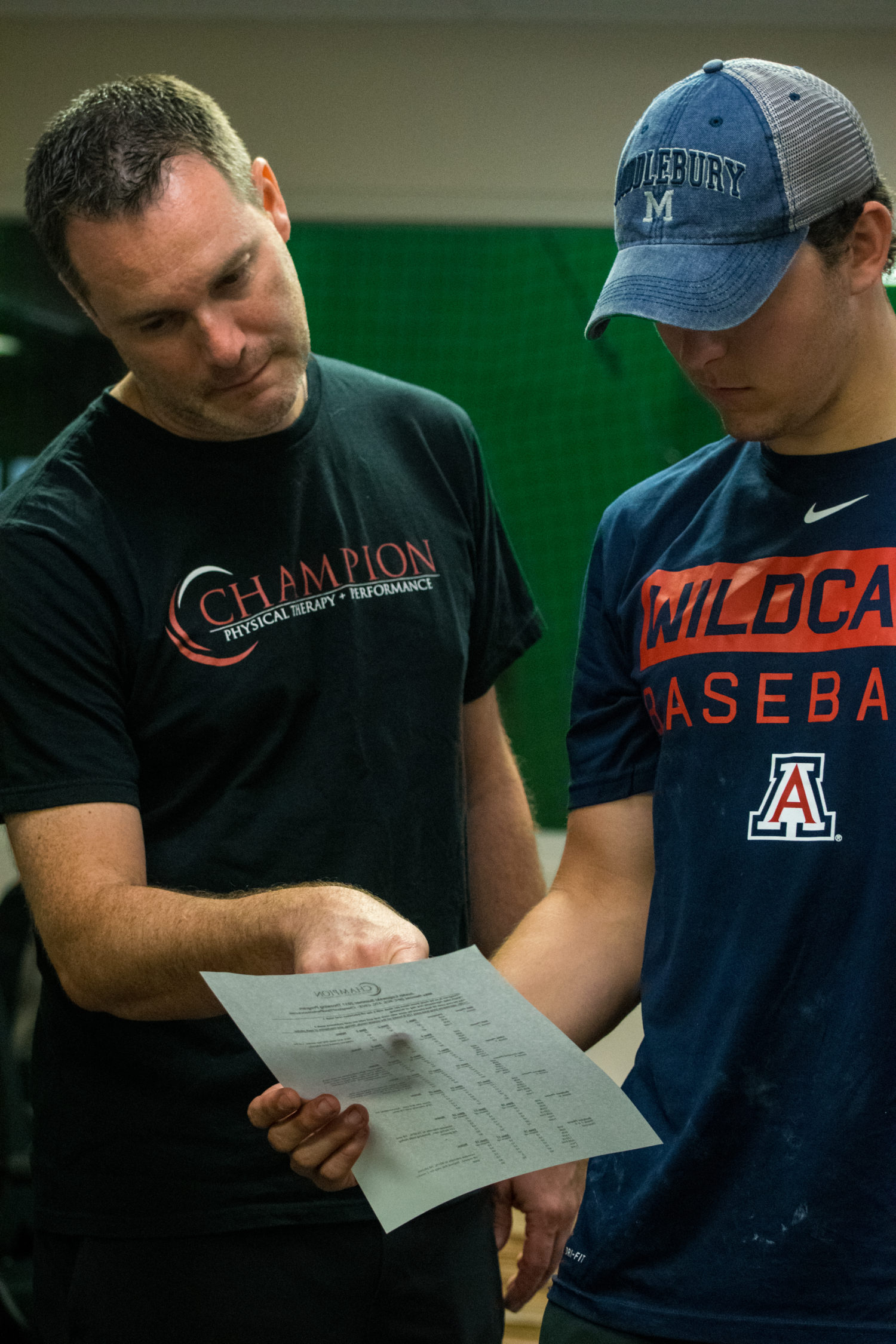
Great ‘Week in Research Review, etc 11-19-18’ that I hope you find helpful to your practice.

I’ve always touted the importance of the subjective portion of the exam so I wanted to share a slide from a recent talk I gave to a group in Canandaigua, NY. Obviously, the squat is a fundamental movement and I wanted to give some basic positions that I use to help assess. So excited that I’ve launched a brand new Medbridge course that helps the rehab specialist better eval and treat the baseball pitcher. On my YouTube channel, I discussed my thoughts on setting the scapula with various upper and lower body exercises. And finally, my co-worker Kiefer Lammi discusses the landmine with exercise.


Importance of the Subjective Exam

Assessing the Squat

My New Baseball Medbridge Course

Set the Scapula with Shoulder Exercises?

6 Ways to use the Landmine by @kieferlammi


💥Subjective the most important aspect of the Evaluation💥

This slide, taken from this past weekend’s course in Canandaigua, NY is always a favorite of mine.

I try to keep a slide like this in all of my lectures because I have found that this portion of the examination can give the rehab specialist a huge look into what is going on with the person in front of them.

Don’t get me wrong, I still consider the biomechanical aspect of what may be causing their symptoms.

It often comes down to a tissue capacity issue but it’s up to me to determine the appropriate course of treatment.

These questions will help build confidence in your client and guide the early stages of rehab.

Do you have any specific questions that you like to ask your clients during their 1st few sessions? Remember, these questions are just not for the evaluation. You should be asking these questions periodically to gauge progress and help guide the next phases of rehab, too!


🔅Assessing the Squat 🔅

Squatting is a fundamental movement that all of us have to do on a daily basis.

Utilizing several different positions can help the rehab specialist better assess the squat and develop a treatment plan that enables their client the ability to improve their squat pattern.

In the above videos, I have utilized 3 different squat patterns and will outline them by the degree of difficulty.

✅The Overhead Squat- by far the most challenging version which challenges the shoulders, thoracic spine, lumbar spine, pelvis, knee and ankles.

A movement limitation at any of these joints will most likely cause the squat pattern to break down. Using overhead resistance would further challenge the system and potentially cause the squat to further breakdown.

✅Arms Crossed Chest Squat- alters the challenge by taking most of the shoulder and thoracic spine out of the equation and isolates the motions to the lumbar spine, hips, knees and ankles.

I often use this position as my fundamental motion because most people don’t have to squat with any weights over their head. This position, in my opinion, should be the most informational and utilized.

✅Counter-weight Squat

This position changes the center of mass by moving some of the weight distribution more anteriorly (front) and making the squat motion slightly easier. I use this position as a regression, for some, which allows them to squat with less stress and potential difficulty.

There are many other variations to the squat that you can make but I wanted to highlight a few of the major changes that you cause successfully. Assessing the squat is essential and can give the rehab specialist a nice picture of the function of multiple joints during a common movement.


My BRAND NEW course on Medbridge’s platform

…that helps the sports and ortho rehab specialist (PT, OT, ATC) better understand the anatomy and biomechanics involved in the baseball pitching motion.

Advanced Rehab for the Baseball Pitcher to Improve ROM & Strength@medbridge_education

The goal of this course was to allow the clinician to be able to evaluate and treat the baseball pitcher using evidence-based guidelines that I use on a daily basis.

Numerous research studies discuss the adaptive changes that occur with the pitching motion followed by numerous videos to help guide the treatment process.

If you’re already a Medbridge subscriber, then you have immediate access today.

If you’re not a Medbridge member, then you can use my promo code “Lenny2018” to save up to 40% off a yearly membership.

This gets you unlimited CEU’s for 1 year and potential access to their online HEP and a lot more!

Students can also get 1 year of unlimited courses (no CEU’s) by using promo code LennySTUDENT2018 and pay only $100.

Check out my other shoulder courses as well by using the Medbridge platform…along with many other great speakers!

Hope you enjoy and good luck!


💥Should you Set the Scapula with your Shoulder Exercise?💥

In this video excerpt from my YouTube channel, I wanted to discuss my opinion on setting the scapula during common exercises.

I think there’s an obvious role for setting the scapula during a heavier lower body lift like a deadlift.

But for a classic upper body exercise like the Full Can (Scaption Raises) or prone T (horizontal abduction), prone Y (Prone full can), etc then I definitely want the scapula to freely move along the rib cage.

I did a quick literature search and didn’t see anything obvious that helped to guide my thoughts so most of this is anecdotal. Check out the video and comment below.

Do you coach your clients to set their scapulae before a rotator cuff workout? If so, why? If not, do you think we should reconsider?


6 WAYS TO USE THE LANDMINE!⁣

Great post from our own @kieferlammi at @championptp on various ways to use the landmine in your client’s workout routine.

If you don’t have one, then I’d highly recommend you try to obtain one because they are highly versatile and can be used in many stages of rehab. See Kiefer’s original post below 👏🏼

_____________

6 WAYS TO USE THE LANDMINE!⁣

The landmine attachment is a super versatile tool for loading that is traditionally known for being used for angled pressing variations. While that’s probably my most programmed use for it, it also provides benefit to a ton of other movements by placing the load and direction of force at a bit of an angle, which can help to promote a particular path of movement, like sitting back more in a squat or lunge. Here are 6 of my favorite ways to use the landmine:⁣

1️⃣1-Leg RDL⁣

2️⃣Split Stance Row⁣

3️⃣Reverse Lunge⁣

4️⃣Deadlift⁣

5️⃣Squat⁣

6️⃣Russian Twist⁣⠀


Save 25% off our OnLine Knee Seminar Course…all this week!

Expires Sunday, November 25th at midnight ET

If you want to learn more about how I treat ACL’s or the knee in general, then you can check out our all online knee seminar at www.onlinekneeseminar.com and let me know what you think.

We cover the anatomy, rehab prescription, ACL, meniscal injuries knee replacements and patellofemoral issues. Furthermore, the course covers both the non-operative and post-operative treatment.t

This is an awesome course if you’re interested in learning more about rehabilitating the knee joint. And if you’re a PT, there’s a good chance you can get CEU’s as well.

The Week in Research Review, etc 11-12-18

This week in research review for 11-12-18 we focused a bit more on assessment and also dabbled in some basic treatment strategies for the back and shoulder. Check out the topics below and like them or comment on Instagram to keep the conversation going…thanks all!


  • A quick fix for a sore low back?
  • Knee Fat Pad Testing and Diagnosis
  • How to Assess the Elbow for a Tommy John (UCL) Sprain
  • Lumbopelvic control on shoulder and elbow kinetics in elite baseball pitchers
  • Full Can or Empty Can? – by @mikereinold


Looking for a quick fix for a sore low back?

I’m speaking from personal experiences when I post a few of the common exercises that have helped me tremendously in the past.

I’m not saying that this is all you have to do but I do think that new onset of low back soreness, you know that tightness that you feel on either side of your spine, can be somewhat alleviated with some foam rolling and active range of motion.

I would definitely include more focal strengthening of the core like deadbugs and bird dogs, squats, deadlifts (when they’re ready), etc.

But for the purpose of this post, I think some foam rolling and motion to the area can take the edge off of someone’s soreness and get them feeling a little better. That’s my goal for many and hopefully those small gins can add up to big gains in the long run!

Do you utilize these techniques as well? If you don’t, then I suggest that you try! They’ve helped me numerous times and continue to help me when my soreness gets a bit out of control.

Tag a friend who may want to check out this post…thanks!

Thanks @corrine_evelyn for the demos!


Knee Fat Pad Testing and Diagnosis

Here’s an excerpt from a previous blog post where I talked about anterior knee pain fat pad irritation. Link in bio!

Keep in mind, my differential diagnosis is all over the place at times. With knee pain you need to consider:

Meniscus (see my previous blog post)⠀

ITB

Osteochondral lesion

Patella tendonitis

Pes anserine bursitis

MPFL sprain

Hamstring strain

Plica syndrome

MCL/LCL

Tumor

Infrapatellar fat pad irritation can be functionally debilitating. I believe it presents itself pretty often in the clinic, more than most PT’s realize.

Use this test to see if it truly is a fat pad issue.


How to Assess the Elbow for a Tommy John (UCL) Sprain

In this excerpt from my YouTube channel, I discuss the tests that I use to help identify an elbow sprain, typically seen in the baseball players that I treat.

In the full video, I discuss:

✅Joint Palpation

✅Seated Milking Sign

✅Prone Valgus Test (maybe a new one for you!)

✅Supine end range External Rotation with Valgus Extension Overload (VEO)

I also wrote a blog post about this topic so hopefully you’ll go to my site and read a bit more about this.

If you treat baseball players of all ages, then you should know how to diagnose a UCL sprain.


The influence of lumbopelvic control on shoulder and elbow kinetics in elite baseball pitchers

Laudner et al JSES 2018.

This study looked at 43 asymptomatic, #NCAA Division I and professional minor league baseball pitchers. They measured the bilateral amount of anterior-posterior lumbopelvic tilt during a single-leg stance trunk stability test.

The Level Belt Pro (Perfect Practice, Columbus, OH, USA) was used to assess anterior-posterior lumbopelvic control. The LevelBelt Pro consists of an iPod–based digital level secured to a belt using hook-and-loop fasteners.

This test has been used and studied previously by Chaudhari et al (JSCR 2011) and he showed that pitchers with less lumbopelvic control produced more walks and hits per inning than those with more control.

Also, pitchers with less lumbopelvic control have been shown to have an increased likelihood of spending more days on the disabled list than those with more control (Chaudhari et al AJSM 2014).

“The results of our study show that as lumbopelvic control of the drive leg decreases, shoulder horizontal abduction torque and elbow valgus torque increase.”

Have you tried this simple test? I will say that having the ability to detect millimeters of motion is clinically difficult.

It is good to see such a simple test utilized clinically can help aid in determining the need for more core/hip exercises for our pitchers. In all, I think it’s a safe bet to incorporate these exercises in all pitchers’ programs.


Full Can or Empty Can?

– by @mikereinold 

Great Post by @mikereinold on which motion is BEST to isolate the supraspinatus during arm elevation. I know you can’t isolate the supraspinatus but numerous studies have (Kelly et al 1996, Reinold et al 2004) shown that the full can (or thumb up position) is better than the empty can position.

Check it out below! 👇🏼

Full Can or Empty Can? – by @mikereinold⠀⠀
-⠀⠀
🧠 WANT TO LEARN MORE FROM ME? Head to my website MikeReinold.com, link in bio.⠀⠀
-⠀⠀
I’m still surprised after all these years that I still see the empty can exercise kicking around. I analyzed these two movements many years ago in an article in JOSPT and showed that the full can exercise (thumbs up 👍) had similar EMG of the supraspinatus with lower levels of deltoid EMG, while the empty can (thumbs down 👎) had higher levels of deltoid EMG.

Why does this matter?

Well, think about it. If you are performing this exercise you probably are trying to strengthen the rotator cuff. And if you are weak and performing an exercise with more deltoid, the ratio of cuff to deltoid will be lower and you’ll have more potential for superior humeral head migration.

Plus, let’s be honest, the empty can just hurts… It’s also a provocative test, and I don’t like to use provocative tests as exercises. 😂😂😂⠀


The Week in Research Review, etc 11-5-18

The Week in Research Review, etc 11-5-18 was filled with more informative and eye-opening posts! Lots of visually stimulating posts to help clarify what exactly is going on in the hip joint with PROM. Another post that shows the suction effect from an intact hip labrum… amongst other great posts.  Just some great stuff..hope you enjoy!


  1. Manual Forearm Resistance Drills
  2. ACL Graft Healing Times to Maturation
  3. Hip Capsule Stress with PROM External Rotation
  4. Muscle Activation Affected by Hip Thrust Variation
  5. Hip Thrust Form by Bret Contreras
  6. Hip Joint Suction Affected by labral Status


Manual Resistance Forearm Exercises

In this post, I wanted to show you guys some of the manual resistance drills we use @championptp on our shoulder and elbow clients, especially our baseball players. We love to use these drills because we can control so many variables with each athlete and tailor it for their specific needs.

We can control the speed and tempo, the direction of forces (eccentric, concentric), and the magnitude of the forces. Plus it’s a great way to interact with our clients. It’s also a great way to feel how well they’re progressing in their programs instead of just giving them dumbbells.

I have found these manual resistance drills to be very helpful with my overhead athletes and hope you give them a try on your clients soon! Let me know what you think or tag a friend below who may like to use these drills too.

In my course that I teach around the US, I try to include these concepts so you can practice and be able to utilize these drills for your clients…thanks!


ACL Graft Harvesting and Healing times

In this post, I wanted to show some research studies on graft healing times and why we need to respect tissue biology.

The systematic review from AJSM 2011 looked at ‘The ‘‘Ligamentization’’ Process in Anterior Cruciate Ligament Reconstruction.’

They essentially looked at 4 different biopsy studies on BPTB and Hamstring autograft reconstructions. They concluded that maturation of the graft, as determined by mainly vascularity and cellularity, was not complete until 12 months at the earliest. The healing time even extended to 24+ months as well.

The ligamentization endpoint is defined as the time point from which no further changes are witnessed in the remodeled grafts. The surgical procedure is quite involved, as you can see in the video that I took from @drlylecain on #YouTube.

As I’m rehabbing my clients, my decision making and post-op progressions often take into account:

✔️Healing biology

✔️Graft harvesting

✔️Graft Type

✔️Bone bruise presence (often!)

✔️Other concomitant issues (meniscus, articular cartilage).

So, respect the tissue and allow natural healing to occur before you add more exercises or are concerned that they’re not making the gains you’d expect.⠀


Hip Capsular Closure: A Biomechanical Analysis of Failure Torque

Chahla et al AJSM 2016

Interesting look at tissue failure, albeit in a cadaver graft, that should help to guide the physical therapist or ATC early in the rehab process after a hip scope.

The purpose of this study was to determine the failure torques of 1-, 2-, and 3-suture constructs for hip capsular closure to resist external rotation and extension.

The 3-suture construct withstood a significantly higher torque (91.7 Nm) than the 1-suture construct (67.4 Nm) but no significant difference was found between the 2- and 3- suture construct.

The hip external rotation degree in which the capsule failed was:

✅1-suture construct: 34 degrees

✅2-suture construct: 44.3 degrees

✅3-sutures: 30.3 degrees (yes, smaller than 2-suture construct)

I think as a #PT, we need to keep this study in mind and respect the healing tissues after a hip scope.

Love when we can get this information and put it into practice, similar to RTC repairs, ACL, etc.

Obviously, this was on a cadaver where there’s no guarding, pain or muscle contraction. We still need to know that there MAY be enough tension on the capsule to create potential issues (like tissue failure).

If you treat patients after hip scopes, then I recommend you read this cadaveric study.


Barbell Hip Thrust Variations Affect Muscle Activation

COLLAZO GARCIA et al JSCR 2018

This study looked at the EMG activity of various lower body muscles while performing the hip thrust in various positions.

Their results showed that by varying the foot position into more external rotation, you can recruit the glute max and medius more than by the traditional hip thrust.⠀ …”the activity of the gluteus maximus increases significantly reaching up to 90% MVIC with only 40% of 1RM” with this hip ER variation.

Also, ‘when the distance between the feet is increased, the activity of knee flexors increases. Therefore, this is a very recommendable option to increase hamstring: quadriceps co-activation ratio.’

I like this study because it helps guide our rehab if we’re targeting a specific muscle group a bit more because of an injury or surgery.

It’s one of my go exercises for anyone with a lower body injury, especially after an ACL reconstruction. But I do use this exercise for most of my clients rehabbing from any injury, including the upper body.

It’s a great way to recruit the gluteus maximus and medius, which we know are hugely? (is that a word?) important to help produce and dissipate forces during athletic movements.

The exercise was widely researched by @bretcontreras1 and should be a staple in your rehab programs.

Check it out and add this to your go-to exercise list…thanks!


Hip Thrust Form

[REPOST] and a great one from @bretcontreras1 talking hip thrust form, which is perfectly coinciding with my post earlier today on variations to the hip thrust and how they affect muscle activation. Check out his original post below…highly recommended!

Teaching optimal hip thrust form is complicated. While the occasional lifter prefers and functions better staying fairly neutral in the head, neck, and spine, the vast majority of lifters do best maintaining a forward head position, which leads to ribs down and a posterior pelvic tilt.

It’s not just the forward eye gaze; the whole head has to maintain its forward position. You’re not hinging around the bench; the body mass above the bench stays relatively put, while the body mass below the bench is where the movement occurs.

The astute science geeks out there will rightfully point out that posterior pelvic tilt is associated with some lumbar flexion, and that lumbar flexion under load can be problematic. However, lumbar flexion is only dangerous when the discs are simultaneously subjected to compressive forces. With this style of hip thrust, the glutes are driving hip extension and posterior pelvic tilt, and erector spinae activation is greatly diminished. Core activation is what creates the bulk of the compressive forces, so with the erectors more “silenced,” the discs aren’t as compressed. This makes the exercise very safe. In fact, it’s safer than the “neutral” technique because as you rep to failure or go a bit too heavy, you will inevitably arch the chest and hyperextend the spine, which can lead to lower back pain. ⁣

We have 200 members at Glute Lab hip thrusting day in and day out, and there have been zero injuries to date. Considering how heavy we go, this is astounding.⠀
⁣⠀
#gluteguy #glutelab #thethrustisamust


Hip Joint Suction and Stability

[REPOST] From @chicagosportsdoc and a very cool look at the suction within the hip joint that contributes to its stability. As the video progresses, they have simulated a labral tear that shows how easily the joint can dislocate. Once the labrum is repaired, the suction effect is recreated, and joint stability is re-established.

That’s 2 posts this week on the hip…if you want to see some awesome posts, then follow him. He just got on Instagram but his visual posts really aid in learning the mechanics of the various joints…see below!

An impressive demonstration of the powerful hip suction seal. When the hip labrum is injured, the seal is disrupted which can potentially produce microinstability. A labral reconstruction can restore the suction seal #labrum #sportsmedicine #hip #anatomy#orthopedicsurgery #medicine


The Week in Research Review, etc 10-22-18

That was a milestone week as my Instagram account finally hit 10k followers, whatever that means! I’ve really been pushing a daily post to help other rehab professionals better simplify the research. One milestone hit but I still want to keep publishing good quality research reviews. The Week in Research Review, etc 10-22-18 included:

  1. Do baseball Pitchers really have a tight posterior capsule?
  2. ACL strain curve during the squat
  3. Does the pec minor length influence shoulder pain?
  4. What does the literature say about the EMG activity of the rotator cuff, particularly of the supraspinatus, with ROM
  5. Classification of Meniscus Tears and Osteoarthritis

Do baseball Pitchers really have a tight posterior capsule?

My guess is emphatically no based on what I see on a daily basis, the general anatomy of the glenohumeral joint and some research studies.

Anatomy
1️⃣When I stretch a baseball pitcher’s shoulder, it is usually very mobile. I find this in both symptomatic and asymptomatic individuals.

If I try to assess their posterior capsule with a joint play technique, I can often translate the humeral head pretty far over the glenoid rim. Sometimes, I can even sublux the humerus!

2️⃣Anatomically, the posterior capsule is relatively thin compared to the anterior and inferior capsule (see the post).

In general, that capsule is thinner probably because of the glenoid position that is not strictly in the frontal plane.
Because of that, it is theorized that the capsule evolved to have less of a role in stabilizing the humerus.

3️⃣There are a couple of research studies that have specifically looked at baseball pitchers to determine their humeral head translation.

Borsa et al AJSM 2005 reported that posterior translation was actually greater than anterior translation in both the dominant and non-dominant shoulders of professional baseball pitchers.

Crawford et al J Ath Train 2006 found no significant differences in posterior glenohumeral laxity and stiffness between the throwing and non-throwing shoulders.

I understand why the theory exists and think it could be plausible but just don’t think it’s truly responsible for what we think.

We just don’t think we can stretch the posterior capsule with any joint mobilization or contract-relax procedure, including a sleeper stretch. I often giggle at all of those MD prescriptions that say ‘#GIRD, posterior capsule tightness’. I just treat what I find on my examination and ignore the script.


ACL strain curve during the squat

As you can see, the strain curve from the Beynnon et al study is very similar to the strain curve during resisted knee extension in a full ROM (90-0).

We argue all of the time about anterior tibial translation during the open chain exercises but often ignore the other side of the story.

The strain on the ligament is barely 4%, which is in line with many functional activities like walking, descending steps, etc. The argument that we’re going to stretch the ligament out just has not been proven in the literature.

I wanted to show the closed chain strain curve so you could compare it to the open chain strain curve. I know the n=8 argument is present but we really don’t have much more data on the ligament in vivo that shows the true effects of open vs closed chain exercises on the ACL.

Again, as @barbhoogie mentioned, you need to monitor the PF joint, especially after a patella tendon autograft but as long as we’re not aggravating that joint, then I begin early 90-0’s and mini squats as tolerated.

Do you agree with this? Do you prevent squats early on during the ACL rehab process? If you don’t, then why do you hold back on full active knee extension exercises?


Does the pec minor length influence shoulder pain

Does the pectoralis minor length influence acromiohumeral distance, shoulder pain-function, and range of movement? Navarro-Ledesma et al Phys Ther Sport Aug 2018.

Their conclusion: Pectoralis minor length is not a distinguishing factor in shoulder⠀
assessment when a chronic condition exists, and it seems not to play a key role in pain perception and ROM.

54 participants with chronic shoulder pain in their dominant arm were recruited, as well as fifty-four participants with a pain-free shoulder.

The resting muscle length is measured between the caudal edge of the 4th rib to the inferomedial aspect of the coracoid process with a sliding caliper.

The acromiohumeral distance (AHD) was defined as the shortest linear distance between the most inferior aspect of the acromion and the adjacent humeral head, measured by ultrasound.

An interesting study that used an asymptomatic control group along with the contralateral shoulder of the symptomatic subject. A pretty clean study that is very interesting. I’m not going to say that the pec minor doesn’t play a role in shoulder pain but maybe its role is not as prominent as we think.

What do you think? Do you find pec minor length has a substantial role in your patients with shoulder pain?⠀


EMG of the rotator cuff during rehab exercises

What does the literature say about the EMG activity of the rotator cuff, particularly of the supraspinatus, with ROM?

Many PT’s and doctors are uncertain when to safely begin physical therapy after a shoulder surgery, particularly after a rotator cuff repair. In my 15+ years as a PT, I’ve seen docs begin PT post-op day 1 or wait as long as 6 weeks (which drives me bonkers!!)

In this snippet that I’ve taken from an upcoming blog post at LennyMacrina.com. I discuss the research that’s helping to guide best practice, in particular, the research that looks at PROM and AAROM and how much EMG activity is actually going on in the supraspinatus with each movement.

As you can see in the video, there’s minimal supraspinatus activity (<20% is considered minimal) for all motions. Keep in mind, many of these studies are done on healthy individuals but who in their right mind would volunteer their newly repaired RTC repair to have fine-wire EMG done on them?

So, I can only draw my conclusions from a limited body of evidence and my own anecdotal evidence (which consists of 12+ years of immediate PROM POD 1). Many still think it’s safe to get a RTC repair patient’s shoulder moving early for many reasons that I will describe in this blog post.

I just wanted to get this early point out there to get another discussion going. I think our patients can do much better after a RTC repair and this is one of the reasons.

Do you agree? Do you advocate for early PROM after a RTC repair, especially a small-medium repair?


Classification of Meniscus Tears and Osteoarthritis

Great post by @physicaltherapyresearch talking about the various types of meniscus tears. Nice visual & description of each type and the incidence of OA. Take a look! 👇🏼
_______________
Meniscus Tears and Osteoarthritis

💡

Prevalence of meniscal tears is estimated as ~24-31% of some populations, increasing with age and ranging from 19% in women aged 50–59 years to 56% among men between 70 and 90 years and is markedly higher in established OA subjects.
💡

Medial meniscus and/or the posterior horn tears make up 66% of cases, with horizontal and complex tears being the most common.
💡

Most subjects with a meniscal tear are asymptomatic.
💡

Regardless of morphologic type, meniscal tears are strongly associated with OA cross-sectionally and predict OA longitudinally and are considered to be part of the spectrum of early or pre-radiographic disease

📝📝📝

TEAR TYPES INFO:

Often enough, meniscal tear types are categorized into varying groups for comparison rather than separately compared to each other.
📝

There is a striking lack of data on the relevance of different morphologic types of meniscal tears in OA.

📝

Horizontal and complex tears are common findings in knees with OA

📝

Posterior radial tears of the medial meniscus are associated with a high degree of cartilage loss and meniscal extrusion, and appear to be a highly relevant event in the progression of OA in the knee. 📝

Lateral meniscus radial tears affect younger individuals and are considered post-traumatic.

📝
Despite their suggested high relevance, radial tears are more commonly misdiagnosed on MRI than any other type of tear.

📝

While medial meniscus posterior root tears are of “radial” morphology, there is growing interest in regarding them as a separate entity.
📝

Longitudinal and bucket handle tears affect younger individuals and are highly associated with ACL injuries, favoring a traumatic etiology.
📝

MRI is important to detect and locate a possible displaced tear.
📝

Further epidemiologic studies should focus on the morphology of specific meniscal tears to better understand their relevance in the genesis and progression of knee OA.
📚📚📚
SOURCE:
Jarraya et al. 2017 Semin Arthritis Rheum


The Week in Research Review, etc 10-8-18

Hey all, the Week in Research Review, etc 10-8-18 has some great articles that really got some good discussion going. I highly recommend reading each post and chiming in. Looking forward to the new comments and discussions!

  1. PT Continuity of care
  2. Fatigue effects on ACL tears
  3. Measuring IR in a baseball pitcher
  4. Lever sign to diagnose an ACL tear
  5. Immediate or delayed ROM after a rotator cuff repair


Longitudinal continuity of care is associated with high patient satisfaction with physical therapy. Beattie et al Phys Ther 2005.

I saw a FB post the other day and it reminded me of a study that I had seen about continuity of care and physical therapy.⠀

This study looked to provide ‘preliminary information regarding the association between longitudinal continuity and reports of patient satisfaction with physical therapy outpatient care.’

What they showed was “Subjects who received their entire course of outpatient physical therapy from only 1 provider were approximately 3x more likely to report complete satisfaction with care than those who received care from more than 1 provider.”

All too often, I hear my current clients talk about their past PT sessions and often complain about seeing a tech/aide or a different PT for each session.

I always thought that was such a wrong concept for the client. Throughout my career, I have strived to connect with each client in an attempt to help them overcome an injury.

We did this at @championsportsm in Birmingham and we do it now in Boston at @championptp.

It is such a game changer for the client when they have complete faith in their treatment, can connect with their PT and their PT can connect with them.

Just my little soapbox rant on continuity of care. Are you able to maintain a good continuity of care with your patients or are you constantly sharing and/or just doing evals?

Tag a friend or colleague who may benefit from this post…thanks!⠀


Fatigue affects quality of movement more in ACL-reconstructed soccer players than in healthy soccer players. van Melick et al Knee Surgery, Sports Traumatology, Arthroscopy 2018.

This study looked at the influence of neuromuscular fatigue on both movement quantity and quality in fully-rehabilitated soccer players after ACLR and to compare them with healthy soccer players.

They showed ACL-reconstructed soccer players had a significantly decreased performance when comparing the non-fatigued with the fatigued state.

For movement quantity, they used a single-leg vertical jump, a single-leg hop for distance, and a single-leg side hop.

For movement quality, they used a double-leg countermovement jump with frontal and sagittal plane video analyses. The Borg Rating of Perceived Exertion (RPE) scale was used to measure fatigue after a soccer-specific field training session. In addition to soccer-specific drills, exercises focussing on speed, stability, and coordination were included in this session.

Seems like a pretty neat study that may help to show us that the fatigued state influences quality of movements and not the quantity of movements. I know Tim Hewett has said that there’s no evidence that fatigue influences ACL tears but maybe this study is the 1st step.

Do you agree with this study? Anecdotally it makes sense but there’s little evidence to support the notions.⠀


Measuring internal rotation in the baseball player

If you treat baseball pitchers, then you should have a good understanding of how to measure internal rotation of the shoulder joint.

Measuring internal rotation of the shoulder is one part of the equation when obtaining total rotational range of motion (TROM). Total rotational range motion is the sum of external rotation plus internal rotation. I use this equation weekly, if not daily when assessing my baseball players’ shoulders.

In a study in 2009 Sports Health Journal titled “Glenohumeral internal rotation measurements differ depending on stabilization techniques”, we looked at 3 different ways to measure IR. We determined that the scapula stabilized method had the best intra-rater reliability.

We also felt this was the best method to measure pure internal rotation of the glenohumeral joint.

Is this how you measure IR in your baseball pitchers? Do you consider TROM when making treatment recommendations?

Let’s talk it out and discuss the concept of TROM and how to measure it.


Accuracy of the Lever Sign to Diagnose Anterior Cruciate Ligament Tear: A Systematic Review with Meta-Analysis. Reiman et al IJSPT Oct 2018

This study was a systematic review with meta-analysis that hoped to summarize the diagnostic accuracy of the Lever sign for use during assessment of the knee for an ACL tear.

They showed that based on limited evidence, the Lever sign can moderately change posttest probability to rule in an ACL tear.

I’m a bit surprised by the limited studies because I’ve had a more difficult time getting consistent results compared to the Lachmans test (definitely my go-to!).

For those not familiar with the Lever test, it was 1st published by Dr Lelli in Knee Surg Sports Traumatol Arthrosc. 2016.

From the review, ‘The test requires the evaluator to place their fist under the calf muscle to create a “fulcrum” extending the knee while applying a moderate downward force to the distal part of the femur.

In an intact knee, the ACL completes a lever mechanism, making the heel rise in response to the force applied to the femur. In an ACL-deficient knee, the heel does not rise indicating a positive Lever sign.’ I have personally struggled to get consistent accuracy using the test. My results have been inconsistent with MRI results.

I’ve also struggled to do the test on a plinth that has padding and often have patients lie on a firm surface like the floor (which is very weird) in order to get a better test result.

Some people are freaked out by the method of the test. The clinician has to apply force to the knee in order to create the fulcrum. Many have not liked that force applied to the knee.

In general, this is not my go-to for a suspicious ACL tear. I have tried and still ty to use it but my results have been less than stellar.

Have you used this test for an ACL tear? Do you like it to supplement your Lachmans?


Should we delay PROM after a rotator cuff repair?

It seems as if we’re all over the place, which usually says the research is not cut and dry. There are so many factors that are considered when trying to figure out the best time to initiate motion.

I’m not talking active ROM or strengthening…I”m talking about passive ROM by a rehab specialist like a #PT#OTor #ATC. Obviously, the docs weigh in heavily with this decision. I feel as if patients are restricted for the wrong reasons and could potentially begin PT earlier than we often see.

This is going to be a beast of a blog post and may alter my thinking, we’ll see.

As of now, I fully embrace immediate PROM for most post-op rotator cuff repairs, including Large and Massive repairs.

For revisions, we may need to think it through but I still feel as if most benefit from early PROM. We did it for years and with very good results during my time in Birmingham but feel as if maybe the pendulum is swinging in the conservative direction (for the wrong reasons).

What do you guys do? Do you have any input with your docs and can influence their rehab decisions? Let’s talk it out now and get prepped for my blog release in the coming days, weeks, months…whenever I can make it the best!⠀


Testing the elbow for a UCL sprain in baseball players

I have assessed hundreds if not thousands of elbows for various injuries. One of the most common ailments that I see in my practice is a UCL (ulnar collateral ligament) sprain, aka ‘Tommy John ligament’, especially in a baseball player. This post discusses the typical presentation of a UCL sprain, testing the elbow for a UCL sprain and how I rule in/out with a few simple tests.

Factors contributing to UCL sprains

Injuries to the elbow UCL continue to grow due to many factors. Some think it may be due to:

  • current training regimens,
  • sports specialization,
  • overuse/fatigue,
  • weighted ball training or
  • increased velocity.

(Seems like a great outline for future blog posts!)

Whatever the thought, the clinician needs to be able to differentially diagnose the issue and come up with a game plan that meets the goals and desires of the athlete. No algorithm is going to fit perfectly into each individual’s current/future baseball plans.

That’s why I carefully consider each factor and review it with my baseball players so we can come up with a good game plan in case they get diagnosed with the dreaded UCL sprain!

The Typical UCL Story

Most of the pitchers that come to me with elbow pain have a similar story… and it’s probably not what you would think. Often times, it’s not a dramatic blowout where the ball goes flying into the stands while the pitcher is writhing in pain. Most commonly, it’s a slow onset of elbow soreness, loss of velocity and/or location, or tingling into their fingertips.

The epidemic of Tommy John injuries has freaked out most pitchers. They automatically think they’ve blown their elbow out if they even feel a slight hint of pain or soreness.

It’s up to the clinician to determine what structures may be involved and to have a firm plan in place that allows for a gradual return to throwing or to refer out to a surgeon that you trust.

Unfortunately, it’s not always clear-cut. We need to be able to sift through the stories and recognize that there are many potential causes for elbow pain in a baseball player.

Differential Diagnosis

There are many structures that could become injured in a baseball pitcher. Let’s quickly run through the most common injuries and give my thoughts on each:

  • Flexor-pronator strain– often accompanies a UCL sprain because static stability compromised; painful resisted wrist flexion and sometimes extension. Palpable tenderness in muscle belly not close to the UCL insertion (sublime tubercle.) Often pain-free UCL special tests.
  • Loose bodies: pain-free UCL special tests, pain with late follow-through phase of throwing; significant posterior elbow pain with bounce home test that replicates their symptoms
  • Ulnar neuritis: tingling in the 4th-5th fingers (ring and pinkie fingers); often accompanies a UCL sprain due to increased medial elbow laxity; rarely an isolated event in my opinion.
  • Little Leaguer’s elbow: younger pitcher/player less than 14 years of age; may have painful UCL tests; pain at similar location but often closer to epicondyle; MRI to confirm; rest for at least 3 months; don’t mess with these (would’ve been a UCL if a couple years older and has a higher propensity for a UCL issue later in life.)
  • Thoracic Outlet Syndrome: pain-free UCL tests; loss of control (some call it the yips); vague heaviness and weakness with loss of velocity and location; TOS sees us more than we see it
  • Cervical spine: negative UCL tests; need to consider myotomes and dermatomes; not as common in younger-aged baseball players but may be seen in older pitchers

My go-to UCL tests

There are many tests out there but after many years of playing around with lots them, I have narrowed it down to 4 tests that I feel are the best to help diagnose a UCL sprain. Check out the video below.

 UCL Treatment Options

The treatment options will vary case by case and highly dependent on many variables. The clinician and client need to consider:

  • How much rest, if any has occurred- should try AT LEAST 4-6 weeks of no throwing and rehab to restore ROM (GH flexion, external rotation, and horizontal adduction) then attempt an ITP if pain-free on the clinical exam.
  • Time of year- If it’s the end of the baseball season, may shut it down to give the athlete plenty of time to rest. If clearly needs surgery, plan surgery and rehab, including throwing programs, to last 12-18 months before return to competition.
  • Previous injury history- previous history of elbow issues, especially Little Leaguer’s elbow sets off many bells and whistles; had tingling into fingers a previous time but it went away with some rest; Rest may help but usually leans toward surgery.
  • Tommy John Surgery- reconstruct the ligament; need at least 9-12 months if a pitcher…the longer the better it seems
  • Internal brace UCL repair- a new procedure that repairs the ligament then braces it with a collage-dipped fiber sewn into the joint; currently a quicker rehab but no long-term outcomes and not for all.

To Tommy John or not…that is the question

Wrapping it up, I’d just like to add that there are probably many more scenarios that could play out. My goal was to give you some sort of framework and guidelines for someone presenting with a medial elbow issue and looking for answers.

The cluster of tests seems to be pretty straightforward and simple to understand, hopefully! There are a bunch of possibilities but Tommy John issues should be diagnosed with a good history and careful examination…good luck!

Anterior Knee Pain- A Test for Fat Pad Irritation

We as physical therapists are constantly seeing patients with anterior knee pain with a very vague history. Often times, there’s not a specific onset or mechanism of injury. With that, it seems as if the retro patellar fat pad is a common source of pain in many people and is commonly overlooked.

What actually hurts in the knee?

Whenever I am evaluating someone with knee pain, I always keep in mind the Dye et al AJSM article from 1998. In my opinion, this is a keeper for all PT’s and future PT’s to have in their arsenal of top literature papers.

For those that are not familiar, let me explain it a bit.

Basically, San Francisco orthopaedic surgeon Dr. Scott Dye had his knee scoped without any anesthesia. That’s right, classic beast mode!

He did have local anesthesia (for the record), so they could make the incisions…otherwise, he was awake and alert for the whole procedure.

This allowed him to report back to his colleagues (one being his brother) an actual pain response as they were poking away at the different structures within the knee. He ranked the pain on a 0-4 scale with 0 being pain-free (patella cartilage) and 4 being a severe pain (fat pad, anterior synovium, joint capsule.)

You can read the article to get the full gist (and I HIGHLY RECOMMEND this!) but my point is to let you realize that there are only certain structures capable of causing severe pain in the anterior knee.

Their Findings

Cartilage is avascular and aneural. It has no blood supply or nerves that innervate it. None! So the whole chondromalacia patella diagnosis attempting to implicate a maltracking patella is often hogwash.

But the fat pad, anterior synovium, and anterior capsule are extremely painful and are often involved, I believe, in patients that we see on a daily basis.

In my opinion, this is a diagnosis that sees us more than we see it. Like thoracic outlet syndrome… we can talk about that a different day though.

Back to the point of the blog.

1 Test for Fat Pad Assessment

Assessing the knee and asking the right questions is critical. Besides a tremendously great subjective, there’s one test that I use to rule in or out a fat pad irritation. Check out the video below:

I promise you this test is a great way to establish a pretty clear diagnosis and reassure the patient that a specific structure may be the issue.

When it’s painful, its pretty obvious and the patient can immediately report back to you their symptoms. I test for this a bunch in a given week and it’s not always present. But when someone presents with a positive finding, it’s pretty relieving to them (believe it or not.)

Differential Diagnosis

Keep in mind, my differential diagnosis is all over the place at times.  With knee pain you need to consider:

  1. Meniscus (see my previous blog post)
  2. ITB
  3. Osteochondral lesion
  4. Patella tendonitis
  5. Pes anserine bursitis
  6. MPFL sprain
  7. Hamstring strain
  8. Plica syndrome
  9. MCL/LCL
  10. Tumor

That’s a bunch to consider for someone who can’t really explain why or when their injury occurred. That’s why I still believe this overuse injury sees us more than we see it.

Treatment of Fat Pad Irritation

This one can get a bit tricky. Again, it’s often due to an overuse injury. In others, it’s attributed to kneeling on it for too long or banging it against something. In those more acute cases, the answer is pretty straightforward. Ice, motion and progressive return to their function.

In the overuse group, which is more prevalent in my opinion, we need to figure out which stimulus is causing the issue. It’s often due to starting a new program or ramping up too quickly during some training event.

In these people, I need to modify their volume of training or destress the area by giving them new activities that they can do pain-free…but only for a short period of time.

Along with activity modification, a course of some form of modality (yup, the M-word was used) can help expedite the pain control. I still have a special place in my heart for iontophoresis with dexamethasone. I’ve had tremendous results in patients with fat pad irritation.

I also like to use a low-level laser to help with the healing process (future blog post alert!) I’ve used it on my self for various ailments and even my dog after her ACL surgery. I’m definitely a believer in the healing power of the laser when applied in the correct situation.

Besides all of that, I would also want to assess the person’s movements and attempt to adjust any motions or movements that I thought could contribute to the knee pain. Often times, there’s an underlying weakness or misconception of a weakness that needs to be explained to the patient.

These things run their course for a few weeks but should improve pretty quickly if handled correctly. Patient gratification is pretty obvious because their pain diminishes after a session or 2 and they buy into the program pretty quickly.

Fat Pad Conclusions

Infrapatellar fat pad irritation can be functionally debilitating. I believe it presents itself pretty often in the clinic, more than most PT’s realize. Use my test above to see if it truly is a fat pad issue. Here’s a nice open-access article that you can read to learn more about the fat pad.

Remember, the test should be pretty obvious and locally oriented. If pain-free, then move on. If not, then try some of my above recommendations. It’s usually an overuse issue so you need to adjust their volumes and maybe some form of mechanics.

Mike Reinold and I discuss this and much more about how we treat the knee conservatively. Check it out in our acclaimed all online knee course www.onlinekneeseminar.com.