Tag: knee
-
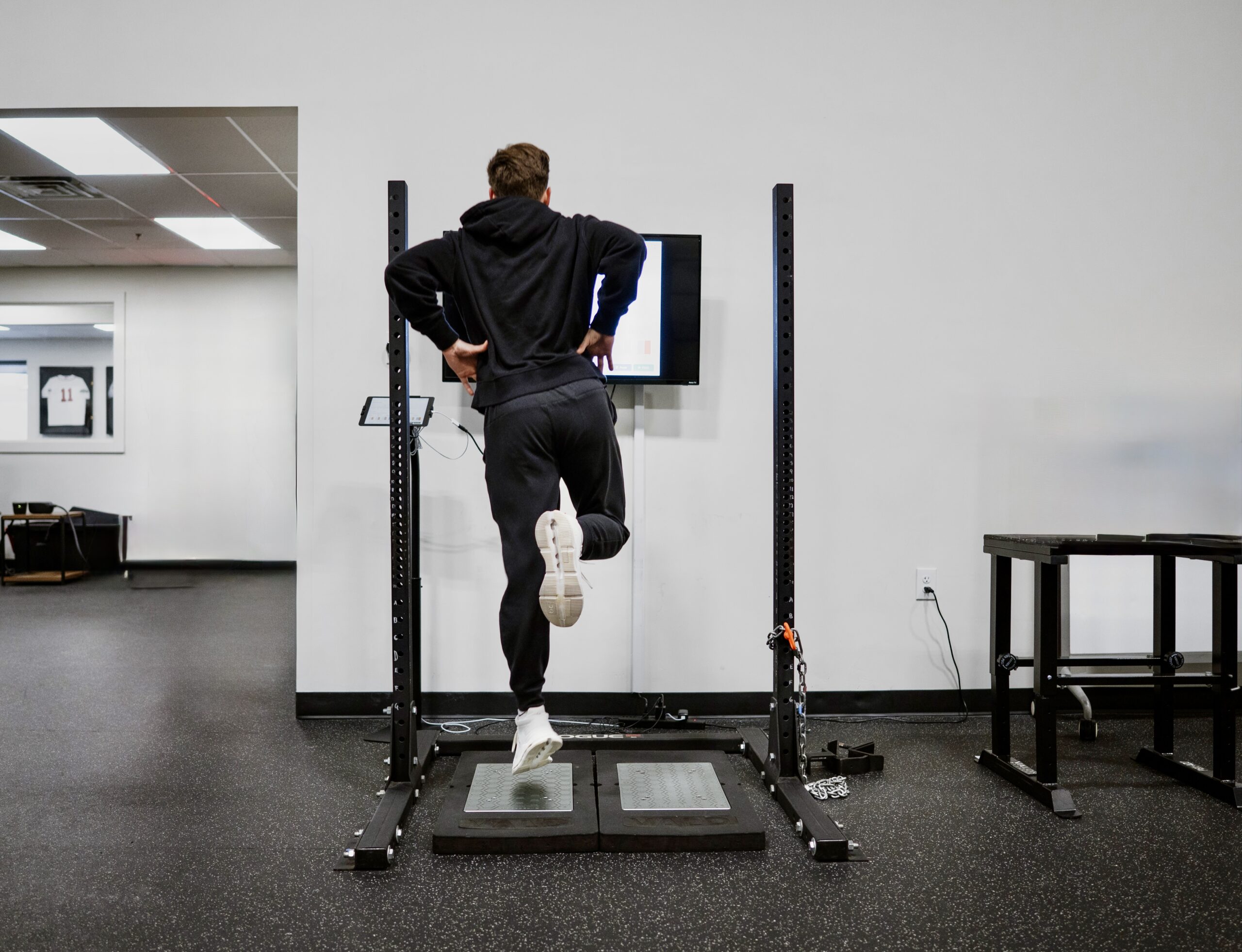
What Actually Changes After ACL Surgery
We recently had a staff meeting and were discussing our ACL Vald metric data and how we interpret it for our patients. As you know, there’s a ton of research on ACL rehab, but not a lot that actually tells you what’s happening week to week and across the full first year in the same…
-

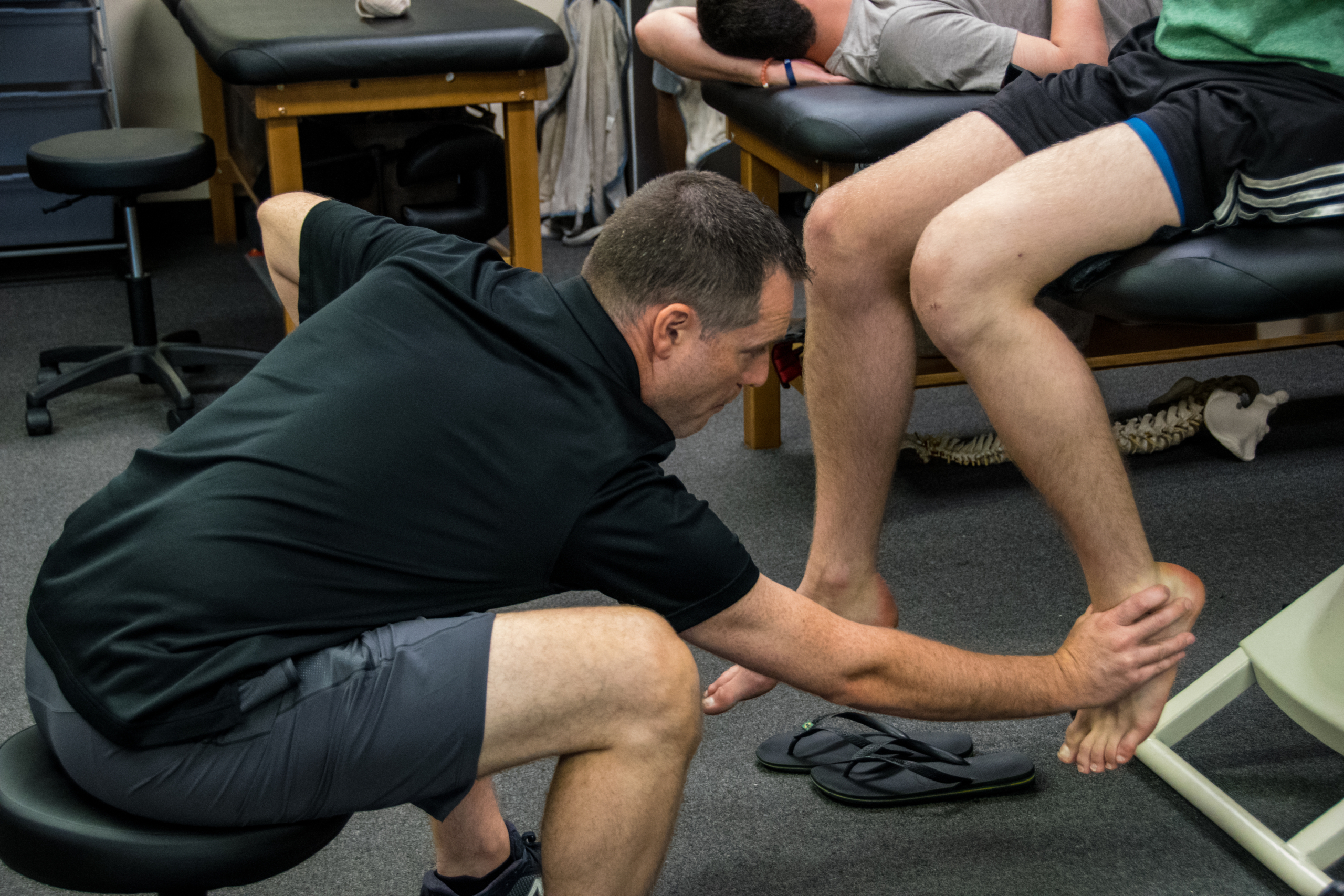
Why Your Quad Tendon ACL Patient’s Quad Is Lagging
By Lenny Macrina, MSPT, CSCS I’ve been publicly skeptical of the quad tendon (QT) graft for ACL reconstruction for a while now. If you follow me on social media, you’ve probably seen me lean toward the patellar tendon as my preferred graft, partly because of the outcomes data, partly because of what I see clinically…
-
Meniscus Repair Rehab: What Does the Evidence Actually Say?
By Lenny Macrina, MSPT, CSCS | Champion PT & Performance I get asked about meniscus repair rehab all the time. And honestly? It’s one of the most variable, most debated areas in sports PT right now (besides ACL graft choices haha). Depending on which surgeon you’re working with, you might see wildly different protocols —…
-
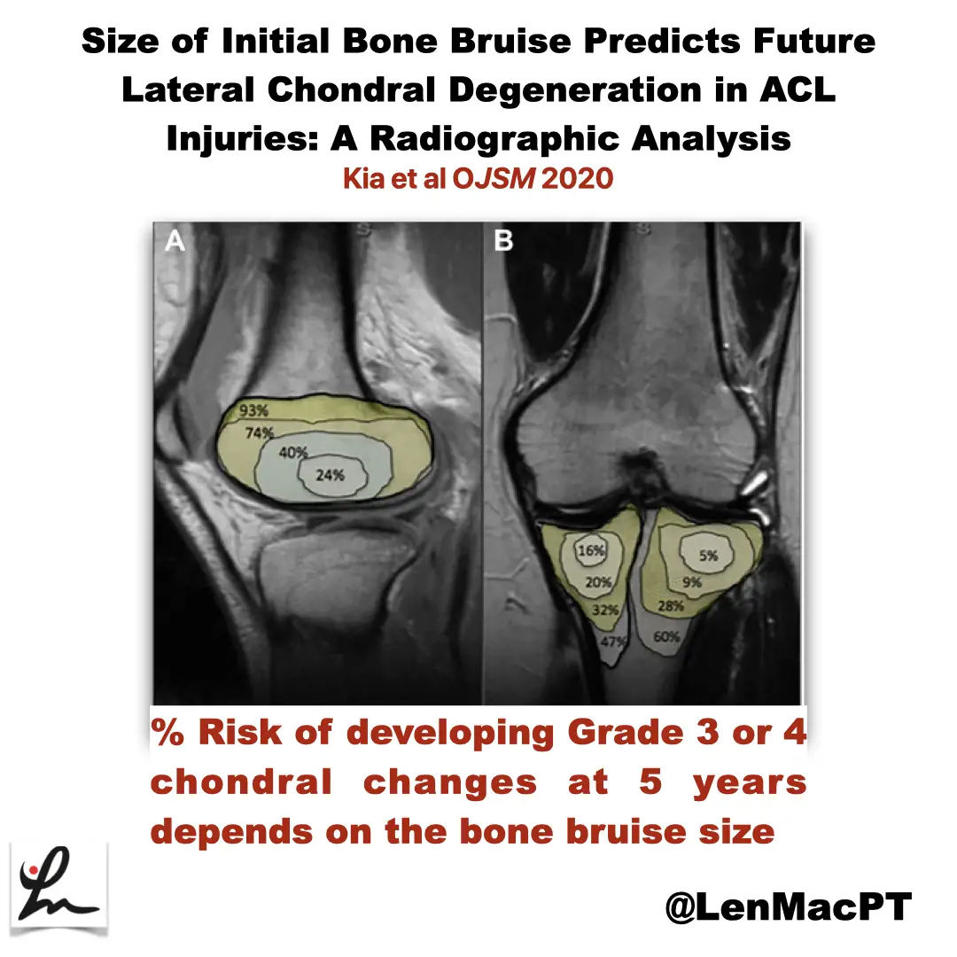
ACL tears and bone bruises
—
by
Research Review Not sure if you saw my recent post on social media about bone bruises after an ACL tear so I wanted to discuss it further here. In this study, the authors looked at the incidence of radiographic chondral changes (without correlation with clinical and functional outcomes) on MRI 5 years after the ACL…
-

ACL Volume Changes over a Women’s Soccer Season
I’m a bit interested, confused and looking to seek more on this open access paper that just came out in March of 2019 looking at the effects of season-long participation on ACL volume in female intercollegiate soccer athletes. The title of the paper is: “Effects of season-long participation on ACL volume in female intercollegiate soccer…
-
Why you need “Feel” as a Physical Therapist
Just thinking…some recent observations of first hand experiences from the field of physical therapy.
-
The Week in Research Review, etc 12-24-18
The Week in Research Review, etc 12-24-18 only had two posts to social media this week but hopefully two very helpful posts for your practice. The back pain post was a repost from a previous time but I thought it was very important to share it again. I also put a new post from my…
-

The Week in Research Review, etc 12-10-18
—
by
This week we’re still playing with formats and learning these Instagram changes. With that, in the week in research review 12-10-18, we discussed many topics that I wanted to share! Surgery vs Physical Therapy for Carpal Tunnel Syndrome View this post on Instagram A post shared by Lenny Macrina MSPT, SCS, CSCS (@lenmacpt) on Dec…
-
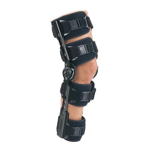
Knee Bracing Immediately After an ACL Reconstruction
I recently came across a Facebook post that discussed bracing immediately after an ACL reconstruction and I was intrigued. I read some of the comments and chimed in with my observations and opinions. In turn, a multi-platform discussion revealed many new details. I wanted to briefly share some of the research and the discussions that…
-
The Week in Research Review, etc 12-3-18
—
by
Hey everyone, The Week in Research Review, etc for this week has a new look, compliments of Instagram’s new algorithm. Hope the new format doesn’t throw you too big of a curveball (maybe you’ll like it better), so here goes… ACL Injury Rates Higher on Synthetic Turf than Natural Grass in the NFL Preventing…