Tag Archive for: physical therapy

The Week in Research Review, etc 10-29-18

This week we started the week off with a couple shoulder posts, specifically the rotator cuff and SLAP tears. As usual, I can’t resist a good ACL paper so included that NM control program that should be in all knee patients’ programs. We ended the week with a recorded knee scope as the surgeon was mobilizing the patella. It was a very informative and fun way to see the patella. We closed the week off with an old school video of myself performing a proprioception drill for the shoulder. I recommend you read these posts and like them on Instagram. Take a look at The Week in Research Review, etc 10-29-18


  1. Topics on the Rotator Cuff including post-op
  2. Classifying SLAP tears
  3. Essential Components of a neuromuscular control program
  4. Live Patellar scope during mobilization
  5. Shoulder Proprioception Drill


Topics on the Rotator Cuff including post-op

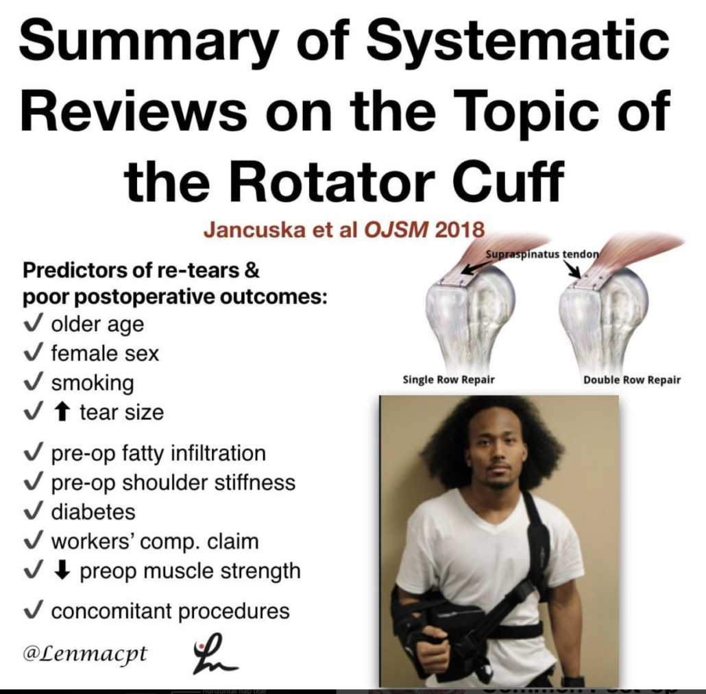
A Systematic Summary of Systematic Reviews on the Topic of the Rotator Cuff- Jancuska et al OJSM 2018

Nice summary of systematic reviews for you guys if you treat patients after a rotator cuff surgery. I’ve been doing a pretty good literature on the topic and wanted to share some of the articles that I have found helpful.

Their conclusions:

❇️There is substantial evidence indicating that the most accurate physical examinations for diagnosing RC tears are a positive painful arc and positive ER lag test

❇️Considerable evidence showing that rehabilitation is better than no rehabilitation for non-op management of RC tears, although RC repair was shown to be superior to rehabilitation alone.⠀

❇️No evidence to support the use of injections for nonoperative management of RC tears.

❇️Double Row repair results in better outcomes and fewer re-tears than Single Row repairs, especially for tears >3 cm.

❇️Predictors of re-tears and poor postoperative outcomes:⠀

✔️older age⠀

✔️female sex⠀

✔️smoking⠀

✔️increased tear size⠀

✔️preoperative fatty infiltration⠀

✔️preoperative shoulder stiffness⠀

✔️diabetes⠀

✔️workers’ compensation claim⠀

✔️decreased preoperative muscle strength⠀

✔️concomitant procedures.

Overall, a good review of the literature on rotator cuffs and anything associated.⠀


Classification of SLAP Tears

If you treat patients with shoulder pain, then you may run into different labral tears of the shoulder.

This post hopes to summarize the 10 different types of #SLAP tears that are currently known.

Type 1️⃣: Fraying but intact biceps

Type 2️⃣: Superior Labrum and biceps detached from the glenoid rim

Type 3️⃣: Bucket handle tear of the superior labrum but biceps anchor attached

Type 4️⃣: Bucket handle tear of the superior labrum that extends up into the biceps tendon

Type 5️⃣: BankartTear and also a detached biceps anchor

Type 6️⃣: an unstable flap of the superior labrum with a detached biceps anchor

Type 7️⃣: Anterior superior labral tear that extends to the middle Glenohumeral ligament; Biceps anchor detached

Type 8️⃣: Superior and posterior labral tear along with detached biceps anchor

Type 9️⃣: 360° labral tear

Type 🔟: Superior labral tear along with reverse Bankart tear and a detached biceps anchor.

That’s a lot and some are pretty rare but it helps to be able to communicate effectively with the medical team or to read an operative report.⠀


Neuromuscular training to reduce ACL injuries in female athletes

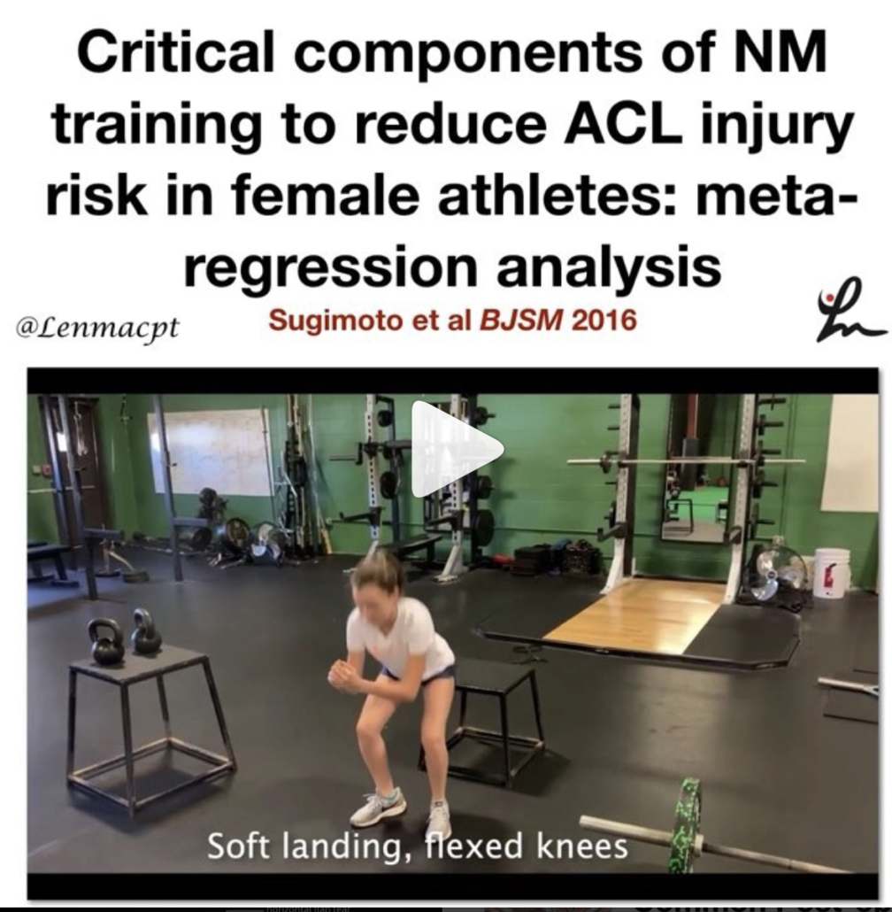
Critical components of neuromuscular training to reduce ACL injury risk in female athletes: meta-regression analysis. Sugimoto et al BJSM 2016.

This meta-regression analysis looked at the effects of combining key components in neuromuscular training (NMT) that optimize ACL injury reduction in female athletes.

They looked at a total of 14 studies that met the inclusion criteria of the current analyses. A total of 23 544 athletes were included.

They showed that there are 4 Key components

✅14-18 years old better than other age groups

✅2x/week for 30 minutes/session

✅Balance, planks, ‘posterior chain’ and plyometrics

✅Verbal cues like ‘Land softly’ or ‘Don’t let knees cave in’

Furthermore, inclusion of 1 of the 4 components in NMT could reduce ACL injury risk by 17.2–17.7% in female athletes. A great look that really specifics what age groups would best benefit from a NMT program. Do you incorporate any of these key concepts into your programs, even 1-2 of them?

I know I try to with most of my clients, whether or not they’re returning from an ACL or not.


Patella mobility during a knee scope

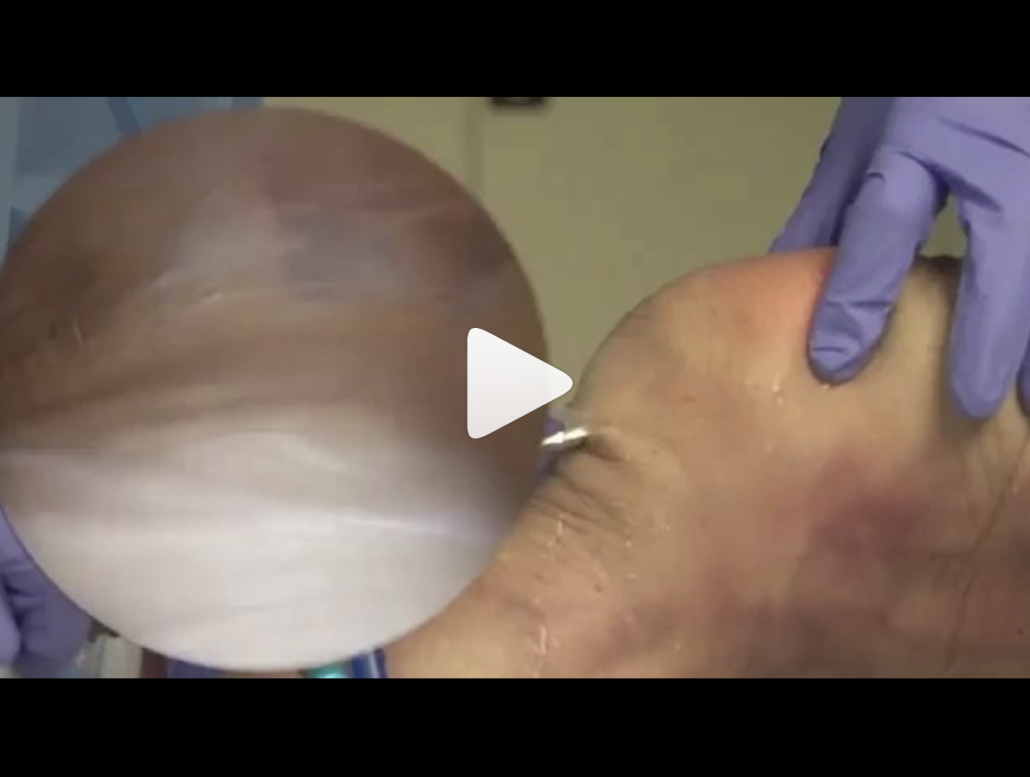
Great video by @physionetwork looking at the patella during a knee scope. This stuff is just exciting to see (in my opinion) because it gives us a little bit of insight into what is exactly going on during a patella mobilization.

In my opinion, the PF joint is often overlooked when it comes to knee surgery and it can affect joint mechanics, quadriceps activation and patient function. You need to mobilize the patella and normalize the motion…can’t stress this enough!

Check out the post below…good stuff!

Patellar mobilization is important to avoid stiffness after surgery. In this video, you can see from an arthroscopic view that little motion outside the knee, translates into a significant motion inside the knee. Mobilization may help prevent the formation of scar tissue and allow for better biomechanics of the knee joint.

We review the latest and most clinically relevant research in physiotherapy. Click link in bio to learn more and boost your knowledge 🔗

Video by Jorge Chahla, MD, PhD – Orthopaedic Surgeon -Sports Medicine Specialist


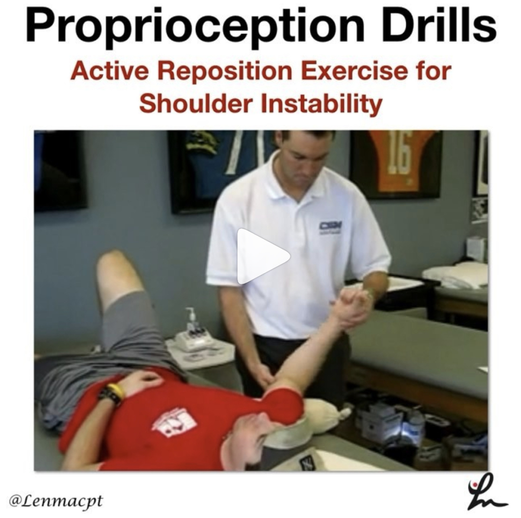
Active Reposition Drill after a Passive Motion

Loss of proprioception after a shoulder injury has been documented numerous times in the literature and can affect long-term function.

This drill may help the rehab specialist to test proprioception by measuring the exact active position difference that the patient attains.

You can also use this drill as a treatment reproduce the exact position that you passively brought them into.

Give it a shot and see what you think…you can use this drill for any joint in which you have assessed proprioception loss.

 

The Week in Research Review, etc 10-22-18

That was a milestone week as my Instagram account finally hit 10k followers, whatever that means! I’ve really been pushing a daily post to help other rehab professionals better simplify the research. One milestone hit but I still want to keep publishing good quality research reviews. The Week in Research Review, etc 10-22-18 included:

  1. Do baseball Pitchers really have a tight posterior capsule?
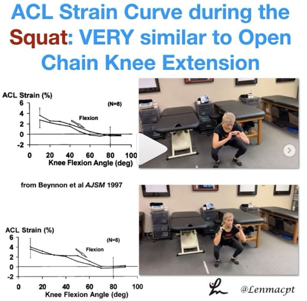
  2. ACL strain curve during the squat
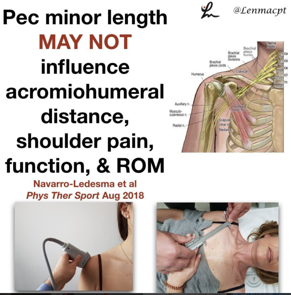
  3. Does the pec minor length influence shoulder pain?
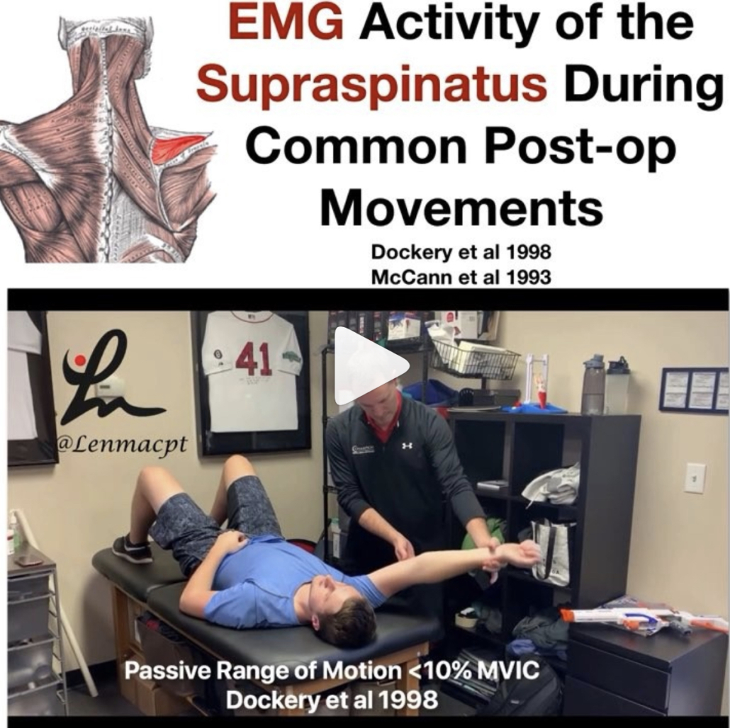
  4. What does the literature say about the EMG activity of the rotator cuff, particularly of the supraspinatus, with ROM
  5. Classification of Meniscus Tears and Osteoarthritis

Do baseball Pitchers really have a tight posterior capsule?

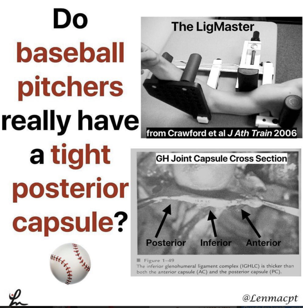
My guess is emphatically no based on what I see on a daily basis, the general anatomy of the glenohumeral joint and some research studies.

Anatomy
1️⃣When I stretch a baseball pitcher’s shoulder, it is usually very mobile. I find this in both symptomatic and asymptomatic individuals.

If I try to assess their posterior capsule with a joint play technique, I can often translate the humeral head pretty far over the glenoid rim. Sometimes, I can even sublux the humerus!

2️⃣Anatomically, the posterior capsule is relatively thin compared to the anterior and inferior capsule (see the post).

In general, that capsule is thinner probably because of the glenoid position that is not strictly in the frontal plane.
Because of that, it is theorized that the capsule evolved to have less of a role in stabilizing the humerus.

3️⃣There are a couple of research studies that have specifically looked at baseball pitchers to determine their humeral head translation.

Borsa et al AJSM 2005 reported that posterior translation was actually greater than anterior translation in both the dominant and non-dominant shoulders of professional baseball pitchers.

Crawford et al J Ath Train 2006 found no significant differences in posterior glenohumeral laxity and stiffness between the throwing and non-throwing shoulders.

I understand why the theory exists and think it could be plausible but just don’t think it’s truly responsible for what we think.

We just don’t think we can stretch the posterior capsule with any joint mobilization or contract-relax procedure, including a sleeper stretch. I often giggle at all of those MD prescriptions that say ‘#GIRD, posterior capsule tightness’. I just treat what I find on my examination and ignore the script.


ACL strain curve during the squat

As you can see, the strain curve from the Beynnon et al study is very similar to the strain curve during resisted knee extension in a full ROM (90-0).

We argue all of the time about anterior tibial translation during the open chain exercises but often ignore the other side of the story.

The strain on the ligament is barely 4%, which is in line with many functional activities like walking, descending steps, etc. The argument that we’re going to stretch the ligament out just has not been proven in the literature.

I wanted to show the closed chain strain curve so you could compare it to the open chain strain curve. I know the n=8 argument is present but we really don’t have much more data on the ligament in vivo that shows the true effects of open vs closed chain exercises on the ACL.

Again, as @barbhoogie mentioned, you need to monitor the PF joint, especially after a patella tendon autograft but as long as we’re not aggravating that joint, then I begin early 90-0’s and mini squats as tolerated.

Do you agree with this? Do you prevent squats early on during the ACL rehab process? If you don’t, then why do you hold back on full active knee extension exercises?


Does the pec minor length influence shoulder pain

Does the pectoralis minor length influence acromiohumeral distance, shoulder pain-function, and range of movement? Navarro-Ledesma et al Phys Ther Sport Aug 2018.

Their conclusion: Pectoralis minor length is not a distinguishing factor in shoulder⠀
assessment when a chronic condition exists, and it seems not to play a key role in pain perception and ROM.

54 participants with chronic shoulder pain in their dominant arm were recruited, as well as fifty-four participants with a pain-free shoulder.

The resting muscle length is measured between the caudal edge of the 4th rib to the inferomedial aspect of the coracoid process with a sliding caliper.

The acromiohumeral distance (AHD) was defined as the shortest linear distance between the most inferior aspect of the acromion and the adjacent humeral head, measured by ultrasound.

An interesting study that used an asymptomatic control group along with the contralateral shoulder of the symptomatic subject. A pretty clean study that is very interesting. I’m not going to say that the pec minor doesn’t play a role in shoulder pain but maybe its role is not as prominent as we think.

What do you think? Do you find pec minor length has a substantial role in your patients with shoulder pain?⠀


EMG of the rotator cuff during rehab exercises

What does the literature say about the EMG activity of the rotator cuff, particularly of the supraspinatus, with ROM?

Many PT’s and doctors are uncertain when to safely begin physical therapy after a shoulder surgery, particularly after a rotator cuff repair. In my 15+ years as a PT, I’ve seen docs begin PT post-op day 1 or wait as long as 6 weeks (which drives me bonkers!!)

In this snippet that I’ve taken from an upcoming blog post at LennyMacrina.com. I discuss the research that’s helping to guide best practice, in particular, the research that looks at PROM and AAROM and how much EMG activity is actually going on in the supraspinatus with each movement.

As you can see in the video, there’s minimal supraspinatus activity (<20% is considered minimal) for all motions. Keep in mind, many of these studies are done on healthy individuals but who in their right mind would volunteer their newly repaired RTC repair to have fine-wire EMG done on them?

So, I can only draw my conclusions from a limited body of evidence and my own anecdotal evidence (which consists of 12+ years of immediate PROM POD 1). Many still think it’s safe to get a RTC repair patient’s shoulder moving early for many reasons that I will describe in this blog post.

I just wanted to get this early point out there to get another discussion going. I think our patients can do much better after a RTC repair and this is one of the reasons.

Do you agree? Do you advocate for early PROM after a RTC repair, especially a small-medium repair?


Classification of Meniscus Tears and Osteoarthritis

Great post by @physicaltherapyresearch talking about the various types of meniscus tears. Nice visual & description of each type and the incidence of OA. Take a look! 👇🏼
_______________
Meniscus Tears and Osteoarthritis

💡

Prevalence of meniscal tears is estimated as ~24-31% of some populations, increasing with age and ranging from 19% in women aged 50–59 years to 56% among men between 70 and 90 years and is markedly higher in established OA subjects.
💡

Medial meniscus and/or the posterior horn tears make up 66% of cases, with horizontal and complex tears being the most common.
💡

Most subjects with a meniscal tear are asymptomatic.
💡

Regardless of morphologic type, meniscal tears are strongly associated with OA cross-sectionally and predict OA longitudinally and are considered to be part of the spectrum of early or pre-radiographic disease

📝📝📝

TEAR TYPES INFO:

Often enough, meniscal tear types are categorized into varying groups for comparison rather than separately compared to each other.
📝

There is a striking lack of data on the relevance of different morphologic types of meniscal tears in OA.

📝

Horizontal and complex tears are common findings in knees with OA

📝

Posterior radial tears of the medial meniscus are associated with a high degree of cartilage loss and meniscal extrusion, and appear to be a highly relevant event in the progression of OA in the knee. 📝

Lateral meniscus radial tears affect younger individuals and are considered post-traumatic.

📝
Despite their suggested high relevance, radial tears are more commonly misdiagnosed on MRI than any other type of tear.

📝

While medial meniscus posterior root tears are of “radial” morphology, there is growing interest in regarding them as a separate entity.
📝

Longitudinal and bucket handle tears affect younger individuals and are highly associated with ACL injuries, favoring a traumatic etiology.
📝

MRI is important to detect and locate a possible displaced tear.
📝

Further epidemiologic studies should focus on the morphology of specific meniscal tears to better understand their relevance in the genesis and progression of knee OA.
📚📚📚
SOURCE:
Jarraya et al. 2017 Semin Arthritis Rheum


The Week in Research Review, etc 10-8-18

Hey all, the Week in Research Review, etc 10-8-18 has some great articles that really got some good discussion going. I highly recommend reading each post and chiming in. Looking forward to the new comments and discussions!

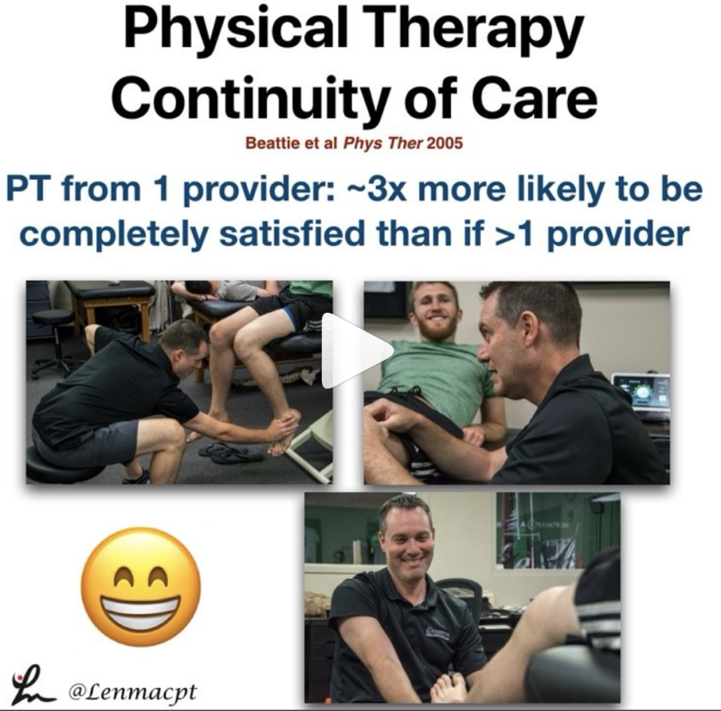
  1. PT Continuity of care
  2. Fatigue effects on ACL tears
  3. Measuring IR in a baseball pitcher
  4. Lever sign to diagnose an ACL tear
  5. Immediate or delayed ROM after a rotator cuff repair


Longitudinal continuity of care is associated with high patient satisfaction with physical therapy. Beattie et al Phys Ther 2005.

I saw a FB post the other day and it reminded me of a study that I had seen about continuity of care and physical therapy.⠀

This study looked to provide ‘preliminary information regarding the association between longitudinal continuity and reports of patient satisfaction with physical therapy outpatient care.’

What they showed was “Subjects who received their entire course of outpatient physical therapy from only 1 provider were approximately 3x more likely to report complete satisfaction with care than those who received care from more than 1 provider.”

All too often, I hear my current clients talk about their past PT sessions and often complain about seeing a tech/aide or a different PT for each session.

I always thought that was such a wrong concept for the client. Throughout my career, I have strived to connect with each client in an attempt to help them overcome an injury.

We did this at @championsportsm in Birmingham and we do it now in Boston at @championptp.

It is such a game changer for the client when they have complete faith in their treatment, can connect with their PT and their PT can connect with them.

Just my little soapbox rant on continuity of care. Are you able to maintain a good continuity of care with your patients or are you constantly sharing and/or just doing evals?

Tag a friend or colleague who may benefit from this post…thanks!⠀


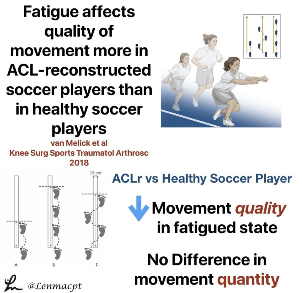
Fatigue affects quality of movement more in ACL-reconstructed soccer players than in healthy soccer players. van Melick et al Knee Surgery, Sports Traumatology, Arthroscopy 2018.

This study looked at the influence of neuromuscular fatigue on both movement quantity and quality in fully-rehabilitated soccer players after ACLR and to compare them with healthy soccer players.

They showed ACL-reconstructed soccer players had a significantly decreased performance when comparing the non-fatigued with the fatigued state.

For movement quantity, they used a single-leg vertical jump, a single-leg hop for distance, and a single-leg side hop.

For movement quality, they used a double-leg countermovement jump with frontal and sagittal plane video analyses. The Borg Rating of Perceived Exertion (RPE) scale was used to measure fatigue after a soccer-specific field training session. In addition to soccer-specific drills, exercises focussing on speed, stability, and coordination were included in this session.

Seems like a pretty neat study that may help to show us that the fatigued state influences quality of movements and not the quantity of movements. I know Tim Hewett has said that there’s no evidence that fatigue influences ACL tears but maybe this study is the 1st step.

Do you agree with this study? Anecdotally it makes sense but there’s little evidence to support the notions.⠀


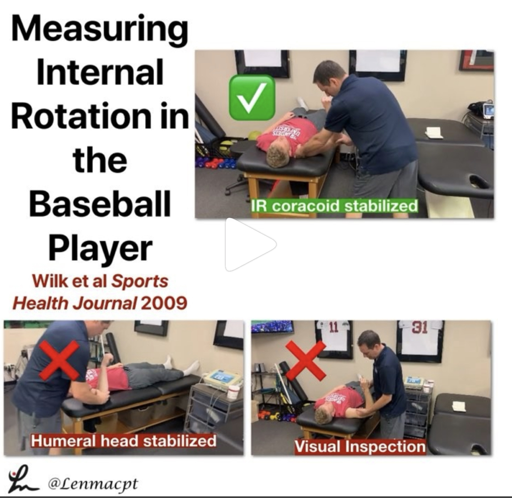
Measuring internal rotation in the baseball player

If you treat baseball pitchers, then you should have a good understanding of how to measure internal rotation of the shoulder joint.

Measuring internal rotation of the shoulder is one part of the equation when obtaining total rotational range of motion (TROM). Total rotational range motion is the sum of external rotation plus internal rotation. I use this equation weekly, if not daily when assessing my baseball players’ shoulders.

In a study in 2009 Sports Health Journal titled “Glenohumeral internal rotation measurements differ depending on stabilization techniques”, we looked at 3 different ways to measure IR. We determined that the scapula stabilized method had the best intra-rater reliability.

We also felt this was the best method to measure pure internal rotation of the glenohumeral joint.

Is this how you measure IR in your baseball pitchers? Do you consider TROM when making treatment recommendations?

Let’s talk it out and discuss the concept of TROM and how to measure it.


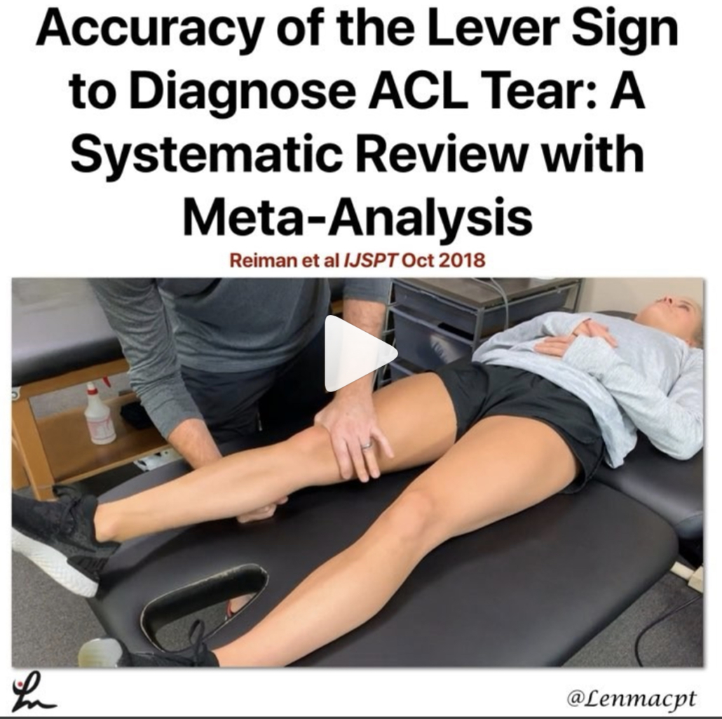
Accuracy of the Lever Sign to Diagnose Anterior Cruciate Ligament Tear: A Systematic Review with Meta-Analysis. Reiman et al IJSPT Oct 2018

This study was a systematic review with meta-analysis that hoped to summarize the diagnostic accuracy of the Lever sign for use during assessment of the knee for an ACL tear.

They showed that based on limited evidence, the Lever sign can moderately change posttest probability to rule in an ACL tear.

I’m a bit surprised by the limited studies because I’ve had a more difficult time getting consistent results compared to the Lachmans test (definitely my go-to!).

For those not familiar with the Lever test, it was 1st published by Dr Lelli in Knee Surg Sports Traumatol Arthrosc. 2016.

From the review, ‘The test requires the evaluator to place their fist under the calf muscle to create a “fulcrum” extending the knee while applying a moderate downward force to the distal part of the femur.

In an intact knee, the ACL completes a lever mechanism, making the heel rise in response to the force applied to the femur. In an ACL-deficient knee, the heel does not rise indicating a positive Lever sign.’ I have personally struggled to get consistent accuracy using the test. My results have been inconsistent with MRI results.

I’ve also struggled to do the test on a plinth that has padding and often have patients lie on a firm surface like the floor (which is very weird) in order to get a better test result.

Some people are freaked out by the method of the test. The clinician has to apply force to the knee in order to create the fulcrum. Many have not liked that force applied to the knee.

In general, this is not my go-to for a suspicious ACL tear. I have tried and still ty to use it but my results have been less than stellar.

Have you used this test for an ACL tear? Do you like it to supplement your Lachmans?


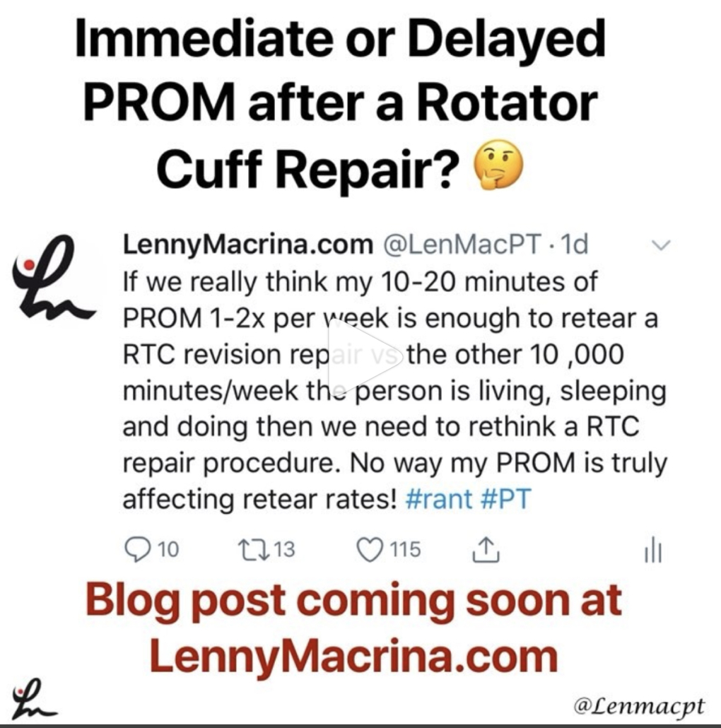
Should we delay PROM after a rotator cuff repair?

It seems as if we’re all over the place, which usually says the research is not cut and dry. There are so many factors that are considered when trying to figure out the best time to initiate motion.

I’m not talking active ROM or strengthening…I”m talking about passive ROM by a rehab specialist like a #PT#OTor #ATC. Obviously, the docs weigh in heavily with this decision. I feel as if patients are restricted for the wrong reasons and could potentially begin PT earlier than we often see.

This is going to be a beast of a blog post and may alter my thinking, we’ll see.

As of now, I fully embrace immediate PROM for most post-op rotator cuff repairs, including Large and Massive repairs.

For revisions, we may need to think it through but I still feel as if most benefit from early PROM. We did it for years and with very good results during my time in Birmingham but feel as if maybe the pendulum is swinging in the conservative direction (for the wrong reasons).

What do you guys do? Do you have any input with your docs and can influence their rehab decisions? Let’s talk it out now and get prepped for my blog release in the coming days, weeks, months…whenever I can make it the best!⠀


Documenting Knee Extension Range of Motion

I’ve talked a lot about the importance of regaining knee extension range of motion (ROM) after a knee injury or surgery. In this post, I want to talk about how exactly I believe we should be documenting knee extension range of motion.

I think it’s important because I hear many other medical professionals and students document differently. Hope this post clears the air and gets everyone on the same page.

Knee Extension after ACL

I’ve written about getting knee extension back after an ACL and how to figure out if it was a cyclops lesion or not. You can read this recent post if you like..it should help you gather more information on diagnosis and treatment of a cyclops lesion.

Therefore, I can’t stress the importance of obtaining not just knee extension, but symmetrical hyperextension after a knee injury or surgery.

With that, I feel as if many practitioners are all over the place with their documentation. This makes it difficult to communicate with each other and with the patients.

Documenting Knee Extension Range of Motion

In this video, I discuss the rationale for how I document knee hyperextension. I think it;’s important that we’re all on the same page to avoid confusion.


Does this make sense to you? Is this how you document knee hyperextension?

Let’s discuss in the comments section or on social media. You can find me on Twitter or Instagram @lenmacPT.


If you want to learn more about how I treat ACL’s or the knee in general, then you can check out our all online knee seminar at www.onlinekneeseminar.com and let me know what you think.

We cover the anatomy, rehab prescription, ACL, meniscal injuries knee replacements and patellofemoral issues. Furthermore, the course covers both the non-operative and post-operative treatment.t

This is an awesome course if you’re interested in learning more about rehabilitating the knee joint. And if you’re a PT, there’s a good chance you can get CEU’s as well.

The Week in Research Review, etc 8-19-18

We posted a lot of information this week to review so hopefully you were able to keep up with it all. If not, here’s a bunch of it from the week. Check it out and comment as you want. Lots of good information on:

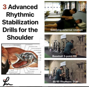
  1. Advanced Rhythmic Stabilization Drills
  2. Our ACL rehab paper from 2012
  3. PT usage for Frozen Shoulder
  4. Comparison of posterior shoulder stretching techniques
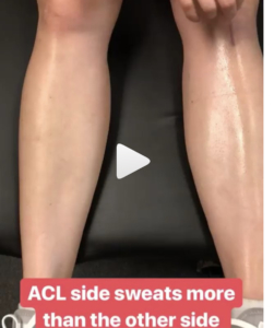
  5. Increased Sweating after an ACL surgery? Seems pretty common in the comments section
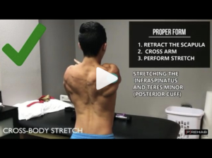
  6. @theprehabguys show how to do a posterior shoulder stretch


Advanced stabilization drills

These advanced stabilization drills are a great way to progress the patient once they’ve shown improved neuromuscular control with the basic drills from the other day.

I use these for most of my clients that need improved dynamic stability, especially those hypermobile athletes that play baseball, swim, or do gymnastics.

You can modify the speed and amount of force that I give during the drill based on how they are responding that day and how they have progressed overall. You can also increase the time of the drill to bring in an endurance component, as well.

The goal is to use these to prep the shoulder for higher level activities such as plyometrics, throwing, etc.

I want the shoulder joint to be as strong as possible. And most importantly, I want the client to perceive that their shoulder is stronger so that they are more confident!

Do you use these rhythmic stabilization drills with your patients?

Tag a friend who may benefit from these drills and try them on your patients the next time they’re ready to progress their dynamic stability drills!


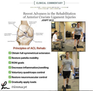
Recent advances in the rehabilitation of anterior cruciate ligament injuries. @wilk_kevin et al JOSPT 2012 @drlylecain @dugasmd1

We wrote this paper in JOSPT to help clinicians better understand the rehab involved after their patient undergoes an ACL reconstruction.

The paper discussed the principles of ACL rehab, including:
✅obtaining full symmetrical extension
✅restoring patella mobility
✅ROM goals
✅Decrease inflammation/swelling
✅voluntary quadriceps control
✅restore neuromuscular control
✅Gradually apply loads

We also discuss special rehab implications for:
✅The Female Athlete
✅Concomitant injuries (MCL, meniscus, LCL, cartilage lesions)
✅Implications of Graft type

Our Accelerated ACL-PTG protocol is included to help guide the rehab process and give the rehab specialist some goals to achieve.

Again, a protocol is just a guide and by no means meant to place aggressive limitations on a patient.

Hope this paper helps you to better understand ACL rehab. Tag a friend or colleague who may benefit from this post.


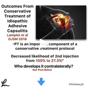
Outcomes From Conservative Treatment of Shoulder Idiopathic Adhesive Capsulitis and Factors Associated With Developing Contralateral Disease Lamplot et al 2018 OJSM 2018

A minimum 2-year follow-up of patients diagnosed with idiopathic adhesive capsulitis.

They were treated with a single intra-articular glenohumeral injection of local anesthetic and corticosteroid as well as 4 weeks of supervised PT.

Physical therapy reduced the use of a second injection as part of treatment. Contralateral disease was more likely in patients with diabetes and those younger than 50 years.

PT is an important component of a conservative treatment protocol, as PT decreased the likelihood of receiving a second injection from 100% to 27.3%.

In my opinion, a cortisone injection + PT is the best treatment ‘cocktail’ for someone with frozen shoulder, particularly in the freezing phase.

Do you guys agree? Let’s discuss this very debilitating pathology. Tag a friend who may want to discuss this further.


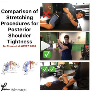
A Randomized Controlled Comparison of Stretching Procedures for Posterior Shoulder Tightness McClure et al JOSPT 2007

This study looked to compare changes in shoulder internal rotation range of motion (ROM), for 2 stretching exercises, the “cross-body stretch” and the “sleeper stretch,” in individuals with posterior shoulder tightness.

From their results, “The improvements in IR ROM for the subjects in the ✅cross-body stretch group (mean ± SD, 20.0° ± 12.9°) were significantly greater than for the subjects in the control group (5.9° ± 9.4°, P = .009). The gains in the ❌sleeper stretch group (12.4° ± 10.4°) were not significant compared to those of the control group (P = .586) and those of the cross-body stretch group (P = .148).”

Just be aware of the large standard deviations and low number of subjects…plus the subjects were all asymptomatic.
With that, I still believe clinically that the cross-body stretch is one of the better stretches for the shoulder. I have gotten away from the sleeper stretch because I don’t believe the risk/reward presents favorably.

✅✅I personally prefer the supine horizontal adduction stretch with the scapula stabilized. I feel like this best isolates the posterior soft tissue (mainly muscle, in my opinion) and that’s my target tissue.

Do you use the sleeper stretch with your patients or do you prefer the cross-body stretch?

Comment below and let’s talk about it. Also, tag a friend who may benefit from this post…thanks!


Sweating Leg after an #ACL

Had this in my story and posted to Twitter and got some pretty good discussion going. Is this some altered autonomic nervous system response after #ACLsurgery? I’ve seen it a bunch, where the area inferior to the incision sweats significantly more than the contralateral leg. Anyone else see this phenomenon and have an opinion? Have had people say that maybe it was compartment syndrome or CRPS but it happens with many many ACL patients so it seems very normal. What do you think? #ACL #ACLrehab


Horizontal Adduction Stretching

Perfect timing by @theprehabguys with their horizontal adduction post from the other day. Blends perfectly with the McClure 2007 et al study I posted the other day that showed horizontal adduction is the best way to obtain internal rotation mobility at the shoulder. I often tell people to wedge their lateral scapula against a wall to help stabilize but this way may also benefit them. Check out their post below!⠀
👇🏻⠀
Are You Performing The Arm Across Body Stretch Correctly⁉️⠀
[How to Stretch Your Posterior Cuff]

A tight posterior cuff is associated with a handful of shoulder dysfunctions like subacrominal impingement syndrome, posterior impingement, anterior instability, etc. And thus, ⬇️ tone/increasing extensibility of the posterior cuff is part of the treatment protocol for many with shoulder pain. The cross-body stretch is a fantastic way to target the posterior cuff but far too often it is done INCORRECTLY.

‼️In order to effectively stretch the posterior cuff, you need to keep your SCAPULA STABILIZED ie your scapula CANNOT MOVE!‼️

❌If you pull your arm across your body and your scapula comes with it into horizontal abduction, the only stretch your getting is of your mid-scapular muscles like your rhomboids or traps. Furthermore, in this position there’s more of a distraction force on the glenohumeral joint than a true stretch of the posterior cuff – aka not as specific as it can be.

✅First pull your shoulder blades back. This will keep your scapula in a retracted position. Only WHILE MAINTAINING THE POSITION OF YOUR SHOULDER BLADES BACK can you effectively target the posterior cuff. Pay attention to WHERE you feel the stretch, as you should feel a “deep stretch” in the back of your shoulder in the highlighted area on the video. If you feel a stretch or anything else not in the back of the shoulder, you’re either doing the stretch incorrectly or abutting other structures in your shoulder due to pathology (ie don’t do the stretch anymore and seek out a physio if you’re in pain).

Try it out and let us know how it feels! Tag a friend who NEEDS this stretch! #shoulderstretch #posteriorcuff#posteriorcapsule


The Week in Research Review, etc 7-29-18

Last week was the 1st of my research review that summarized my social media posts from the previous week. It seemed to be well received so I figured I’d continue it. My goal is to help summarize some of the research that I found interesting and package it nicely for my readers.

Each photo contains a link back to a social media feed where you can see the conversation that ensued and maybe chime in…or just be a passive reader and see where the conversation went. You never know where the conversation may go on social media…so be ready! haha!


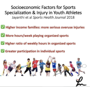
Socioeconomic Factors for Sports Specialization and Injury in Youth Athletes Jayanthi et al Sports Health Journal 2018.

This study looked at the effect of socioeconomic status (SES) on rates of sports specialization and injury among youth athletes.

They looked at injured athletes between the ages of 7 to 18 years that were recruited from 2 hospital-based sports medicine clinics. They compared these with uninjured athletes presenting for sports physicals at primary care clinics between 2010 and 2013.

They concluded that:
✅High-SES athletes reported more serious overuse injuries than low-SES athletes
✅More hours/wk playing organized sports
✅Higher ratio of weekly hours in organized sports to free play
✅Greater participation in individual sports

I applaud the authors for attempting to bring this very difficult collection of data into a formal research paper. I will say some of the statistics and standard deviations may not make the conclusions as powerful.

I do think this is a good paper to help educate our athletes on injury rates, especially in those that specialize in 1 sport.

What do you think? Tag a friend that may benefit from this article!


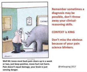
From #Twitter’s @retlouping that perfectly sums up what I’ve observed recently on social media with many PT’s.

For some reason, pain science has overtaken most diagnosis and treatment conversations.

It’s as if you get bullied into talking pain science and ignoring our clinical judgment and diagnosis skills. I understand there’s a constant tug-of-war between the biomechanical PT’s and the pain science PTs.

But as usual, the answer usually lies somewhere in between and both groups are correct. The biomechanics of an injury are often important as well as the language we use to explain these tissue biomechanics.

To my fellow clinicians, especially the newer grads and #dptstudent, remember this little cartoon for every future encounter.

Yeah, speak to people in non-threatening tones (in my world it’s just being respectful) but trust me, they WANT to hear what could be going wrong or what may be causing their pain.

Don’t blow off their symptoms and don’t go into depth about pain science because they won’t understand.

Trust me, the clinicians that try to do that often end up losing their patients in the long run.

I hear these stories day after day of people coming to me because the last PT either only talks to them or made them ONLY do strength exercises and it didn’t help their pain.

The PT didn’t listen to them and was so blinded by their pain science background that they ignored the person sitting right in front of them. Remember, the person sitting there will tell you what is going on and what treatment will most help them feel/move better.


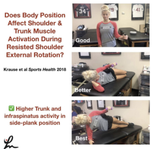
Influence of Body Position on Shoulder and Trunk Muscle Activation During Resisted Isometric Shoulder External Rotation Krause et al Sports Health 2018.

The purpose of this study was to examine ER torque and electromyographic (EMG) activation of shoulder and trunk muscles while performing resisted isometric shoulder ER in 3 positions:
✔️Standing
✔️Side-lying
✔️Side plank

Using surface EMG and a hand-held dynamometer, the researchers tried to determine EMG activity of the:
✔️infraspinatus
✔️Posterior Deltoids
✔️Mid traps
✔️Multifidi
✔️External/internal obliques (dominant side)
✔️External/internal obliques (non-dominant side)

EMG values for the infraspinatus were greatest in the side plank position. In general, EMG values for the trunk muscles were also greatest in the side plank position.

✅Their Conclusions: If the purpose of a rehabilitation program is to strengthen the rotator cuff, in particular, the infraspinatus, the side plank is preferred over standing or side lying. If the goal is to simultaneously strengthen both the rotator cuff and trunk muscles, the side plank position again is preferred.

Makes sense but good to see the research and have concrete evidence to back up what we think actually goes on.

Tag a friend who may be interested in this research paper!


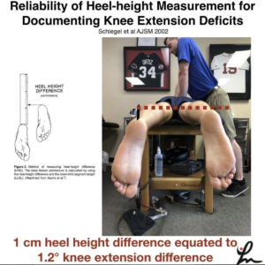
Reliability of heel-height measurement for documenting knee extension deficits. Schlegel et al AJSM 2002

Prone heel-height difference of 1cm equates to 1.2 degree difference in knee extension ROM.

Do you use this method to assess knee ROM? I still measure knee extension ROM is supine but find this method helpful as well.

I know my friend and colleague @wilk_kevin has measured this way for many years. i originally saw his use this technique at @ChampionSportsM

I don’t want people to confuse this with prone hangs for knee extension ROM. I am not a fan of that method as I’ve stated in the past.

This is a method to assess knee extension differences, particularly after an ACL reconstruction. I have gone back to using this method for some people that have subtle ROM differences side-to-side.

The patella position (on the plinth or off) did not matter in the study and thigh girth did not appear to make a difference.

I would recommend stabilizing the pelvis to prevent excess ROM from occurring at that region and to better isolate the knee joint.

Have you tried this method? Tag a friend who may benefit from using this ROM method…thanks!


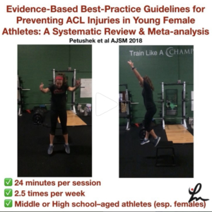
Evidence-Based Best-Practice Guidelines for Preventing #ACL Injuries in Young Female Athletes: A Systematic Review and Meta-analysis Petushek et al AJSM 2018.

Injury prevention neuromuscular training (NMT) programs reduce the risk for anterior cruciate ligament (ACL) injury.

Eighteen studies were included in the meta-analyses, with a total of 27,231 participants, 347 sustaining an ACL injury.

The overall mean training amount was 57 sessions totaling 18.17 hours (roughly 24 minutes per session, 2.5 times per week).

They concluded:

✔️Interventions targeting middle school or high school–aged athletes reduced injury risk to a greater degree than did interventions for college or professional-aged athletes.

✔️Continued exposure to neuromuscular training throughout the sport season seems to enhance prophylactic effects of NMT.

✔️NMT interventions were effective for female basketball, and handball athletes and interventions including various athletes were potentially effective (eg, soccer, basketball, and volleyball).

✔️ Interventions included some form of implementer training (eg, instructional workshop, video, or brochure) on proper program implementation.

✔️Programs including more landing stabilization and lower body strength exercises during each session were most effective.

🤔Programs including balance, core-strengthening, stretching, or agility exercises were no more effective than programs that did not incorporate these components.

✔️ Specifically, programs that included more landing stabilization exercises (eg, drop landings, jump/hop and holds), hamstring strength (eg, Nordic hamstring), lunges, and heel-calf raises reduced the risk for ACL injury to a greater degree than did programs without these exercises.

✅ Wow, lots of great information here. Please share this with a friend or colleague who may benefit from knowing this information.


Hope that helped to catch you up on my posts from this week.

Do you like these weekly updates? Let me know if I should continue…love your feedback!

Thanks for reading!

The Week in Research Review, etc 7-22-18

The Week in Research Review, etc 7-22-18

I’m trying out this new concept of publishing my social media posts into a nice package for a weekly delivery to my subscribers.

  1. Knee Case Study
  2. Contralateral ACL Strengthening
  3. Shoulder Static Stabilizers
  4. Weighted Ball Research
  5. Glute Activation


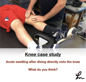
This kid came to me the other day with L knee swelling after sliding headfirst into 2nd base during a baseball game.⠀

Continued to play in the game and even pitched the next day, all without pain or loss of motion.⠀

As you can see from the video, he has a bunch of fluid in his knee, medial ecchymosis (bruising) but full pain-free ROM.

Ligamentous tests appear negative and he has absolutely no pain or stiffness with anything.

I took this video to show what appears to be a bursal sac disruption from the impact of his knee into the ground as he was sliding.

The mechanism fits the presentation and clinical exam.

I advised him to monitor his swelling, wear a knee sleeve and continue his activities per his tolerance.

He is going to touch base with me next week to make sure the fluid is dissipating (and not worsening) and he remains asymptomatic.

What do you think? Am I missing anything? What’s your diagnosis? Tag a friend who may be interested in this case.

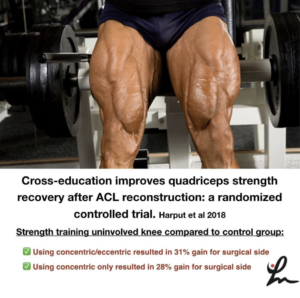
Cross-education improves quadriceps strength recovery after ACL reconstruction: a randomized controlled trial. Harput et al Knee Surg Sports Traumatol Arthrosc. 2018

This study looked at a group of ACL reconstructed patients that were divided into 3 groups.

All 3 groups performed the same standardized ACL rehab, but one group was the control group that performed the standardized rehab only.

The other 2 groups did either 3x per week extra concentric knee extensions on their uninjured leg for 2 months (beginning at 1-month post-op through 3-months post-op) or additional eccentric knee extensions on their uninjured leg 3x per week for 2 months between months 1-3 post-op.
💪🏼
They found that the quads strength for the concentric group was 28% greater compared to the control group. 💪🏼
The eccentric group was 31% greater when compared to the control group.

Conclusion: Concentric and eccentric quadriceps strengthening of healthy limbs in early phases of ACL rehabilitation improved post-surgical quadriceps strength recovery of the reconstructed limb.

Pretty crazy stuff and one more reason to work on bilateral strengthening with most of our patients, especially when they’re post-op ACL reconstruction.

Do you work on bilateral strengthening? if not, why? If you do, what other studies have you seen that show similar results?
Tag a friend who may benefit from this study or let’s discuss in the comments section!

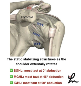
This picture shows a simplified view of the static stabilizers of the shoulder joint. I highly recommend reading a classic paper by Wilk et al 1997 JOSPT that talks about this and cites a paper from Bowen et al Clin Sports Med 1991 @wilk_kevin

When one is picturing these stabilizers, the superior glenohumeral ligament (SGHL) is most taut when the shoulder is externally rotated at 0 degrees of abduction.

As we progress to 45 degrees of GH abduction, we stress the middle glenohumeral ligament (MGHL) as we externally rotate the humerus.

Finally, at 90 degrees of GH abduction, we stress the inferior glenohumeral ligament (IGHL) as we externally rotate. More specifically, the anterior band of the IGHL.

As we internally rotate at 90 degrees of abduction, we stress the posterior band of the IGHL.

These concepts have rehab implications and should be kept in mind when we’re rehabbing people after an injury or surgery.

For example, if someone has an anterior Bankart lesion (front labral repair), then we need to progress them slowly into external rotation, especially at 45 and 90 degrees of abduction.

Another example would be a rotator cuff repair, like the supraspinatus. We would want to progress them slowly at lower degrees of abduction 0-45 degrees but maybe we can progress them a bit quicker at 90 degrees of abduction.

Hope these concepts make sense because they are very important to understand for many patients with shoulder injuries.

Does this make sense? Have you heard this info before? Tag a friend who may benefit from this post!

Effect of a 6-Week Weighted Baseball Throwing Program on Pitch Velocity, Pitching Arm Biomechanics, Passive Range of Motion, and Injury Rates. Reinold et al Sports Health Jul-Aug 2018. @mikereinold

Our 1st of potentially 3 research articles looking at the effects of weighted balls on youth baseball pitchers.

High school baseball pitchers performed a 6-week weighted ball training program.

Players gradually ramped up over the 6 weeks to include kneeling, rocker, and run-and-gun throws with balls ranging from 2oz to 32 oz.

🤔After 6 weeks, the weighted ball group did increase velocity by 3.3%, 8% showed no change, and 12% demonstrated a decrease in pitch velocity. Also of note, 67% of the control group also showed an increase in pitch velocity.⠀

The weighted ball group had a 24% injury rate although half of the injuries occurred during the study, and the other half occurred the next season. There were no injuries observed in the control group during the study period or in the following season.

The weighted ball group showed almost a 5-degree increase in passive shoulder external rotation, also known biomechanically as the late cocking position or layback position.

There were no statistically significant differences between pre- and post-testing valgus stress or angular velocity in either group.

✅Our conclusion: Although weighted-ball training may increase pitch velocity, caution is warranted because of the notable increase in injuries and physical changes observed in this cohort.

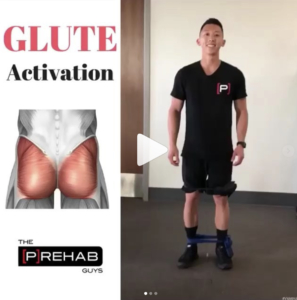
Some great Glute 🍑thoughts buy the @theprehabguys. Check out their videos and content for some great ideas that you can add to your practice!⠀
👇🏼⠀
___________________________________________________________________⠀
Episode 705: “Hip Prep for Glute Activation”⠀
.⠀
Tag a friend looking for a glute🍑 killer!⠀
Hip prep is a series of 6 exercises I’ve adopted from my girlfriend @smenzz and her clinic @eliteorthosport. I use it with my patients to prime the glutes and lower body in general before getting into more dynamic and plyometric activities. I will make the statement right now: if done RIGHT, it’s an absolute glute killer & I promise you that you will feel your glutes!⠀
.⠀
I like these 6 exercises in particular for a variety of reasons.⠀
✅They challenge the glutes in all 3 planes of motion.⠀
✅They hit all types of muscle contractions: isometric, concentric, and eccentric⠀
✅They are performed upright in a functional position⠀
✅There is a variety of double leg, single leg, and split stance variations⠀
✅They train proper lower extremity alignment in a variety of hip and trunk flexed/neutral/extended positions⠀
.⠀
The 6 exercises are:⠀
1️⃣3 way clams: 5 per leg per position⠀
2️⃣Side steps: Alternating steps to the left and right starting with 1 step all the way to 5 steps⠀
3️⃣Monster Walks: 10 steps forward, 10 steps backwards⠀
4️⃣W’s: 10 steps to the left, 10 steps to the right⠀
5️⃣Squats: 10 squats⠀
6️⃣Single leg fire hydrants: 30s per side⠀
.⠀
💡Understand that you first need to teach these exercises in isolation first, before throwing someone all 6 at once⠀
.⠀
Have fun!⠀


Hope this helps you keep up to date and fulfill my goal of this website…simplify the literature and bring great content to you so you can apply it 1st thing Monday morning! Happy Reading! 👊🏼

Follow me on Social Media here:

Image result for instagram logo vectorImage result for twitter iconImage result for facebook logo

The Evolution of a Physical Therapist

I’ve been a practicing Physical Therapist since 2003. I’ve observed a lot, talked to a bunch and read a lot. By all means, I am no expert! The evolution and growth of a physical therapist can take many roads.

I am always learning and listening but at times I do become complacent (that’s human nature). I’d be the 1st to admit that. I sometimes get stuck in my ways despite what others are saying in the literature or on social media.

I think that’s the great thing about social media…it keeps me listening. It has helped me to evolve and keep me on my game.

Ultimately, what has kept me on my game has been my desire to give my patients the best care that I can give them. I would expect the same from my own personal healthcare provider (I have a PCP, dermatologist, and a rheumatologist).

We have a responsibility to be the best for our patients. There are a lot of people chirping their opinions all over the place s I wanted to take this time to reflect on how I’ve seen many PT’s grow.

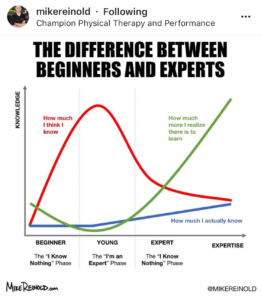
I’ve always wanted to write this post but I was inspired by my friend and co-owner of Champion Physical Therapy Mike Reinold when he posted this graphic on his Instagram feed.

I’d like to briefly chat about the evolution of a physical therapist through my eyes.

There seems to be a general development that occurs in the PT world- most are good but I’m beginning to see a side that is a bit disheartening. Maybe it’s a social media thing but I think we need to take a step back and re-evaluate for a second.

We’re always looking for a protocol to guide our patients. Here’s my attempt at the phases of a PT…Hope you enjoy (some of it is tongue-in-cheek so don’t get all crazy on me!)

The New Grad DPT student

This is the hungry, newly crowned physical therapist looking to break into the profession. Their eyes finally on the prize but probably scared to death (I hyperbolize). No more clinical instructors to guide you. No more reliance on someone else to lead the way. The plan of care is all yours!

Looking at your schedule for the next day or week, you may see that eval that worries you. Someone on the schedule with a diagnosis of “LBP” or a post-op ACL.

It was easier to treat these when your CI called the shots and you could observe, help and chime in with your thoughts and treatments. Accountability was minimal but the rewards seemed grandiose when the patient emerged with better function.

  • When is it safe to push an ACL?
  • How fast should one start strengthening after a rotator cuff repair?
  • When is it safe to start a throwing program after a Tommy John surgery?
  • What do you tell the patient when they come in with their 1st episode of acute low back pain and how do you treat it?

These are just a few of the challenges a new grad has to face.

Insurances are daunting. People can be daunting. You greatly influence the functional outcomes of that person sitting in front of you. Your words and actions matter but you don’t know that yet.

I often compare this stage to a new NFL quarterback who struggles to read defenses and rushes the ball when he throws…oftentimes to a defensive back waiting for an easy interception. He wasn’t anticipating that defense and got nervous. The game was moving too quickly and he can’t keep up with the schemes.

This is the new grad, a simplified version, but one that tries to do a lot but has minimal experiences and abilities to “read the defense.” The game is moving quickly and your decisions often come with little confidence.

But don’t worry, the game will slow down a bit.

2-5 years out and Feeling Confident

At this point, you’ve seen a bunch. You better understand the complexities of people, the medical system and how to kinda manipulate your way through. You realize that you can do it but your school studying was only a small prep for reality.

You’re motivated, finding your groove and beginning to get comfortable. There are still some questions but you don’t have to rely on the other PT’s in your group to help with progressions.

Pubmed has hopefully become your greatest ally, hopefully.

Although I do run into many that rely on Facebook and Twitter for their ‘research’. There’s always a post looking for advice on progressing a meniscus repair or return to a sport after an ACL.

I’ll often direct them to PubMed because just feeding people research is not helping them in the long run. They need to know where to find the information and learn how to interpret it.

The game is slowing down and your confidence is growing. Some even think they’re super-confident and try to ‘take on the world’. They are the ones out on social media leading the charge for change. A revolution of sorts…that their way is better than what has been done the past 5, 10, 20, 40 years!

They’re seeing their practice through rosy glasses with blinders. Blind to the fact that there are many before them who equally tried to champion a cause only to find out there’s more to it. The journey, although it seems triumphant and vigilant, falls short.

There will always be a new treatment technique, new modality a new system that is promising better outcomes. Your words, although seemingly loud, fall on so many deaf ears because the ship is going to steer itself. You’ve tried to lead the charge only to learn that the profession of PT is bigger than you.

You can only control what happens within your practice, or the few people that follow you on Instagram.  Although those ‘followers’ are often bots of some sort, pretending to like your content.

It’s a strange world out there, your words are seemingly wise, but there are so many out there shouting similar words that it gets drowned out. You think your experiences, although limited in the grand scheme of things, should guide your practice and the people that ‘follow’ you.

This, my friend, is where you’ve gone wrong. You have a ways to go. In fact, you’ll never get there. You’ll realize that each day presents a new challenge that doesn’t fall into a predefined mental algorithm.

When you have this revelation, then I think you’re ready to explore the next phase of your growth curve.

5+ years- beyond

The chart above calls you an expert but I’m not 100% a fan of this. No one in our field is truly an expert because there are challenges way above anything we could ever control.

Each personality that enters into our facilities presents with life stories that have shaped their pain, their expectations, and their outcomes.

But you know what, it’s at this stage that you realize that you are only a small piece of the puzzle. You can only help guide the process based on your plethora of experiences.

You’ve stayed on top of the literature and have altered how you practice. You no longer think that your way is the best but have dabbled in many other systems and taken a bit from all of them. Your way is NOT the best way.

You also have come to realize that there are always outliers out there. You know the ones that think the extreme positions are the best for all patients.

For example, there’s a huge social media push that says “manual therapy sucks”. No one should use manual therapy and you’re only wasting your time.

The flip side arguments say that there are many people that have a shifted inominate (whatever the hell that means) or a rib that is out of place. That therapist has been pounding on that pelvis or relocating that rib 1x per week for 52 weeks and has that patient convinced that they need more visits.

These are the outlier PT’s (I’m not speaking for other professions so don’t try to sucker me in) that are loud on social media but don’t necessarily represent the majority.

You see, the majority are trying to do it correctly (at least I think they are). The young PT that is 2-5 years out only sees those outliers as a challenge to his/her practice and is trying to yell at them. When in reality, you’re speaking to the minority, the group that barely exists.

You should be speaking to everyone else. The ones on social media that have taken a middle-of-the-road approach. They are doing their best, are on facebook looking for advice and busting their butts in the clinic.

They are limited by resources, time and updated knowledge. These are the people hungry to learn but are stuck somewhere in the 3 categories above.

These therapists are the ones you should be trying to chat with. You recognize that your vast experiences can help them.

It’s when you have this breakthrough that I think you’re ready to enter that last growth phase. You’re confident in your practice and willing to share. You speak to other groups, you publish clinical research, and you review research papers for journals.

To me, this is the utmost level and should be where most of the PT’s strive to get. Your knowledge continues to grow as you read. Each patient experience and interaction is another mental data point that sharpens your practice. These data points will blend in with your research readings and produce a so-called ‘expert’.

I invite you to challenge yourself by aligning with a clinician or group that produces clinical research, reviews journals and stays on top of the literature. Until then, don’t talk the talk unless you can walk the walk.

Some will think I’m being harsh, but I think you’ll have the realization, like I did back in the day, that our PT profession is bigger than us. Control what you can control and keep the ultimate goal in mind- THE PRIORITY IS TO GIVE THE BEST CARE FOR OUR PATIENTS, ALWAYS!

I’ve written about this before…about empowering the patient and keeping them in the driver’s seat. Check it out here.

I’d love to hear your comments. Please share with your friends, new grads and experienced PT’s. Social media has given many a voice but the loudest voices are not always the wisest voices!

Loss of extension after ACL surgery: How to assess for a cyclops lesion

Loss of extension after an ACL reconstruction can be debilitating for the patient. It’s not as common as you would think but I see it enough in the clinic from people that are months out from surgery. Usually, this loss of knee extension after an ACL reconstruction is caused by a cyclops lesion. Let’s dive deeper into this!

Often times, they’ll present with anterior knee pain, posterior knee soreness and a relatively weakened quadriceps muscle that just won’t return. No matter what they do to get the motion back, the knee just never feels normal.

I’ve written about the loss of extension after an ACL reconstruction in the past. In this post, I discussed how I like to work on knee extension immediately after an ACL surgery. There are a few ways that I think are most effective and with minimal patient efforts.

What is a Cyclops Lesion?

For those not familiar, a cyclops lesion is a wad of scar tissue in the anterior aspect of the knee joint. It is believed to be a remnant of the previous ACL stump that had remained during the reconstruction surgery. At least that’s one theory. Another theory states that it may be fibrocartilage as a result of drilling the tibial tunnels.

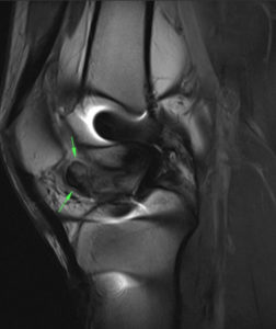
Whatever the case, this arthrofibrosis (scar tissue) physically blocks the knee joint from locking out into full extension. Check out this MRI that shows the scar tissue in the anterior knee.

Cyclops lesion in the anterior knee blocking full (hyper) extension

How do I assess for a cyclops lesion after ACL surgery?

In this video, I describe why and how I assess for a cyclops lesion. Check it out.

Is it a Cyclops lesion or just a tight knee?

From the video, you can clearly note that anterior pain, in my experiences, is most often related to a cyclops lesion. Other factors to consider include:

  1. temporary/transient gains in extension
  2. anterior knee pain after increasing activity
  3. poor patella mobility
  4. quads just won’t come back
  5. continued hamstring/calf soreness

These are tell-tale signs that there’s more going on and you should refer back to the doctor so they can order an MRI to rule in/out the anterior scarring. If diagnosed, the best (and only) option is to have a knee scope and remove that scar tissue.

There’s nothing else that can be done. No PT, injections or manual therapy can restore full symmetrical knee extension.

The scar tissue needs to be removed by surgical excision. Aggressive PT should commence immediately after surgery to restore the extension range of motion.

The Best and Easiest Way to Restore Knee Extension after an ACL

Rehab after an ACL is never easy. There are many things that could affect a patient’s outcome. I’ve treated hundreds of patients after an ACL reconstruction and each one is a unique challenge. I wrote about this in a previous post here..check it out and let me know what you think.

I put this video together for Mike Reinold’s website so you could see what I exactly do to gain knee extension back….and why I’m not a fan of prone hangs. Hope it helps with some of your knee patients.

Final Cyclops Thoughts

As a PT or athletic trainer, don’t blame yourself if the patient needs another surgery to remove the scarring. It seems as if it was inevitable and was going to occur no matter the efforts to work on knee extension. I think the lesson here is:

  • work on extension early and often
  • maintain good compliance at home
  • assess/measure each visit to determine gains or losses
  • early patella mobility and knee PROM
  • get the pain and swelling out as quickly as possible
  • refer back to the doctor if the ROM not improving despite your best efforts

Hope this post helps you get better outcomes for your ACL patients!


If you want to learn more about how I treat ACL’s, then you can check out our all online knee seminar. If interested, check it out at www.onlinekneeseminar.com and let me know what you think. We cover the anatomy, rehab prescription, ACL, knee replacements and patellofemoral issues both non-operative treatment and post-operative treatment. This is an awesome course if you’re interested in learning more about rehabilitating the knee joint. And if you’re a PT, there’s a good chance you can get CEU’s as well.

Anterior Knee Pain- A Test for Fat Pad Irritation

We as physical therapists are constantly seeing patients with anterior knee pain with a very vague history. Often times, there’s not a specific onset or mechanism of injury. With that, it seems as if the retro patellar fat pad is a common source of pain in many people and is commonly overlooked.

What actually hurts in the knee?

Whenever I am evaluating someone with knee pain, I always keep in mind the Dye et al AJSM article from 1998. In my opinion, this is a keeper for all PT’s and future PT’s to have in their arsenal of top literature papers.

For those that are not familiar, let me explain it a bit.

Basically, San Francisco orthopaedic surgeon Dr. Scott Dye had his knee scoped without any anesthesia. That’s right, classic beast mode!

He did have local anesthesia (for the record), so they could make the incisions…otherwise, he was awake and alert for the whole procedure.

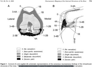
This allowed him to report back to his colleagues (one being his brother) an actual pain response as they were poking away at the different structures within the knee. He ranked the pain on a 0-4 scale with 0 being pain-free (patella cartilage) and 4 being a severe pain (fat pad, anterior synovium, joint capsule.)

You can read the article to get the full gist (and I HIGHLY RECOMMEND this!) but my point is to let you realize that there are only certain structures capable of causing severe pain in the anterior knee.

Their Findings

Cartilage is avascular and aneural. It has no blood supply or nerves that innervate it. None! So the whole chondromalacia patella diagnosis attempting to implicate a maltracking patella is often hogwash.

But the fat pad, anterior synovium, and anterior capsule are extremely painful and are often involved, I believe, in patients that we see on a daily basis.

In my opinion, this is a diagnosis that sees us more than we see it. Like thoracic outlet syndrome… we can talk about that a different day though.

Back to the point of the blog.

1 Test for Fat Pad Assessment

Assessing the knee and asking the right questions is critical. Besides a tremendously great subjective, there’s one test that I use to rule in or out a fat pad irritation. Check out the video below:

I promise you this test is a great way to establish a pretty clear diagnosis and reassure the patient that a specific structure may be the issue.

When it’s painful, its pretty obvious and the patient can immediately report back to you their symptoms. I test for this a bunch in a given week and it’s not always present. But when someone presents with a positive finding, it’s pretty relieving to them (believe it or not.)

Differential Diagnosis

Keep in mind, my differential diagnosis is all over the place at times.  With knee pain you need to consider:

  1. Meniscus (see my previous blog post)
  2. ITB
  3. Osteochondral lesion
  4. Patella tendonitis
  5. Pes anserine bursitis
  6. MPFL sprain
  7. Hamstring strain
  8. Plica syndrome
  9. MCL/LCL
  10. Tumor

That’s a bunch to consider for someone who can’t really explain why or when their injury occurred. That’s why I still believe this overuse injury sees us more than we see it.

Treatment of Fat Pad Irritation

This one can get a bit tricky. Again, it’s often due to an overuse injury. In others, it’s attributed to kneeling on it for too long or banging it against something. In those more acute cases, the answer is pretty straightforward. Ice, motion and progressive return to their function.

In the overuse group, which is more prevalent in my opinion, we need to figure out which stimulus is causing the issue. It’s often due to starting a new program or ramping up too quickly during some training event.

In these people, I need to modify their volume of training or destress the area by giving them new activities that they can do pain-free…but only for a short period of time.

Along with activity modification, a course of some form of modality (yup, the M-word was used) can help expedite the pain control. I still have a special place in my heart for iontophoresis with dexamethasone. I’ve had tremendous results in patients with fat pad irritation.

I also like to use a low-level laser to help with the healing process (future blog post alert!) I’ve used it on my self for various ailments and even my dog after her ACL surgery. I’m definitely a believer in the healing power of the laser when applied in the correct situation.

Besides all of that, I would also want to assess the person’s movements and attempt to adjust any motions or movements that I thought could contribute to the knee pain. Often times, there’s an underlying weakness or misconception of a weakness that needs to be explained to the patient.

These things run their course for a few weeks but should improve pretty quickly if handled correctly. Patient gratification is pretty obvious because their pain diminishes after a session or 2 and they buy into the program pretty quickly.

Fat Pad Conclusions

Infrapatellar fat pad irritation can be functionally debilitating. I believe it presents itself pretty often in the clinic, more than most PT’s realize. Use my test above to see if it truly is a fat pad issue. Here’s a nice open-access article that you can read to learn more about the fat pad.

Remember, the test should be pretty obvious and locally oriented. If pain-free, then move on. If not, then try some of my above recommendations. It’s usually an overuse issue so you need to adjust their volumes and maybe some form of mechanics.

Mike Reinold and I discuss this and much more about how we treat the knee conservatively. Check it out in our acclaimed all online knee course www.onlinekneeseminar.com.