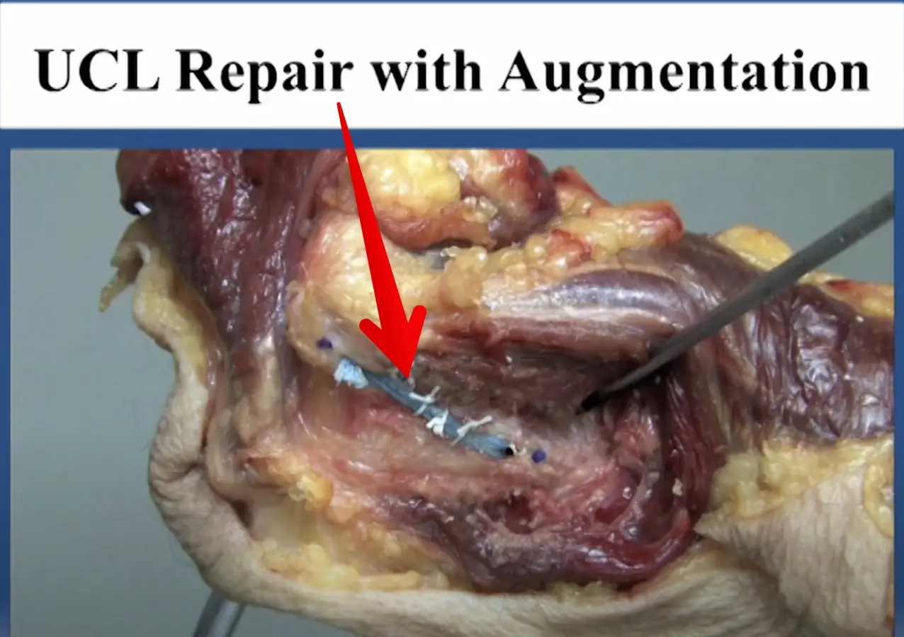
Diagnosed with an elbow UCL Tear- Reconstruction or Internal Brace surgery?

So, you’ve been diagnosed with a UCL tear in your elbow and your world has been turned upside down. Have no fear, many have been there before you and have done pretty well.

But now, there’s a new option for elbow surgery and you’re not sure if it’s right for you. I hope this post can help you decipher the jargon and ease your mind a bit.

Tommy John Surgery

Tommy John surgery has been around since 1974, when legendary Frank Jobe performed the 1st surgery on pro baseball pitcher, Tommy John (shocker!)

It was a pretty epic failure and required a subsequent surgery to fix some of the original issues (massive claw hand due to ulnar nerve issues). Tommy John did return after numerous surgeries to have an amazing MLB career. This set the precedent and baseball has not been the same since.

Fortunately, we have done much better overall. We have improved our surgical techniques and rehab outcomes. Nearly 80%+ of baseball players that have the reconstruction surgery can return to a pretty high level of function. By definition, they will play at the same level or higher compared to their pre-surgery level.

If you don’t believe me, then I suggest you read this article right here. I can attest that the TJ patients that I have rehabbed over the years have done very well. The road is long but most can get back to nearly 100% at around 9-12 months.

Tommy John Surgery Video

Curious to know what the reconstruction surgery looks like? My colleague Dr. Chris Ahmad (who we work with a bunch) presented on his surgery technique recently. Watch this video (as long as you’re not squeamish!) and enjoy!

UCL Docking Technique by Dr Chris Ahmad

I usually tell my patients to fast forward 12 months and we can count backwards to figure out the path that we’re going to take.

But that’s not why you are here, right?

You want to know about the internal brace surgery that has taken the TJ world by storm!

Elbow Internal Brace Surgery

This is a relatively new procedure that has been around since about 2013. My friend and colleague Dr Jeff Dugas began doing this procedure in Birmingham, AL when I was down there. I got to see the early results 1st hand and was excited but skeptical.

Baseball players were returning to their sport in 6-8 months versus the 1 year that we had seen in a full TJ surgery.

Why a quick return after surgery?

Why have them return sooner if the surgeon is not using the patient’s native tissue?

Who should get this surgery anyway?

We didn’t necessarily know, but like any other orthopaedic surgery, we had to wing it a bit. We had to figure out a protocol that was appropriate for the tissues involved.

I was there in Birmingham with another friend and colleague, physical therpaist Kevin Wilk. I helped put together the early protocols and was excited by the potentials.

The thought is that the collagen dipped tape that is re-enforcing the repaired ligament is stornger than the native ligament. Its fixation to the bone is strong. A quicker return to throwing and sport is possible, because of those reasons.

Its worked for the ankle and so why can’t it work for the elbow too?

Internal Brace Surgery Specifics

This internal brace surgery was developed by surgical company Arthrex. It’s pretty neat to see how it has taken on a whole world of uses, including in the knee and ankle. Its many uses has helped numerous athletes return back from their injuries quicker than ever.

See Tua at the University of Alabama, for examaple. He had a high ankle sprain and retuned to the field ~3 weeks after his ankle surgery. Again, surgery performed by my friends Dr Norman Waldrop and Dr Lyle Cain, of Andrews Sports Medicine and Orthopaedic Center. They’re studs and surgeons that i’d highly recommend!

Who benefits the most from Internal Brace surgery?

For this elbow surgery, the internal brace is most appropriate for the athlete that has a UCL sprain that is not complex. Most times, they won’t know until they’re in the surgery if the internal brace is appropriate.

If it is appropriate, then surgical consent probably happened before the surgery. The doctor won’t know if the repair is appropriate until he/she can visualize the tissue directly. A decision is made on the spot even if the MRI said something differently.

Why try the internal brace?

I basically recommend this surgery for my athletes who don’t have a lot of time.

Let’s say they sprain the ligament in the offseason, like in November of their junior year in high school, for example. If a full-blown TJ reconstruction was done, then they’d be out until at least the following November (remember my 12-month comment earlier). That would mean no junior year baseball or Summer ball. That would also mean no exposure for college recruiting!

But wait, there’s an alternative! Internal Brace repair surgery…see the video here!

…or here by Dr Jeff Dugas:

Elbow Internal Brace Procedure by Dr Jeff Dugas

In the internal brace situation, you could have the surgery in November and be back for some of your High School season and most likely for that important Summer travel season.

But remember, the ligament can’t be chewed up a lot. That decision will be made intraoperatively. Be ready to wake up from surgery with news that a full TJ reconstruction had to be done.

But for many, an internal brace repair is possible. And a quicker return may be possible too.

Should you do this surgery?

For those considering it and fit the requirements, then I’d recommend it. Just keep in mind that we really don’t have too many long term outcomes.

But for the High School or College athlete looking to play a few more years, then I’d say go for it.

For the HS or college pitcher who has aspirations of playing pro ball, then I’d recommend the full reconstruction. We just know more about the surgery and long term outcomes. it’s tried and true in every way.

That’s not to say that the internal brace procedure cannot be the gold standard surgery in a few years. That is quite possible. I really hope to update this blog post in the future and say that I was wrong.

But as of now, I would recommend the reconstruction for the pro athlete or amateur athlete looking to play pro ball. Otherwise, the internal brace procedure is a very strong option for many pitchers (or even positional players looking to get back quicker).

Summary- Who should get this surgery?

Pitchers or positional players that don’t have much time before their next season and NEED to play. But the tissue needs to be repairable and not beat up (this is the key!)

Consult your surgeon to discuss this but they won’t know until they’re in your elbow and you’re out cold from anesthesia.

Good luck…it’s a long and winding road but most do well. I’ve treated a lot of these cases and no 2 are ever the same. There’s always a glitch and a freak out period but it often works out in the end!

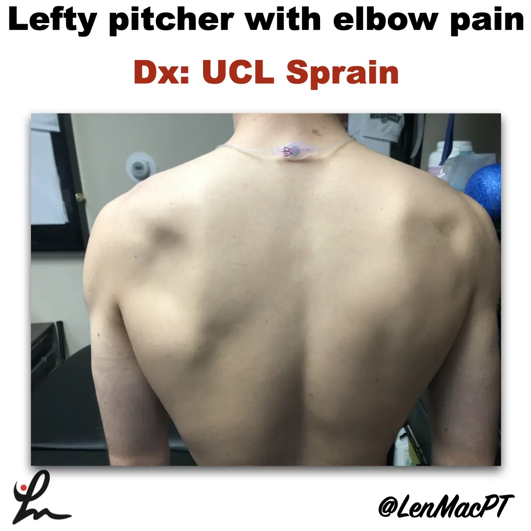
Infraspinatus atrophy in a baseball pitcher with a UCL sprain

Infraspinatus atrophy due to a spinoglenoid notch cyst

This was an incidental finding of mine in a collegiate baseball pitcher. He presented with a UCL sprain during his senior year and was struggling.

He came in looking to salvage his senior year and attempt to pitch at some point. UCL surgery would’ve been warranted if he was anything but a senior.

UCL Evaluation

We evaluated his elbow and he presented with typical findings of painful valgus stress tests. I’ve discussed my UCL exam previously in a post that you can find here.

UCL Tommy John Assessment

His shoulder PROM was slightly limited compared to what we normally see in our overhead throwers, which may have contributed to his elbow issues.

He only had 120 degrees of external rotation compared to 110 degrees on the right side. If you’re wondering how I measure external rotation, then this video below may be a good one to review.

External rotation passive range of motion (PROM)

We use these concepts for our research studies that we have published on weighted ball usage the past few years.

For those that will ask about GIRD, he had 60 degrees of IR on the L side and 50 degrees on the R side. I’m not a big GIRD guy like has been written by others. I still think GIRD is pretty normal and our research has shown that too.

Remember, we showed that differences in total rotational range of motion greater than 5 degrees was significant. This study was published in AJSM back in 2011.

Furthermore, we showed a connection between the loss of passive flexion and elbow injury.

This result was also shown by Camp et al in a separate study, as well.

I had him remove his shirt and noticed a large defect in the infraspinatus area that showed obvious atrophy (see picture below).

Infraspinatus atrophy due to a spinoglenoid notch cyst

The cyst was compressing the suprascapular nerve as it coursed along the spine of the scapula.

His external rotation strength was significantly weaker on his throwing side compared to his right side. This would make complete sense considering the amount of atrophy present.

Often times surgery is warranted to decompress the nerve. There’s often an underlying labral tear that has resulted in a cyst to form. The surgery would remove the cyst and repair the labrum to prevent future issues.

Quick Literature Review on Surgical Outcomes

Reports of improved outcomes are in the literature, here and here and here.

Obviously, this may have contributed to his elbow issues as the posterior cuff muscles are tremendous stabilizers of the arm, especially during the deceleration phase of throwing.

Treatment for infraspinatus muscle atrophy

Our goal was to calm the elbow down through active rest and focus on strengthening of his forearm and shoulder musculature.

We used neuromuscular electrical stimulation (NMES) to the infraspinatus. We felt as if the electrical stimulation would allow for a better muscle contraction than what he could do actively. This is a similar concept to using NMES to the quads after a knee surgery.

NMES to the infraspinatus to help with muscle recruitment

We have previously shown in AJSM that NMES to the infraspinatus was beneficial to patients after a rotator cuff repair. It helped improve their muscle contraction by 22% in the group with the NMES applied during testing.

Outcomes and final thoughts

Believe it or not, we were able to salvage his senior year despite all of the issues that presented.

He made numerous pitching outings and was pretty successful for his team.

The lesson here: Don’t take anything for granted, observe and test.

Make sure you look distal and proximal. It could’ve been very easy to just treat the elbow with some exercises and massage. Don’t get me wrong, we did a bunch of strengthening exercises to his forearm.

His secondary finding was most likely a leading cause for his elbow dysfunction.

This isn’t the 1st time that I’ve run across an issue like this. I’ve found numerous scapula dysfunctions like this involving the infraspinatus. But also a winging issue due to a long thoracic nerve palsy.

You must look at the whole picture… shirts off for a guy and a sports bra for a female to make sure we’re not missing anything obvious!

Lumbar Spine Stress Injuries in Baseball Players

Low back pain can be very debilitating in an athlete, especially in an adolescent baseball player trying to make it through his season. More specifically, I have noticed an increase in lumbar spine stress fractures in baseball players and it is quite disturbing and frustrating!

I wanted to dive deep into this rabbit hole, as I always do. Low back pain is way too common in youth athletes. I wanted to know why it’s happening so often and try to figure out a solution for my clients.

It can get complicated so I will try to simplify the process.

My goals:

  1. What is a stress fracture or a stress reaction?
  2. How common is this injury in youth sports compared to other sports and age groups?
  3. What is the typical clinical presentation?
  4. What are some clinical tests that PT’s can use in the clinic?
  5. Demonstrate a typical treatment of a young athlete with a low back stress injury?
  6. What are the expected outcomes?

Pain is Complicated

Low back pain can take on many etiologies. Pain, in and of itself, is extremely complicated and very personal.

It can manifest in different ways for each person. In this post, I want to talk about the athlete with an acute injury. Specifically, the youth and adolescent baseball player.

Many factors can contribute to someone’s pain including stress, anxiety, medical history, their environment, support system, etc.

This topic is a bit more personal because of what do on a daily basis. I see a ton of youth athletes with a variety of injuries. Plus, I was diagnosed with spondylolisthesis (bilateral fracture in the spine and some shifting of the vertebral body) while a junior in college so I’ve lived it myself!

Recently, I’ve started to see a lot more athletes with low back pain as a result of a stress reaction or fracture. It really made me wonder what was truly causing this in the 12-18 year old athlete.

Video example

courtesy of pitching coach Anthony Meo of The Farm Baseball Waltham, MA

As you can see in the video, the pre-injury video shows how long the pitching motion is for this athlete. His throwing arm is wrapped around his body which forces him to keep his weight back.

His arm is trailing his body significantly and he tries to make up for it by hyperextending his back. His timing is way off and his arm is not keeping up with his forwardly moving body.

You can see in the post-injury video that he is much more balanced. His arm also has better timing with his body. He is driving toward home plate with his lower body while maintaining a better upper body sequence

There are many issues going on here but you can see the significant changes that were made after the injury to help him improve his mechanics and hopefully prevent a future injury.

LUMBAR STRESS INJURIES DEFINED

Stress Reaction Defined

Basically, a stress reaction is the early breakdown of the bone without a clinical diagnosis of a true fracture. Often times, a suspected fracture will be diagnosed as a stress reaction. On MRI, there is only edema noted in the area and not a true fracture.

I hear a lot of people talk about fractures in different terms. Simply stated, a fracture is a crack, break, defect in the bone. Whether it’s a hairline fracture or a gross disruption of the bone may determine prognosis but the bone has been compromised.

taken from: Link
taken from: Link

Obviously pain is an early sign but it needs to fit the clinical presentation. Has there been a history of excessive activity or a change in the volume of playing?

Factors to consider in a baseball player with low back pain

I usually have to dive a bit deeper into the player’s life to figure out the root cause. It’s often something very overlooked but is often the main contributor.

These are just some, off the top of my head, that may be involved in the athlete’s predicament and that may nee to be changed.

It is an early indicator that something is wrong and it can progress to a stress fracture if not carefully treated. We’ll talk treatment later on so be patient!

How common is a stress reaction in youth sports?

As with any research, it is quite mixed and will heavily depend on the sport that you are tracking. From what I can see, if a kid has low back pain for greater than a week, then that’s a pretty good sign that a more serious injury may be present.

For example, this retrospective case study from 2017 showed that 30% of pediatric cases with low back presenting to this hospital were diagnosed with spondylolysis.

Specializing in a single sport was not associated with an increased risk of spondylolysis when compared with multisport athletes. Male athletes were 1.5 times more likely to have a spondylolysis than female athletes.

Males with LBP who participated in baseball had the highest risk of being diagnosed with a spondylolysis, followed by soccer and hockey.

Females with LBP who participated in gymnastics had the highest risk of spondylolysis, followed by marching band and softball. I will refer to my good friend Dave Tilley and his website for his professional judgment on this one. Based on what I see at our facility, I’m not surprised.

As I usually tell my clients and their families…I’m going to treat this as a spondy until proven otherwise.

More Research on Spondylolysis Prevalence

According to recent research, it looks as if persistent pain for 1-2 weeks is associated with a positive MRI diagnosis for spondylolysis in 33-40% of the kids scanned. You can read the abstracts here and here if you’re interested in more.

This study out of New York showed that the most common athletic activities associated with spondylolysis were:

  • Soccer (19.3%)
  • Basketball (17.2%)
  • Lacrosse (9.4%).

Of note, 71% of the children in this study were male and the most common level to have the spondy was at L5.

Do keep in mind that false positive results are pretty prevalent in patients getting an MRI for low back pain.

Don’t treat the MRI (or x-ray!)

This systematic review looked at the incidence or prevalence of incidental findings on MRI in asymptomatic pediatric patients. The results are pretty eye-opening too!

  • Degenerative disc disease (prevalence 19.6%)
  • Disc herniation/protrusion (prevalence 2.9%)
  • disc height/narrowed disc space (prevalence 33.7%)
  • Spondylolisthesis/spondylolysis (prevalence 2.3%)
  • annular tear and/or nerve root compression (prevalences ranging from 4.5-51.6%)

Pretty amazing to see the prevalence of false positives read on an MRI in a pediatric population. We need to be able to clinically diagnose these first then use the MRI scan to further determine a course of action.

Baseball specific injury rates

In this study from 2015, the proportion of High School baseball players with a low back stress fracture was 3.3%. So, a typical AAU baseball club of 100 kids will have 3 kids that have a stress fracture.

They also reported that High School girls sustained more stress fractures (63.3%) than did boys (36.7%) but for overall injuries and not just in the low back. This seems consistent with what I see in my practice, as well.

In college, 12.1% of the stress fractures noted in this NCAA study from 2017 were in the low back. Women experienced stress fractures at higher rates than men, more often in the preseason, and predominantly in the foot and lower leg.

Baseball Specific low back pain

This nice review of the literature discusses low back pain in general athletes. They went on to further discuss baseball and “the asymmetric baseball postures and motions lead to asymmetric spondylosis in right and left sides and cause mechanical degeneration in intervertebral discs.”

In this study, “students who played baseball were 3.2x more likely to experience LBP in their lifetime only behind volleyball which had an odds ratio of 3.8. 

The repetitive nature of the baseball specific movements and a preponderance to specialize in one sport can definitely affect these injury rates.

The role of mental stress and low back pain

The role of stress, particularly mental stress on bone development, has been studied too. Our teenage athletes are under a tremendous amount of stress at home and socially. This study from 2017 discussed the emerging concept of mental stress as an important player in bone adaptation and its potential cross-talk with physical stress.

Essentially, mental stress can have a negative impact on normal hormone production. Hormones are needed to maintain normal bone health. A shift in normal hormone regulation can negatively affect a youth’s growth and healing capabilities.

They went on to say that there is “compelling evidence has recently emerged that biochemical and psychoneuroendocrinological maladaptations caused by mental stress are not only also relevant for bone quality, but may furthermore considerably interact with physical stress.”

Clinical Presentation of a Spondylolysis or Spondylolisthesis

Clinically, pain is the guide here. It’s often described as a pretty sharp pain that is localized to the site of the injury.

There is rarely a referred pain down the leg or the buttocks. The pain is often close to the spine at the level of the injury.

In this study, activities involving repetitive hyperextension and/or extension rotation of the lumbar spine were described as painful in 98% of the patients.

Pretty powerful statement. In my examination, I look to recreate these symptoms with these exact movements.

Many use a single-legged position, also known as the Stork Test, to test for spondylosis. I’ve simplified the test to just include regular standing on 2 feet to simulate regular daily movements.

A standing extension test can also be used to assess end range extension and rotation irritability. I find this test (see video below) is a great way to begin to hone in on a diagnosis.

I find that pain in full hyperextension and rotation is a pretty powerful indicator of a stress reaction or fracture. It is similar to the standing position that I described above. However, the prone position takes the postural component out of the picture and may help to better isolate the location of the lesion.

I also like to have the patient get in the prone position and use different levels of active extension to help guide my clinical examination.

Again, people will report pain that is very close to the spine and that replicates their pain.

Any young athlete with low back pain for greater than 1 week should be assessed for some kind of bony issue in their low back.

That’s not to say that we will always find something.

But I just think that in younger athletes, this prevalence is all too common and needs to be ruled out to prevent further low back issues.

Treatment for Spondylosis in Baseball Players

Muscle tenderness too

Patients will usually have tenderness right at the region in which they have pain. I suspect it’s the muscles that stabilize those lumbar segments.

The multifidi and most likely quadratus lumborum are often implicated. Soft tissue work of any kind can have positive effects on muscle tightness. The athlete should perform some self-myofascial release with a foam roller or light pressure with a baseball in the affected muscles.

Manual therapy can also be utilized to feel tissue density and help to pinpoint the exact location of their muscle soreness. By utilizing this, I can help to better educate the athlete for their home exercise programs.

Manual Therapy can be helpful

In this photo below, you can see I am applying direct pressure with my elbow right into the muscles of the low back. The patient will usually give me feedback and tell me if I am in the right area. Muscle soreness is often detected and after a short amount of time, the symptoms should decrease.

In the short term, manual therapy can help with some of the acute symptoms. For long-term changes to occur, activity modification and exercises that involve low back, hip and core strengthening are critical.

Soft tissue work to the muscles of the low back in an athlete with low back pain.
Soft tissue work to the low back in an athlete with low back pain.

My treatments for kids with low back pain are pretty simple. I want to rest them from the repetitive activities, such as swinging a bat or even throwing a baseball.

Brace or no brace?

Also during this time, the athlete will most likely have to wear some form of a back brace to prevent excessive lumbar extension and rotation. One particular brace is called a Boston Brace. It was developed by doctors at Children’s Hospital in Boston, MA.

The research is unclear and can favor both methods of treating with or without bracing. A study in 1986 showed that bracing ‘restricted at least some gross body motion to approximately ⅔ to ½ of no-orthosis values.’ The restrictions we’re most pronounced when wearing a larger Thoracolumbosacral orthosis (TLSO) versus just a corset.

If the goal is to restrict motion, then it seems as if a brace will accomplish that. Most research shows about 40-65% restriction in motion no matter the brace. This paper showed similar findings as well.

Here’s a version that our local docs have been using that is less cumbersome and not as embarrassing to wear as a teenager!

Brace used to help restrict motion during a low back stress fracture

These are just some of my quick and easy exercises to help promote movement in the spine.

Obviously, I want to avoid painful motions like lumbar hyperextension and rotation. I still want to promote some sort of motion to build confidence and promote muscle recruitment in that area.

Specific treatment ideas for low back pain

I like foam rolling or some form of self-myofascial release to the area. This will help the muscles to feel better if done multiple times per day. See the video below

Some would say motion is lotion and I tend to agree! I like to use active range of motion through the cat-cow video below.

Cat-Cow exercise to promote active motion in the low back

Some general guidelines for returning to baseball

From what I’ve seen, most baseball players need at least 3 months of active rest.

I’ve written about my general low back treatment philosophies in a previous blog post.

You can find that post here .

Active Rest is key

I generally want to begin a general strength training program at around 6-8 weeks following the diagnosis. It Is critical to include restricted range of motion exercises to avoid painful hyperextension. These exercises are restricted to avoid excessive extension that may place extra stress on the low back. I prefer to bias flexion type exercises like squats, lunges and step-ups.

I like to closely monitor their form and symptoms. We don’t want any pain at all in that region…a very important concept!

I like to perform these exercises for at least six weeks to get a good base of strength in the lower body and core.

Lose the brace at 12 weeks

At about 12 weeks, they will come out of the brace and we can begin to be a bit more aggressive in our strengthening. Again, we are avoiding hyperextension type movements in the low back. We prefer to stay in a flexed or neutral spine as much as possible.

I also like a supine dead bug exercise to promote a flexed spine with active motion. See the video below.

Throughout this time, we encourage cardiovascular exercises that involve the stationary bike. This seems to be a safe modality that allows the athlete to work up some sweat but also maintain a relatively flexed spine, which is usually pain-free.

Unrestricted activities at 5-6 months

At around 5 to 6 months post-diagnosis, we encourage for pain-free activities that involve strength training for the whole body. There are no limits in range of motion in the athlete is free to work out. We then encourage more functional activities that involve rotation. This will allow them to replicate the motions involved with hitting and throwing.

Return to sports at 6-8 months

On average, it seems as if a patient recovering from a low back stress fracture will need at least 5 to 6 months before I would feel comfortable having them return to their support. These injuries usually happen in a younger population so the risk for re-injury is pretty high if they return to their sport too quickly.

I believe the risk is high because of their underdeveloped muscular system. Their bodies are growing and tremendous stress may be placed upon their joints with excessive activities. A slow progression back to their sport is critical.

Concluding thoughts on stress fracture injuries in youth athletes

Baseball players face numerous challenges during their training.

As you can see, numerous injuries can result and not just to the shoulder and elbow.

Low back pain injuries are all too common but are often preventable. It seems as if the volume of throws and hits may play a huge role. It is up to the coach, physical therapist, or athletic trainer to monitor this workload for each athlete.

There is no exact science but listening to the athlete is critical. A well-balanced training program that addresses some of the obvious weaknesses is key.

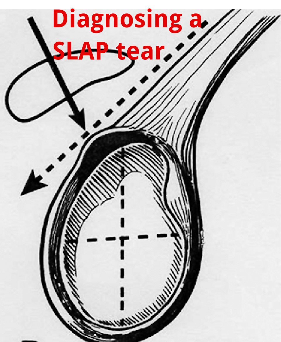
An Update on Diagnosing SLAP tears

Diagnosing a SLAP tear is not easy

We hear a lot about trying to diagnose shoulder pain and to be as specific as possible. It’s often difficult to differentiate SLAP (superior labrum anterior to posterior) tears from other soft tissue injuries of the shoulder.

For a review of the different types of SLAP tears, check out this old blog post that classifies the 10 different types of tears.

from https://commons.wikimedia.org/wiki/File:SLAP-Lesion-front-2.jpg

This paper in IJSPT by Clark et al 2019 attempts to help out the process and recommend a few special tests that MAY aid in diagnosing a SLAP tear.

What do they Recommend to diagnose a slap tear?

They recommend that a combination of at least 3 positive SLAP lesion tests may be clinically useful in diagnosing a shoulder SLAP lesion with greater diagnostic accuracy.

Combo of Tests

The combination of the Biceps Load I/II and O’Brien’s showed the highest sensitivity and specificity.

I have found similar results with this set of special tests so maybe this paper just hits my biases correctly.

In this video at my YouTube channel, I wanted to let you hear my thoughts and small tweaks to the evaluation process.

It’s not easy to diagnose a SLAP tear.

Differential Diagnosis is Critical

Furthermore, does it really matter and will it change the treatment plan much at all? I think it may a little but overall it will remain a pretty similar treatment approach to other similar pathologies like:

  • rotator cuff tendonopathy
  • Biceps strain
  • Latissimus strain
  • Subscapularis strain
  • internal impingement
  • pectoralis major strain

I think one also needs to consider the cervical spine and to make sure the pain is not referred from the neck.

Otherwise, a well thought out program should be implemented that addresses the strain on the shoulder and any strength issues.

I talked about this in an article that I wrote for Medbridge a while back so check out that post here:

You can also check out a snippet of one of my courses at this YouTube video where I discuss rotator cuff and labral issues. Hope it helps too!

Will this Change your Practice?

How ever you look at it, I wanted to use this paper to let you know that there MAY be a cluster of tests that better diagnose a suspected SLAP tear in your next patient’s shoulder.

Check out the paper and comment so we can talk it through. Are these tests similar to what you use in your clinical practice? Will this paper change what you do in your clinical practice?

The Week in Research Review, etc 12-10-18


This week we’re still playing with formats and learning these Instagram changes. With that, in the week in research review 12-10-18, we discussed many topics that I wanted to share!


Surgery vs Physical Therapy for Carpal Tunnel Syndrome

Cost-Effectiveness Evaluation of Manual Physical Therapy Versus Surgery for Carpal Tunnel Syndrome: Evidence From a Randomized Clinical Trial Fernández-de-las-Peñas et al JOSPT 2018.

This Level 1b study looked to evaluate cost-effectiveness differences of manual physical therapy versus surgery in women with carpal tunnel syndrome (CTS).

Performed in Spain, 120 women with a clinical and electromyographic diagnosis of CTS were randomized through concealed allocation to either manual physical therapy or surgery.

They concluded that manual PT including desensitization maneuvers of the central nervous system has found to be equally effective but less costly, i.e., more cost-effective than surgery for women with CTS.

From a cost-benefit perspective, the proposed manual PT intervention of CTS can be considered.

Interesting results but 2 obvious limitations to this study:
1️⃣No control group. What if the symptoms could spontaneously improve over time
2️⃣ They only looked at 1-year improvement and not short-term improvements. I would’ve liked to have seen 3 months and 6 months results as well to see the acute effects.

Not sure what to make of this study but it does seem as if a population of Spanish women may respond to Rx of CTS without surgical intervention.

This could be a huge cost/time saver for society!


Return to Sport Criteria and Reinjury Rates

The Association Between Passing Return-to-Sport Criteria and Second ACL Injury Risk: A Systematic Review With Meta-Analysis  Losciale et al JOSPT 2018.

Not going to lie, this study caught my attention because the results match my confirmation bias.⠀

I’ve been saying for years that hop tests, even combined with other tests, just don’t cut it.⠀

I wrote a blog post about this too for @mikereinold. This study, although with its limitations, did show that passing RTS criteria did not show a statistically significant association with risk of a second ACL injury. 

This review also determined that 12% of those who failed RTS testing suffered a graft injury, compared to 5.9% of patients who passed. 

It seems as if quadriceps strength measured via isokinetic testing or isometric testing may be an important factor to consider for RTS decision making.


Also, hamstring-quadriceps strength ratio symmetry should also be considered.

So with this review demonstrating that current objective criteria-based RTS decisions did not show an association with the risk of a second ACLI, how does this affect your practice?


Physical Therapy vs Knee Scope for Meniscus Tears

💥PT vs Scope for Meniscus Tear 💥
.
Effect of Early Surgery vs Physical Therapy on Knee Function Among Patients With Non-obstructive Meniscal Tears: The ESCAPE Randomized Clinical Trial. van de Graaf VA et al JAMA Oct. 2018

Among patients with non-obstructive meniscal tears, PT was equal to arthroscopy for improving patient-reported knee function over a 24-month follow-up period. 

They went on to say that “Based on these results, PT may be considered an alternative to surgery for patients with non-obstructive meniscal tears.”

So basically, if there’s no bucket handle tear present that may be blocking joint range of motion, then it is highly encouraged that the patient #GetPT1st and not do surgery.

Without going out on a limb, I’d say this is a much more cheaper treatment option as well and would save society many costs associated with the surgery and lost time from work.

I do note a couple limitations: the surgical group did not get PT after surgery if they did ‘as expected’ but they could get PT to help improve their symptoms.

The PT group did pretty basic exercises although leg press, lunges, and balance type exercises were included.

Have you read this paper? It was a multi-center, randomized controlled trial performed in 9 hospitals in the Netherlands.

So, are we encouraged or surprised? Let me know by commenting below…thanks!


Shoulder Health Accessory Exercises

by @kieferlammi

Want strong and healthy shoulders!?

Shoulder strength is about more than pushing big lifts like strict pressing, push pressing, etc.

If you want a robust, healthy, well moving shoulder you should be including lower level drills that more specifically address scapular and RTC strength and control. –

I will always be a fan of traditional exercises like side like ERs, Prone Ys, Ts, etc.

Lately, I’ve thrown in more band work because it’s easy for me to do for higher volumes on a frequent basis and I enjoy the constant tension that the band provides. 

Give these two exercises a try:

✅ Band Front Raise Pull-Apart

✅ Band Overhead Y Raise

I find that these two do a great job of targeting my mid back and posterior shoulder without much compensation through a big range of motion.

Give them a try either in a warm-up for 1-3 sets of 10-15 reps or at the end of a training session for 2-4 sets of 10-25 reps depending on the difficulty of your band and your capacity. 


Should we Brace after an ACL Surgery?

View this post on Instagram

[NEW BLOG POST]⠀ 💥Knee Bracing Immediately After an ACL Reconstruction 💥⠀ In this post, I review some recent Twitter, Facebook and Instagram discussions about the usage of a hinged knee brace after an ACL surgery.⠀ .⠀ You'd be surprised what I found in the literature and the differences that exist throughout the US and the world.⠀ .⠀ Go to my website <LINK in my BIO> and read/share with your friends/colleagues.⠀ .⠀ It was a bit eye-opening and I'm curious to hear what others have to say.⠀ .⠀ I still like to recommend a brace for my clients because it seems to give them an added security after a pretty painful surgery.⠀ .⠀ I typically keep them braced 4-6 weeks, depending on their quadriceps activity and if they can do an active straight leg raise without a lag.⠀ .⠀ Many others don't even bother bracing at anytime post-op, which was surprising.⠀ .⠀ What do you think? Read the blog post and let me know. Let's try to educate and come to a better consensus...thanks!⠀ .⠀ #kneerehab #knee #kneepain #kneesurgery #acl #aclsurgery #ROM #physio #physiotherapist #crossfit #exercise #deadlift #physiotherapy #physicaltherapy #physicaltherapist #athletictraining #athletictrainer #ATC #PT #teamchampion #dptstudent #lenmacpt #instagram

A post shared by Lenny Macrina MSPT, SCS, CSCS (@lenmacpt) on

You can also get to the blog post by clicking this link

Let me know what you think about this new blog post or any of my social media posts…thanks!

The Week in Research Review, etc 12-3-18

Hey everyone,  The Week in Research Review, etc for this week has a new look, compliments of Instagram’s new algorithm. Hope the new format doesn’t throw you too big of a curveball (maybe you’ll like it better), so here goes…


ACL Injury Rates Higher on Synthetic Turf than Natural Grass in the NFL

Preventing low back pain by @joegambinodpt

Female Soccer Players have a 5x Increased Risk of a Second ACL injury

Anatomy of the Proximal Humerus


ACL Injury Rates Higher on Synthetic Turf than Natural Grass in the NFL


View this post on Instagram


A post shared by Lenny Macrina MSPT, SCS, CSCS (@lenmacpt) on

Preventing low back pain by @joegambinodpt


View this post on Instagram


A post shared by Lenny Macrina MSPT, SCS, CSCS (@lenmacpt) on

Female Soccer Players have a 5x Increased Risk of a Second ACL injury


View this post on Instagram


A post shared by Lenny Macrina MSPT, SCS, CSCS (@lenmacpt) on

Anatomy of the Proximal Humerus


View this post on Instagram


A post shared by Lenny Macrina MSPT, SCS, CSCS (@lenmacpt) on

The Week in Research Review, etc 11-19-18

Great ‘Week in Research Review, etc 11-19-18’ that I hope you find helpful to your practice.

I’ve always touted the importance of the subjective portion of the exam so I wanted to share a slide from a recent talk I gave to a group in Canandaigua, NY. Obviously, the squat is a fundamental movement and I wanted to give some basic positions that I use to help assess. So excited that I’ve launched a brand new Medbridge course that helps the rehab specialist better eval and treat the baseball pitcher. On my YouTube channel, I discussed my thoughts on setting the scapula with various upper and lower body exercises. And finally, my co-worker Kiefer Lammi discusses the landmine with exercise.


Importance of the Subjective Exam

Assessing the Squat

My New Baseball Medbridge Course

Set the Scapula with Shoulder Exercises?

6 Ways to use the Landmine by @kieferlammi


💥Subjective the most important aspect of the Evaluation💥

This slide, taken from this past weekend’s course in Canandaigua, NY is always a favorite of mine.

I try to keep a slide like this in all of my lectures because I have found that this portion of the examination can give the rehab specialist a huge look into what is going on with the person in front of them.

Don’t get me wrong, I still consider the biomechanical aspect of what may be causing their symptoms.

It often comes down to a tissue capacity issue but it’s up to me to determine the appropriate course of treatment.

These questions will help build confidence in your client and guide the early stages of rehab.

Do you have any specific questions that you like to ask your clients during their 1st few sessions? Remember, these questions are just not for the evaluation. You should be asking these questions periodically to gauge progress and help guide the next phases of rehab, too!


🔅Assessing the Squat 🔅

Squatting is a fundamental movement that all of us have to do on a daily basis.

Utilizing several different positions can help the rehab specialist better assess the squat and develop a treatment plan that enables their client the ability to improve their squat pattern.

In the above videos, I have utilized 3 different squat patterns and will outline them by the degree of difficulty.

✅The Overhead Squat- by far the most challenging version which challenges the shoulders, thoracic spine, lumbar spine, pelvis, knee and ankles.

A movement limitation at any of these joints will most likely cause the squat pattern to break down. Using overhead resistance would further challenge the system and potentially cause the squat to further breakdown.

✅Arms Crossed Chest Squat- alters the challenge by taking most of the shoulder and thoracic spine out of the equation and isolates the motions to the lumbar spine, hips, knees and ankles.

I often use this position as my fundamental motion because most people don’t have to squat with any weights over their head. This position, in my opinion, should be the most informational and utilized.

✅Counter-weight Squat

This position changes the center of mass by moving some of the weight distribution more anteriorly (front) and making the squat motion slightly easier. I use this position as a regression, for some, which allows them to squat with less stress and potential difficulty.

There are many other variations to the squat that you can make but I wanted to highlight a few of the major changes that you cause successfully. Assessing the squat is essential and can give the rehab specialist a nice picture of the function of multiple joints during a common movement.


My BRAND NEW course on Medbridge’s platform

…that helps the sports and ortho rehab specialist (PT, OT, ATC) better understand the anatomy and biomechanics involved in the baseball pitching motion.

Advanced Rehab for the Baseball Pitcher to Improve ROM & Strength@medbridge_education

The goal of this course was to allow the clinician to be able to evaluate and treat the baseball pitcher using evidence-based guidelines that I use on a daily basis.

Numerous research studies discuss the adaptive changes that occur with the pitching motion followed by numerous videos to help guide the treatment process.

If you’re already a Medbridge subscriber, then you have immediate access today.

If you’re not a Medbridge member, then you can use my promo code “Lenny2018” to save up to 40% off a yearly membership.

This gets you unlimited CEU’s for 1 year and potential access to their online HEP and a lot more!

Students can also get 1 year of unlimited courses (no CEU’s) by using promo code LennySTUDENT2018 and pay only $100.

Check out my other shoulder courses as well by using the Medbridge platform…along with many other great speakers!

Hope you enjoy and good luck!


💥Should you Set the Scapula with your Shoulder Exercise?💥

In this video excerpt from my YouTube channel, I wanted to discuss my opinion on setting the scapula during common exercises.

I think there’s an obvious role for setting the scapula during a heavier lower body lift like a deadlift.

But for a classic upper body exercise like the Full Can (Scaption Raises) or prone T (horizontal abduction), prone Y (Prone full can), etc then I definitely want the scapula to freely move along the rib cage.

I did a quick literature search and didn’t see anything obvious that helped to guide my thoughts so most of this is anecdotal. Check out the video and comment below.

Do you coach your clients to set their scapulae before a rotator cuff workout? If so, why? If not, do you think we should reconsider?


6 WAYS TO USE THE LANDMINE!⁣

Great post from our own @kieferlammi at @championptp on various ways to use the landmine in your client’s workout routine.

If you don’t have one, then I’d highly recommend you try to obtain one because they are highly versatile and can be used in many stages of rehab. See Kiefer’s original post below 👏🏼

_____________

6 WAYS TO USE THE LANDMINE!⁣

The landmine attachment is a super versatile tool for loading that is traditionally known for being used for angled pressing variations. While that’s probably my most programmed use for it, it also provides benefit to a ton of other movements by placing the load and direction of force at a bit of an angle, which can help to promote a particular path of movement, like sitting back more in a squat or lunge. Here are 6 of my favorite ways to use the landmine:⁣

1️⃣1-Leg RDL⁣

2️⃣Split Stance Row⁣

3️⃣Reverse Lunge⁣

4️⃣Deadlift⁣

5️⃣Squat⁣

6️⃣Russian Twist⁣⠀


Save 25% off our OnLine Knee Seminar Course…all this week!

Expires Sunday, November 25th at midnight ET

If you want to learn more about how I treat ACL’s or the knee in general, then you can check out our all online knee seminar at www.onlinekneeseminar.com and let me know what you think.

We cover the anatomy, rehab prescription, ACL, meniscal injuries knee replacements and patellofemoral issues. Furthermore, the course covers both the non-operative and post-operative treatment.t

This is an awesome course if you’re interested in learning more about rehabilitating the knee joint. And if you’re a PT, there’s a good chance you can get CEU’s as well.

The Week in Research Review, etc 11-12-18

This week in research review for 11-12-18 we focused a bit more on assessment and also dabbled in some basic treatment strategies for the back and shoulder. Check out the topics below and like them or comment on Instagram to keep the conversation going…thanks all!


  • A quick fix for a sore low back?
  • Knee Fat Pad Testing and Diagnosis
  • How to Assess the Elbow for a Tommy John (UCL) Sprain
  • Lumbopelvic control on shoulder and elbow kinetics in elite baseball pitchers
  • Full Can or Empty Can? – by @mikereinold


Looking for a quick fix for a sore low back?

I’m speaking from personal experiences when I post a few of the common exercises that have helped me tremendously in the past.

I’m not saying that this is all you have to do but I do think that new onset of low back soreness, you know that tightness that you feel on either side of your spine, can be somewhat alleviated with some foam rolling and active range of motion.

I would definitely include more focal strengthening of the core like deadbugs and bird dogs, squats, deadlifts (when they’re ready), etc.

But for the purpose of this post, I think some foam rolling and motion to the area can take the edge off of someone’s soreness and get them feeling a little better. That’s my goal for many and hopefully those small gins can add up to big gains in the long run!

Do you utilize these techniques as well? If you don’t, then I suggest that you try! They’ve helped me numerous times and continue to help me when my soreness gets a bit out of control.

Tag a friend who may want to check out this post…thanks!

Thanks @corrine_evelyn for the demos!


Knee Fat Pad Testing and Diagnosis

Here’s an excerpt from a previous blog post where I talked about anterior knee pain fat pad irritation. Link in bio!

Keep in mind, my differential diagnosis is all over the place at times. With knee pain you need to consider:

Meniscus (see my previous blog post)⠀

ITB

Osteochondral lesion

Patella tendonitis

Pes anserine bursitis

MPFL sprain

Hamstring strain

Plica syndrome

MCL/LCL

Tumor

Infrapatellar fat pad irritation can be functionally debilitating. I believe it presents itself pretty often in the clinic, more than most PT’s realize.

Use this test to see if it truly is a fat pad issue.


How to Assess the Elbow for a Tommy John (UCL) Sprain

In this excerpt from my YouTube channel, I discuss the tests that I use to help identify an elbow sprain, typically seen in the baseball players that I treat.

In the full video, I discuss:

✅Joint Palpation

✅Seated Milking Sign

✅Prone Valgus Test (maybe a new one for you!)

✅Supine end range External Rotation with Valgus Extension Overload (VEO)

I also wrote a blog post about this topic so hopefully you’ll go to my site and read a bit more about this.

If you treat baseball players of all ages, then you should know how to diagnose a UCL sprain.


The influence of lumbopelvic control on shoulder and elbow kinetics in elite baseball pitchers

Laudner et al JSES 2018.

This study looked at 43 asymptomatic, #NCAA Division I and professional minor league baseball pitchers. They measured the bilateral amount of anterior-posterior lumbopelvic tilt during a single-leg stance trunk stability test.

The Level Belt Pro (Perfect Practice, Columbus, OH, USA) was used to assess anterior-posterior lumbopelvic control. The LevelBelt Pro consists of an iPod–based digital level secured to a belt using hook-and-loop fasteners.

This test has been used and studied previously by Chaudhari et al (JSCR 2011) and he showed that pitchers with less lumbopelvic control produced more walks and hits per inning than those with more control.

Also, pitchers with less lumbopelvic control have been shown to have an increased likelihood of spending more days on the disabled list than those with more control (Chaudhari et al AJSM 2014).

“The results of our study show that as lumbopelvic control of the drive leg decreases, shoulder horizontal abduction torque and elbow valgus torque increase.”

Have you tried this simple test? I will say that having the ability to detect millimeters of motion is clinically difficult.

It is good to see such a simple test utilized clinically can help aid in determining the need for more core/hip exercises for our pitchers. In all, I think it’s a safe bet to incorporate these exercises in all pitchers’ programs.


Full Can or Empty Can?

– by @mikereinold 

Great Post by @mikereinold on which motion is BEST to isolate the supraspinatus during arm elevation. I know you can’t isolate the supraspinatus but numerous studies have (Kelly et al 1996, Reinold et al 2004) shown that the full can (or thumb up position) is better than the empty can position.

Check it out below! 👇🏼

Full Can or Empty Can? – by @mikereinold⠀⠀
-⠀⠀
🧠 WANT TO LEARN MORE FROM ME? Head to my website MikeReinold.com, link in bio.⠀⠀
-⠀⠀
I’m still surprised after all these years that I still see the empty can exercise kicking around. I analyzed these two movements many years ago in an article in JOSPT and showed that the full can exercise (thumbs up 👍) had similar EMG of the supraspinatus with lower levels of deltoid EMG, while the empty can (thumbs down 👎) had higher levels of deltoid EMG.

Why does this matter?

Well, think about it. If you are performing this exercise you probably are trying to strengthen the rotator cuff. And if you are weak and performing an exercise with more deltoid, the ratio of cuff to deltoid will be lower and you’ll have more potential for superior humeral head migration.

Plus, let’s be honest, the empty can just hurts… It’s also a provocative test, and I don’t like to use provocative tests as exercises. 😂😂😂⠀


The Week in Research Review, etc 10-22-18

That was a milestone week as my Instagram account finally hit 10k followers, whatever that means! I’ve really been pushing a daily post to help other rehab professionals better simplify the research. One milestone hit but I still want to keep publishing good quality research reviews. The Week in Research Review, etc 10-22-18 included:

  1. Do baseball Pitchers really have a tight posterior capsule?
  2. ACL strain curve during the squat
  3. Does the pec minor length influence shoulder pain?
  4. What does the literature say about the EMG activity of the rotator cuff, particularly of the supraspinatus, with ROM
  5. Classification of Meniscus Tears and Osteoarthritis

Do baseball Pitchers really have a tight posterior capsule?

My guess is emphatically no based on what I see on a daily basis, the general anatomy of the glenohumeral joint and some research studies.

Anatomy
1️⃣When I stretch a baseball pitcher’s shoulder, it is usually very mobile. I find this in both symptomatic and asymptomatic individuals.

If I try to assess their posterior capsule with a joint play technique, I can often translate the humeral head pretty far over the glenoid rim. Sometimes, I can even sublux the humerus!

2️⃣Anatomically, the posterior capsule is relatively thin compared to the anterior and inferior capsule (see the post).

In general, that capsule is thinner probably because of the glenoid position that is not strictly in the frontal plane.
Because of that, it is theorized that the capsule evolved to have less of a role in stabilizing the humerus.

3️⃣There are a couple of research studies that have specifically looked at baseball pitchers to determine their humeral head translation.

Borsa et al AJSM 2005 reported that posterior translation was actually greater than anterior translation in both the dominant and non-dominant shoulders of professional baseball pitchers.

Crawford et al J Ath Train 2006 found no significant differences in posterior glenohumeral laxity and stiffness between the throwing and non-throwing shoulders.

I understand why the theory exists and think it could be plausible but just don’t think it’s truly responsible for what we think.

We just don’t think we can stretch the posterior capsule with any joint mobilization or contract-relax procedure, including a sleeper stretch. I often giggle at all of those MD prescriptions that say ‘#GIRD, posterior capsule tightness’. I just treat what I find on my examination and ignore the script.


ACL strain curve during the squat

As you can see, the strain curve from the Beynnon et al study is very similar to the strain curve during resisted knee extension in a full ROM (90-0).

We argue all of the time about anterior tibial translation during the open chain exercises but often ignore the other side of the story.

The strain on the ligament is barely 4%, which is in line with many functional activities like walking, descending steps, etc. The argument that we’re going to stretch the ligament out just has not been proven in the literature.

I wanted to show the closed chain strain curve so you could compare it to the open chain strain curve. I know the n=8 argument is present but we really don’t have much more data on the ligament in vivo that shows the true effects of open vs closed chain exercises on the ACL.

Again, as @barbhoogie mentioned, you need to monitor the PF joint, especially after a patella tendon autograft but as long as we’re not aggravating that joint, then I begin early 90-0’s and mini squats as tolerated.

Do you agree with this? Do you prevent squats early on during the ACL rehab process? If you don’t, then why do you hold back on full active knee extension exercises?


Does the pec minor length influence shoulder pain

Does the pectoralis minor length influence acromiohumeral distance, shoulder pain-function, and range of movement? Navarro-Ledesma et al Phys Ther Sport Aug 2018.

Their conclusion: Pectoralis minor length is not a distinguishing factor in shoulder⠀
assessment when a chronic condition exists, and it seems not to play a key role in pain perception and ROM.

54 participants with chronic shoulder pain in their dominant arm were recruited, as well as fifty-four participants with a pain-free shoulder.

The resting muscle length is measured between the caudal edge of the 4th rib to the inferomedial aspect of the coracoid process with a sliding caliper.

The acromiohumeral distance (AHD) was defined as the shortest linear distance between the most inferior aspect of the acromion and the adjacent humeral head, measured by ultrasound.

An interesting study that used an asymptomatic control group along with the contralateral shoulder of the symptomatic subject. A pretty clean study that is very interesting. I’m not going to say that the pec minor doesn’t play a role in shoulder pain but maybe its role is not as prominent as we think.

What do you think? Do you find pec minor length has a substantial role in your patients with shoulder pain?⠀


EMG of the rotator cuff during rehab exercises

What does the literature say about the EMG activity of the rotator cuff, particularly of the supraspinatus, with ROM?

Many PT’s and doctors are uncertain when to safely begin physical therapy after a shoulder surgery, particularly after a rotator cuff repair. In my 15+ years as a PT, I’ve seen docs begin PT post-op day 1 or wait as long as 6 weeks (which drives me bonkers!!)

In this snippet that I’ve taken from an upcoming blog post at LennyMacrina.com. I discuss the research that’s helping to guide best practice, in particular, the research that looks at PROM and AAROM and how much EMG activity is actually going on in the supraspinatus with each movement.

As you can see in the video, there’s minimal supraspinatus activity (<20% is considered minimal) for all motions. Keep in mind, many of these studies are done on healthy individuals but who in their right mind would volunteer their newly repaired RTC repair to have fine-wire EMG done on them?

So, I can only draw my conclusions from a limited body of evidence and my own anecdotal evidence (which consists of 12+ years of immediate PROM POD 1). Many still think it’s safe to get a RTC repair patient’s shoulder moving early for many reasons that I will describe in this blog post.

I just wanted to get this early point out there to get another discussion going. I think our patients can do much better after a RTC repair and this is one of the reasons.

Do you agree? Do you advocate for early PROM after a RTC repair, especially a small-medium repair?


Classification of Meniscus Tears and Osteoarthritis

Great post by @physicaltherapyresearch talking about the various types of meniscus tears. Nice visual & description of each type and the incidence of OA. Take a look! 👇🏼
_______________
Meniscus Tears and Osteoarthritis

💡

Prevalence of meniscal tears is estimated as ~24-31% of some populations, increasing with age and ranging from 19% in women aged 50–59 years to 56% among men between 70 and 90 years and is markedly higher in established OA subjects.
💡

Medial meniscus and/or the posterior horn tears make up 66% of cases, with horizontal and complex tears being the most common.
💡

Most subjects with a meniscal tear are asymptomatic.
💡

Regardless of morphologic type, meniscal tears are strongly associated with OA cross-sectionally and predict OA longitudinally and are considered to be part of the spectrum of early or pre-radiographic disease

📝📝📝

TEAR TYPES INFO:

Often enough, meniscal tear types are categorized into varying groups for comparison rather than separately compared to each other.
📝

There is a striking lack of data on the relevance of different morphologic types of meniscal tears in OA.

📝

Horizontal and complex tears are common findings in knees with OA

📝

Posterior radial tears of the medial meniscus are associated with a high degree of cartilage loss and meniscal extrusion, and appear to be a highly relevant event in the progression of OA in the knee. 📝

Lateral meniscus radial tears affect younger individuals and are considered post-traumatic.

📝
Despite their suggested high relevance, radial tears are more commonly misdiagnosed on MRI than any other type of tear.

📝

While medial meniscus posterior root tears are of “radial” morphology, there is growing interest in regarding them as a separate entity.
📝

Longitudinal and bucket handle tears affect younger individuals and are highly associated with ACL injuries, favoring a traumatic etiology.
📝

MRI is important to detect and locate a possible displaced tear.
📝

Further epidemiologic studies should focus on the morphology of specific meniscal tears to better understand their relevance in the genesis and progression of knee OA.
📚📚📚
SOURCE:
Jarraya et al. 2017 Semin Arthritis Rheum


The Week in Research Review, etc 10-15-18

This week I posted a lot of research and thoughts on shoulder and knee rehab, particularly after an ACL injury. I also shared some others posts that really complimented my posts so there’s some bonus reading to do too. Hope The Physical Therapy Week in Research Review helps your Monday patients and beyond! Take a read and share with your friends!


  1. Co-morbidities in the first 2 years after arthroscopic hip surgery: substantial increases in mental health disorders, chronic pain, substance abuse, and cardiometabolic conditions. Rhon et al BJSM 2018.⠀
  2. Range of Extension Correlates with Posterior Capsule Length after Knee Remobilization Zhou et al Med Sci Sports Exerc 2018⠀
  3. Sidelying External Rotation- The 1 exercise in all upper body programs
  4. @dr.jacob.harden talking Infraspinatus release.
  5. Do you account for Bone Bruises after an ACL
  6.  @cbutlersportspton bone bruises and the specifics
  7. When is it safe to initiate full AROM knee extension after an ACL-PTG autograft
  8. @mickhughes.physio on when it MAY be safe to initiate full knee extension from 90-0 after an ACL reconstruction.


Comorbidities after Hip Arthroscopy

Co-morbidities in the first 2 years after arthroscopic hip surgery: substantial increases in mental health disorders, chronic pain, substance abuse and cardiometabolic conditions. Rhon et al BJSM 2018.

This is an interesting study on 1870 mainly US Military personnel between 2004-13 (~33% were not active duty).

Relative to baseline, cases of:

❇️mental health disorders rose 84%

❇️chronic pain diagnoses increased by 166%

❇️substance abuse disorders rose 57%

❇️cardiovascular disorders rose by 71%

❇️metabolic syndrome cases rose by 85.9%

❇️systemic arthropathy rose 132%

❇️sleep disorders rose 111%

The comorbidity with the greatest increase of new cases was that of mental health disorders (26% of the entire cohort). Age and socioeconomic status had significant associations on outcomes as well.

Just an eye-opening study that followed each subject 2 years after their respective surgeries. One giant variable that jumped out at me was that they used mainly military personnel only as the subjects.

We certainly can’t extrapolate on non-military personnel but need to keep this study in mind for others treating a similar cohort. Did the surgery cause these disorders? Absolutely not! No causation can be associated and that is very important!

What do you think about this study and how mainly military personnel and civilians that were tracked ending up developing many chronic disorders? I say it is very troubling! Let’s chat…and remember, this is not a causation study but just a reminder to educate and monitor your patients’ well-being after a surgery.


Posterior Capsule Limits Knee Extension after an ACL

Range of Extension Correlates with Posterior Capsule Length after Knee Remobilization Zhou et al Med Sci Sports Exerc 2018

This study is a confirmation bias for me because it showed that the knee’s posterior capsule limits extension after immobilization (in rats!) This is why I’m a huge proponent of low load long duration stretching of most joints when they begin to get stiff.

It seems as if the prolonged stretching is needed to regain collagen length and return the ROM. I know it’s in rats so calm down…but we need to get the data from somewhere.

Take it with a grain of salt but know that LLLD is going to be the best mode to return ROM (and not just hamstring stretching).⠀
.⠀
Do you agree? Do you treat rats with stiff knees? Then this study was created for you!


The Best Exercise for the Rotator Cuff

❗️Sidelying External Rotation- The 1 exercise in all upper body programs❗️

I really think this exercise should be in everyone’s program, whether going through rehab for a painful shoulder or a high level, healthy powerlifter. The role of the infraspinatus and other rotator cuff muscles is crucial to maintaining humeral head stability.

Sidelying external rotation has been shown to elicit the highest amount of EMG activity for the infraspinatus so I give this exercise to everyone, once there are no precautions for tissue healing. The infraspinatus and subscapularis (front rotator cuff muscle) are force couples that help to stabilize the humerus within the glenoid. Weakness of the infraspinatus may affect this force couple and create an inefficient movement within the joint.

My goal for all of my clients is to create an efficient movement that allows them to work at their highest level. The infraspinatus is a critical muscle of the shoulder complex so MOST of my programs include this exercise.


Myofascial Release of the Infraspinatus

Great post by @dr.jacob.harden talking Infraspinatus release. Perfect timing for my earlier post today looking at my go to exercise for the shoulder joint. Check his post out below!👉🏻 🔴 𝙃𝙊𝙒 𝙏𝙊 𝙍𝙀𝙇𝙀𝘼𝙎𝙀 𝙄𝙉𝙁𝙍𝘼𝙎𝙋𝙄𝙉𝘼𝙏𝙐𝙎

Coming at ya with a little #throwbackthursday since I’m about to jump on a plane across the pond to London. So we’re looking at how to do a pin and stretch for the rotator cuff, specifically the infraspinatus. The infraspinatus is the main external rotator of your shoulder, so it’s that muscle we see everyone working when they swing there 5 pound plates side to side in their warm-ups. (Side note: if you do that, please use a band or do it sidelying. Standing with plates does nothing but work the bicep.👍)

This can also help with some those little hypersensitive areas in the back of the shoulder. If you’re feeling those spots or having shoulder pain or just want to improve your internal rotation a bit, this release can help.

𝗛𝗲𝗿𝗲’𝘀 𝗵𝗼𝘄 𝘁𝗼 𝗱𝗼 𝗶𝘁:

🔹️Ball placement is below the spine of the scapula.

🔹️Internally rotate, flex, and adduct the shoulder

🔹️Work back and forth for a minute or so


Bone Bruises after an ACL

Do you even consider a bone bruise after an ACL when progressing your patients? I know I certainly do and one of the major reasons why I have gone a bit slower with my latter stage progression, especially to impact activities like plyometrics and running.

There are a few studies that have shown the presence of a bone bruise after an ACL injury but we are not 100% certain this eventually leads to joint degradation.

Hanypsiak et al included 44 patients (82%) who underwent unilateral ACLR without multi-ligament involvement. Thirty-six (82%) patients had a bone bruise on index MRI. Potter et al reported all patients in their cohort sustained chondral damage at the time of injury.

Faber et al examined 23 patients with occult osteochondral lesion (bone bruise) who underwent ACLR. They found that at 6-year follow-up, a significant number of patients had evidence of cartilage thinning adjacent to the site of the initial osteochondral lesion (13/23 patients).

So as you can see, bone bruises are more common than most people think. This may be one reason why osteoarthritis rates are much higher in ACL reconstructed knees.

Additional factors, such as cartilage and meniscus injury, associated with ACL rupture may play an important role in subsequent outcomes following surgical reconstruction independent of a bone bruise.

Do you consider a bone bruise when progressing your patients back from a knee injury like an ACL reconstruction?


Types of Bone Bruises after an ACL Injury

@cbutlersportspton bone bruises, which fits perfectly with my post earlier today. He talks about the 3 different types of common bone bruises…check it out below!

❗️What is a Bone Bruise❗️We often hear that one of our Fantasy Football players has a Bone Bruise and may be out for a few weeks.

It sounds like something that an NFL athlete should be able to tough out, right?

Here’s why you may need to put in a backup for a few games.

A bone bruise occurs when several trabeculae in the bone are broken, whereas a fracture occurs when all the trabeculae in one area have broken. Trabecular bone is also known as spongy bone.

—-Three Types of Bone Bruises—-⠀
1️⃣Subperiosteal hematoma: A bruise that occurs due to an impact on the periosteum that leads to pooling of blood in the region.⠀
2️⃣Intraosseous Bruising: The bruise occurs in the bone marrow and is due to high impact stress on the bone.⠀
3️⃣Subchondral Bruise: This bruise is bleeding between cartilage and bone such as in a joint.

—-Symptoms of Bone Bruises—-

•Pain and tenderness in the region of injury

•Swelling in the region of injury

•Skin discoloration in the region of injury

Bone bruises often occur with joint injuries, such as ankle sprains and ACL tears, therefore a bone bruise can also coincide with stiffness and swelling in the joint.⠀


When is it safe to initiate full AROM knee extension after an ACL-PTG autograft?

I posted this video in my the other day and had a ton of people message me about the exercise.

Most people wanted to know how far out of surgery the patient was and when I felt it was safe to begin full, active knee extension after an ACL.

I’ve always been relatively conservative with my rehab (at least I think so) but I wanted to dig a little deeper. I recently saw a post by @mickhughes.physio and he was talking about the Fukuda et al study from 2013.

The study looked at 90-40 knee extensions and ‘ACLR patients can perform 3×10 at a 70% 1RM load through a restricted 45-90deg ROM between weeks 4-12 post-op, and then the same load full ROM from 12 weeks post-op. ‘

It made me dive a bit deeper and I went to my trusty Beynnon et al AJSM studies from the late 90’s. You can see the strain on the ACL decreases as we approach 40 degrees and stays low out to 90 degrees…but is 3-4% strain on the ligament significant?

If you look at the study (yes, it’s only on 8 subjects) you’ll see a similar strain curve for closed chain exercises as well…but we do mini squats immediately after surgery without 2nd guessing!

In 2011, Beynnon et al AJSM showed that an accelerated program that initiated full resisted knee extension (90-0) at 4 weeks showed similar knee laxity throughout the study. The other group initiated full resisted knee extension at 12 weeks. Also, those who underwent accelerated rehabilitation experienced a significant improvement in thigh muscle strength at the 3-month follow-up.

So, what do we do with this data? I have begun to do full, resisted knee extensions with my patients between 4-6 weeks post-op, as long as it’s a patella tendon autograft. For allografts or HS autografts, I tend to delay it a bit longer because of the soft tissue healing that is delayed.

What do you think? When do you initiate full AROM after an ACL? Do you know of a study that definitively says the strain on the ACL graft is detrimental to the healing ligament?


How much Resistance Should we Recommend Open Chain Exercises After an ACL

This is the post from @mickhughes.physio that made me dive a bit deeper into the research on when it MAY be safe to initiate full knee extension from 90-0 after an ACL reconstruction. Check out his post below! ⠀
____________________

So if we can safely perform OKC exercises (knee extensions) as part of ACLR rehab; how heavy can lift?⠀
*⠀
*

This is a question I often get asked. Based on the work by Fukuda et al (2013), ACLR patients can perform 3×10 at a 70% 1RM load through a restricted 45-90deg ROM between weeks 4-12 post-op, and then the same load full ROM from 12 weeks post-op. *⠀
*⠀

From then you can progressively load as per what can be tolerated. Usually the first sign that the knee is unhappy with the load is that the underneath the kneecap will be sore/painful. That’s a sign you need to back the load off a little so the exercise is felt in the quads only. *

If you’re still unsure about OKC exercises (knee extensions) during ACLR rehab read my blog by clicking on the link in my bio ⠀
#ACL #Physio #Knee #Rehab