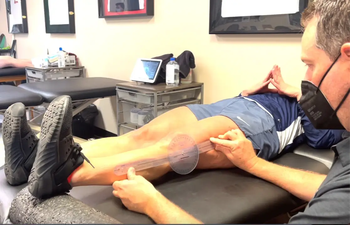
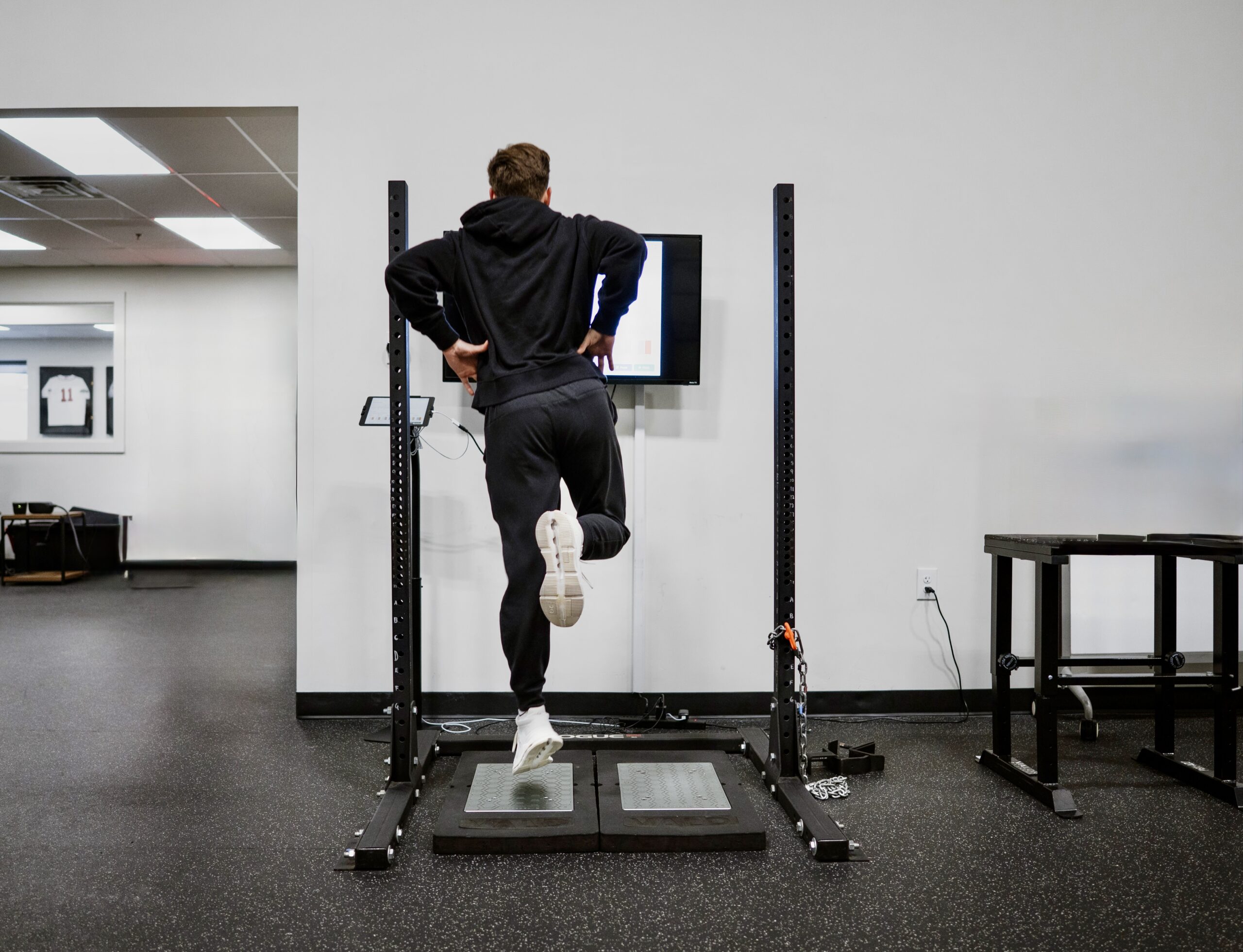
Simplifying ACL Rehab

ACL surgery continues to be a huge focus in the literature and in our outpatient rehabilitation settings. Numerous studies focus on return to play guidelines and retear rates. Social media is all over the place, most times. Let’s try to make things simple and set the stage early.

Paralysis by Analysis- What ACL tests are best for return to play

I previously wrote about return to play guidelines here  and even the neurocognitive challenges that are associated with ACL rehabilitation.

As troubling as it all seems, I almost feel as if the recent research is confusing and often unattainable in a traditional outpatient PT setting. I’m worried about the paralysis by analysis mindset that seems to have overtaken my recent discussions on social media.

Most don’t have an isokinetic device to test.

Hop tests, vertical jump, strength…what really matters?

Do hop tests really give us a complete picture of an athlete’s return to play readiness?

What combinations of tests are appropriate?

Is it more than just quadriceps strength?

What about a vertical jump test to determine lower extremity strength and function for an LSI?

Lots of questions!

Timeframes have shifted from a 4-6 month return to play goal to a more realistic 9+ months before we return people. Studies continue to show that a slower rehab may be better.

ACL retear rates still too high

Yet, our retear rates still remain very high and we’re still missing the boat for many.

I get it. It’s not just the timelines and rehab. Many are limited by equipment, insurance limitations, differing MD protocols, and so much more. It’s easy to blame one concept when it truly is multifactorial. 

That’s why I employ a very simple approach to my ACL rehab programs that seems to work for most and hope this concept can help you too.

It all really occurs at the beginning of rehab when you set the stage for the rest of your planning. If you can establish full motion, remove most of the swelling and restore a pretty normal gait, then you have achieved your goals.

From here, it gets fun and we’ll talk about that later…

An easier approach to ACL rehab

Immediately Post-op Phase

During the earlier stages of rehab, we’ll call it 6 weeks post-op, my main focus is calming the knee down and establishing a normal range of motion.

Range of Motion

It is critical to get the knee as straight as possible and as quickly as possible. For example, most people naturally have some 3-5 degrees of hyperextension. For these individuals, I like to get 0-3 degrees of hyperextension immediately and allow the rest to come through normal functional stresses.

For hypermobile patients, say 10-15 degrees of knee hyperextension, I will only get about 5-7 degrees of hyperextension because I know their underlying tissue mobility will allow the motion to return very easily. 

These individuals will not struggle with ROM and it often comes too easily. I don’t want to put unneeded stress on the graft.

Before you yell at me that knee extension will affect retear rates, a study in AJSM showed that the degree of hyperextension did not affect graft laxity and retear rates.

For flexion ROM, I like to have them seated at the edge of the table as I have shown in this video.

I just feel as if it’s easier on the patient and their knee instead of supine or prone, as I’ve described in the video.

I’m a bit obsessive with measuring ROM early, especially extension. It’s critical to stay on top of it and monitor for subtle changes in the motion and end-feel. Here’s an example of an ACL patient of mine who I recently saw and her post-session ROM.

measuring knee extension after an ACL surgery

Normalize Patella Mobility

You must also normalize patella mobility, especially in a patella tendon autograft. It is critical to regain this mobility in order for the normal motions to occur in the knee and to restore normal arthrokinematics.

I don’t push things too quickly and think this sets the stage for the rest of the rehab. In my hands, slow and steady is the best approach. A spike in volume can slow things down.

When I say a spike in volume, it could simply be a long walk or an extra bike session. The patients are often feeling good and looking for some independence and normalcy. They want to push it and we need to let them know that an increase in swelling or pain can create an issue.

Strengthening

I tend to go pretty slow with this concept as well. I tend to stay conservative for 4-6 weeks and stay with mainly table exercises like leg raises and mini squats.

I do love to use electrical stimulation (I prefer the DJO Global Continuum 2 unit!) for the first 2 weeks then add blood flow restriction training to compliment the NMES.

Not sure the research backs up my thoughts besides this 2015 paper or this one from 2018 but it definitely shows promise and makes sense in my head.

I’ll stay with these particular exercises for 4-6 weeks to ensure that the knee is calming down and my exercise progressions are not causing more pain or swelling.

Let the ACL rehab fun begin

If you’ve made it this far, then the important concepts have been met. No really!

The first 6 weeks set the tone and it can only go up from here.

For my patients, I basically turn into a strength coach and progress them based on muscle capacity and progress their programs based on movements and muscle groups.

I like to really hammer single leg work early but only after I’ve given them a good bout of 2-legged work to establish a base of movement.

Some do it the other way but I want to build confidence with 2-legged squats and/or deadlifts to reinforce a movement then use single leg work to take it to the next level.

When to run after an ACL

As I’ve gotten older in my career, I’ve gotten slower with my progressions. I typically don’t like to start running until at least 4 months after surgery and that’s if their quadriceps muscle is strong enough.

I look to this paper that shows a quadriceps strength to bodyweight ratio (QS/BW) of 1.45 Nm/Kg as a rough estimate to initiate running.

Some may argue that this paper only considers ACL patients that utilized a hamstring autograft and that’s a fair argument. But there’s limited information out there and I wanted something more objective.

So, until they can establish a good quadriceps contraction and their ROM/gait are normalized, I will hold off running.

ACL rehab is mainly about strengthening

The cat’s out of the bag…don’t tell anyone!

It’s pretty simple, once you get through that 6 week interval that we talked about earlier.

If you’re not proficient in this, find someone who is in your area.

Otherwise, program using simple strength training principles that incorporate power, velocity, full ROM and tempo. Work on their aerobic capacity while building strength, power and endurance.

I could write another full blog post on this but we touch upon these concepts in our all online knee course.

Hope this helps!

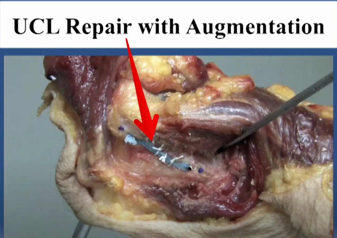
Diagnosed with an elbow UCL Tear- Reconstruction or Internal Brace surgery?

So, you’ve been diagnosed with a UCL tear in your elbow and your world has been turned upside down. Have no fear, many have been there before you and have done pretty well.

But now, there’s a new option for elbow surgery and you’re not sure if it’s right for you. I hope this post can help you decipher the jargon and ease your mind a bit.

Tommy John Surgery

Tommy John surgery has been around since 1974, when legendary Frank Jobe performed the 1st surgery on pro baseball pitcher, Tommy John (shocker!)

It was a pretty epic failure and required a subsequent surgery to fix some of the original issues (massive claw hand due to ulnar nerve issues). Tommy John did return after numerous surgeries to have an amazing MLB career. This set the precedent and baseball has not been the same since.

Fortunately, we have done much better overall. We have improved our surgical techniques and rehab outcomes. Nearly 80%+ of baseball players that have the reconstruction surgery can return to a pretty high level of function. By definition, they will play at the same level or higher compared to their pre-surgery level.

If you don’t believe me, then I suggest you read this article right here. I can attest that the TJ patients that I have rehabbed over the years have done very well. The road is long but most can get back to nearly 100% at around 9-12 months.

Tommy John Surgery Video

Curious to know what the reconstruction surgery looks like? My colleague Dr. Chris Ahmad (who we work with a bunch) presented on his surgery technique recently. Watch this video (as long as you’re not squeamish!) and enjoy!

UCL Docking Technique by Dr Chris Ahmad

I usually tell my patients to fast forward 12 months and we can count backwards to figure out the path that we’re going to take.

But that’s not why you are here, right?

You want to know about the internal brace surgery that has taken the TJ world by storm!

Elbow Internal Brace Surgery

This is a relatively new procedure that has been around since about 2013. My friend and colleague Dr Jeff Dugas began doing this procedure in Birmingham, AL when I was down there. I got to see the early results 1st hand and was excited but skeptical.

Baseball players were returning to their sport in 6-8 months versus the 1 year that we had seen in a full TJ surgery.

Why a quick return after surgery?

Why have them return sooner if the surgeon is not using the patient’s native tissue?

Who should get this surgery anyway?

We didn’t necessarily know, but like any other orthopaedic surgery, we had to wing it a bit. We had to figure out a protocol that was appropriate for the tissues involved.

I was there in Birmingham with another friend and colleague, physical therpaist Kevin Wilk. I helped put together the early protocols and was excited by the potentials.

The thought is that the collagen dipped tape that is re-enforcing the repaired ligament is stornger than the native ligament. Its fixation to the bone is strong. A quicker return to throwing and sport is possible, because of those reasons.

Its worked for the ankle and so why can’t it work for the elbow too?

Internal Brace Surgery Specifics

This internal brace surgery was developed by surgical company Arthrex. It’s pretty neat to see how it has taken on a whole world of uses, including in the knee and ankle. Its many uses has helped numerous athletes return back from their injuries quicker than ever.

See Tua at the University of Alabama, for examaple. He had a high ankle sprain and retuned to the field ~3 weeks after his ankle surgery. Again, surgery performed by my friends Dr Norman Waldrop and Dr Lyle Cain, of Andrews Sports Medicine and Orthopaedic Center. They’re studs and surgeons that i’d highly recommend!

Who benefits the most from Internal Brace surgery?

For this elbow surgery, the internal brace is most appropriate for the athlete that has a UCL sprain that is not complex. Most times, they won’t know until they’re in the surgery if the internal brace is appropriate.

If it is appropriate, then surgical consent probably happened before the surgery. The doctor won’t know if the repair is appropriate until he/she can visualize the tissue directly. A decision is made on the spot even if the MRI said something differently.

Why try the internal brace?

I basically recommend this surgery for my athletes who don’t have a lot of time.

Let’s say they sprain the ligament in the offseason, like in November of their junior year in high school, for example. If a full-blown TJ reconstruction was done, then they’d be out until at least the following November (remember my 12-month comment earlier). That would mean no junior year baseball or Summer ball. That would also mean no exposure for college recruiting!

But wait, there’s an alternative! Internal Brace repair surgery…see the video here!

…or here by Dr Jeff Dugas:

Elbow Internal Brace Procedure by Dr Jeff Dugas

In the internal brace situation, you could have the surgery in November and be back for some of your High School season and most likely for that important Summer travel season.

But remember, the ligament can’t be chewed up a lot. That decision will be made intraoperatively. Be ready to wake up from surgery with news that a full TJ reconstruction had to be done.

But for many, an internal brace repair is possible. And a quicker return may be possible too.

Should you do this surgery?

For those considering it and fit the requirements, then I’d recommend it. Just keep in mind that we really don’t have too many long term outcomes.

But for the High School or College athlete looking to play a few more years, then I’d say go for it.

For the HS or college pitcher who has aspirations of playing pro ball, then I’d recommend the full reconstruction. We just know more about the surgery and long term outcomes. it’s tried and true in every way.

That’s not to say that the internal brace procedure cannot be the gold standard surgery in a few years. That is quite possible. I really hope to update this blog post in the future and say that I was wrong.

But as of now, I would recommend the reconstruction for the pro athlete or amateur athlete looking to play pro ball. Otherwise, the internal brace procedure is a very strong option for many pitchers (or even positional players looking to get back quicker).

Summary- Who should get this surgery?

Pitchers or positional players that don’t have much time before their next season and NEED to play. But the tissue needs to be repairable and not beat up (this is the key!)

Consult your surgeon to discuss this but they won’t know until they’re in your elbow and you’re out cold from anesthesia.

Good luck…it’s a long and winding road but most do well. I’ve treated a lot of these cases and no 2 are ever the same. There’s always a glitch and a freak out period but it often works out in the end!

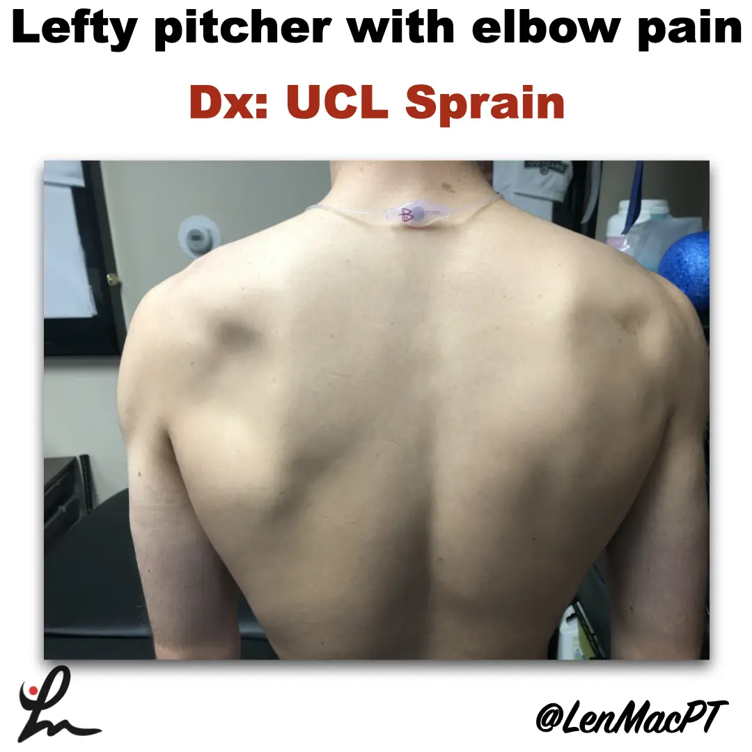
Infraspinatus atrophy in a baseball pitcher with a UCL sprain

Infraspinatus atrophy due to a spinoglenoid notch cyst

This was an incidental finding of mine in a collegiate baseball pitcher. He presented with a UCL sprain during his senior year and was struggling.

He came in looking to salvage his senior year and attempt to pitch at some point. UCL surgery would’ve been warranted if he was anything but a senior.

UCL Evaluation

We evaluated his elbow and he presented with typical findings of painful valgus stress tests. I’ve discussed my UCL exam previously in a post that you can find here.

UCL Tommy John Assessment

His shoulder PROM was slightly limited compared to what we normally see in our overhead throwers, which may have contributed to his elbow issues.

He only had 120 degrees of external rotation compared to 110 degrees on the right side. If you’re wondering how I measure external rotation, then this video below may be a good one to review.

External rotation passive range of motion (PROM)

We use these concepts for our research studies that we have published on weighted ball usage the past few years.

For those that will ask about GIRD, he had 60 degrees of IR on the L side and 50 degrees on the R side. I’m not a big GIRD guy like has been written by others. I still think GIRD is pretty normal and our research has shown that too.

Remember, we showed that differences in total rotational range of motion greater than 5 degrees was significant. This study was published in AJSM back in 2011.

Furthermore, we showed a connection between the loss of passive flexion and elbow injury.

This result was also shown by Camp et al in a separate study, as well.

I had him remove his shirt and noticed a large defect in the infraspinatus area that showed obvious atrophy (see picture below).

Infraspinatus atrophy due to a spinoglenoid notch cyst

The cyst was compressing the suprascapular nerve as it coursed along the spine of the scapula.

His external rotation strength was significantly weaker on his throwing side compared to his right side. This would make complete sense considering the amount of atrophy present.

Often times surgery is warranted to decompress the nerve. There’s often an underlying labral tear that has resulted in a cyst to form. The surgery would remove the cyst and repair the labrum to prevent future issues.

Quick Literature Review on Surgical Outcomes

Reports of improved outcomes are in the literature, here and here and here.

Obviously, this may have contributed to his elbow issues as the posterior cuff muscles are tremendous stabilizers of the arm, especially during the deceleration phase of throwing.

Treatment for infraspinatus muscle atrophy

Our goal was to calm the elbow down through active rest and focus on strengthening of his forearm and shoulder musculature.

We used neuromuscular electrical stimulation (NMES) to the infraspinatus. We felt as if the electrical stimulation would allow for a better muscle contraction than what he could do actively. This is a similar concept to using NMES to the quads after a knee surgery.

NMES to the infraspinatus to help with muscle recruitment

We have previously shown in AJSM that NMES to the infraspinatus was beneficial to patients after a rotator cuff repair. It helped improve their muscle contraction by 22% in the group with the NMES applied during testing.

Outcomes and final thoughts

Believe it or not, we were able to salvage his senior year despite all of the issues that presented.

He made numerous pitching outings and was pretty successful for his team.

The lesson here: Don’t take anything for granted, observe and test.

Make sure you look distal and proximal. It could’ve been very easy to just treat the elbow with some exercises and massage. Don’t get me wrong, we did a bunch of strengthening exercises to his forearm.

His secondary finding was most likely a leading cause for his elbow dysfunction.

This isn’t the 1st time that I’ve run across an issue like this. I’ve found numerous scapula dysfunctions like this involving the infraspinatus. But also a winging issue due to a long thoracic nerve palsy.

You must look at the whole picture… shirts off for a guy and a sports bra for a female to make sure we’re not missing anything obvious!

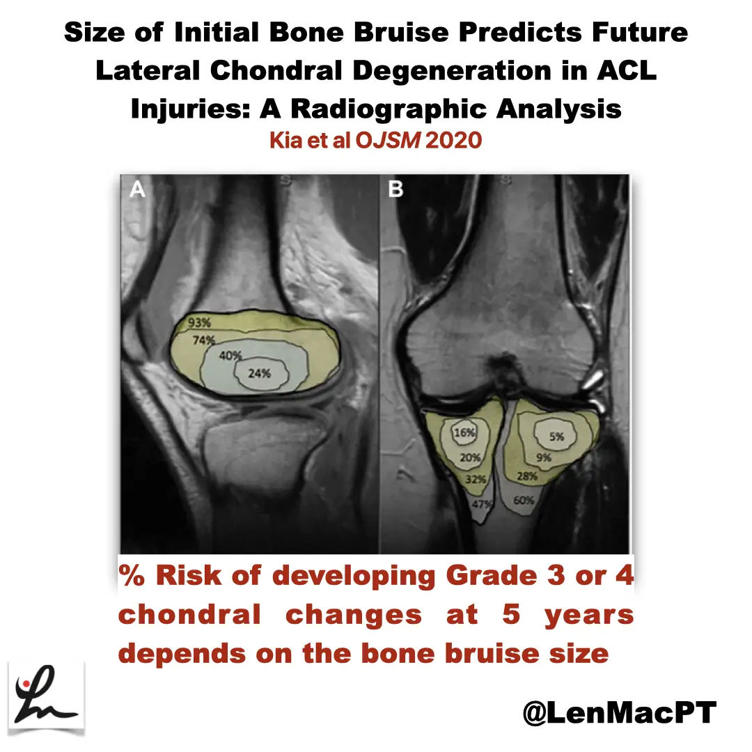
ACL tears and bone bruises

Research Review

Not sure if you saw my recent post on social media about bone bruises after an ACL tear so I wanted to discuss it further here.

In this study, the authors looked at the incidence of radiographic chondral changes (without correlation with clinical and functional outcomes) on MRI 5 years after the ACL tear.

Bone bruises often coincide with an acute ACL tear and can be seen on an MRI. Basically, a larger bone bruise was shown to have a significant influence on chondral changes 5 years post-surgery.

In most of the cases, the lateral side of the knee (both the tibia and the femur) were involved. Whereas the medial side had fewer incidences of a bone bruise after the injury.

Outline of a bone bruise on the distal femur and proximal tibia after an ACL injury

This is very typical after an ACL injury and previously cited by numerous authors.

I’ve always said that these bone bruises need to be communicated throughout the rehab team. These bone bruises should influence the progression back to sport.

Delay impact activities after ACL surgery

It is for this reason that I have delayed most of my athletes’ return to impact activities until 4-5 months after the surgery. I often don’t initiate running and plyometrics until 4-5 months after the surgery.

We need to respect the bone bruise healing times (which are still not truly known). They seem to take months to achieve homeostasis, which means no pain or swelling.

Only another MRI would truly confirm full healing so we need to rely on symptoms, most of the time.

I think this may allow the athlete to achieve better long-term success. But we need to understand these bruises more before we can fully determine the correct rehab process.

I think the long term life of the athlete’s knee can be influenced by our rate of rehab progressions. To me, slower seems to be better in these situations.

Gone are the days of trying to return our athletes back to their sport as quickly as possible.

Take the time to get their motion back, especially knee extension.

We are beginning to better understand the implications of these bone bruises on the long-term health of the athlete’s knee.

Educate the patient fully and build confidence! Rehab after an ACL surgery is never easy so don’t take anything for granted!

Lumbar Spine Stress Injuries in Baseball Players

Low back pain can be very debilitating in an athlete, especially in an adolescent baseball player trying to make it through his season. More specifically, I have noticed an increase in lumbar spine stress fractures in baseball players and it is quite disturbing and frustrating!

I wanted to dive deep into this rabbit hole, as I always do. Low back pain is way too common in youth athletes. I wanted to know why it’s happening so often and try to figure out a solution for my clients.

It can get complicated so I will try to simplify the process.

My goals:

  1. What is a stress fracture or a stress reaction?
  2. How common is this injury in youth sports compared to other sports and age groups?
  3. What is the typical clinical presentation?
  4. What are some clinical tests that PT’s can use in the clinic?
  5. Demonstrate a typical treatment of a young athlete with a low back stress injury?
  6. What are the expected outcomes?

Pain is Complicated

Low back pain can take on many etiologies. Pain, in and of itself, is extremely complicated and very personal.

It can manifest in different ways for each person. In this post, I want to talk about the athlete with an acute injury. Specifically, the youth and adolescent baseball player.

Many factors can contribute to someone’s pain including stress, anxiety, medical history, their environment, support system, etc.

This topic is a bit more personal because of what do on a daily basis. I see a ton of youth athletes with a variety of injuries. Plus, I was diagnosed with spondylolisthesis (bilateral fracture in the spine and some shifting of the vertebral body) while a junior in college so I’ve lived it myself!

Recently, I’ve started to see a lot more athletes with low back pain as a result of a stress reaction or fracture. It really made me wonder what was truly causing this in the 12-18 year old athlete.

Video example

courtesy of pitching coach Anthony Meo of The Farm Baseball Waltham, MA

As you can see in the video, the pre-injury video shows how long the pitching motion is for this athlete. His throwing arm is wrapped around his body which forces him to keep his weight back.

His arm is trailing his body significantly and he tries to make up for it by hyperextending his back. His timing is way off and his arm is not keeping up with his forwardly moving body.

You can see in the post-injury video that he is much more balanced. His arm also has better timing with his body. He is driving toward home plate with his lower body while maintaining a better upper body sequence

There are many issues going on here but you can see the significant changes that were made after the injury to help him improve his mechanics and hopefully prevent a future injury.

LUMBAR STRESS INJURIES DEFINED

Stress Reaction Defined

Basically, a stress reaction is the early breakdown of the bone without a clinical diagnosis of a true fracture. Often times, a suspected fracture will be diagnosed as a stress reaction. On MRI, there is only edema noted in the area and not a true fracture.

I hear a lot of people talk about fractures in different terms. Simply stated, a fracture is a crack, break, defect in the bone. Whether it’s a hairline fracture or a gross disruption of the bone may determine prognosis but the bone has been compromised.

taken from: Link
taken from: Link

Obviously pain is an early sign but it needs to fit the clinical presentation. Has there been a history of excessive activity or a change in the volume of playing?

Factors to consider in a baseball player with low back pain

I usually have to dive a bit deeper into the player’s life to figure out the root cause. It’s often something very overlooked but is often the main contributor.

These are just some, off the top of my head, that may be involved in the athlete’s predicament and that may nee to be changed.

It is an early indicator that something is wrong and it can progress to a stress fracture if not carefully treated. We’ll talk treatment later on so be patient!

How common is a stress reaction in youth sports?

As with any research, it is quite mixed and will heavily depend on the sport that you are tracking. From what I can see, if a kid has low back pain for greater than a week, then that’s a pretty good sign that a more serious injury may be present.

For example, this retrospective case study from 2017 showed that 30% of pediatric cases with low back presenting to this hospital were diagnosed with spondylolysis.

Specializing in a single sport was not associated with an increased risk of spondylolysis when compared with multisport athletes. Male athletes were 1.5 times more likely to have a spondylolysis than female athletes.

Males with LBP who participated in baseball had the highest risk of being diagnosed with a spondylolysis, followed by soccer and hockey.

Females with LBP who participated in gymnastics had the highest risk of spondylolysis, followed by marching band and softball. I will refer to my good friend Dave Tilley and his website for his professional judgment on this one. Based on what I see at our facility, I’m not surprised.

As I usually tell my clients and their families…I’m going to treat this as a spondy until proven otherwise.

More Research on Spondylolysis Prevalence

According to recent research, it looks as if persistent pain for 1-2 weeks is associated with a positive MRI diagnosis for spondylolysis in 33-40% of the kids scanned. You can read the abstracts here and here if you’re interested in more.

This study out of New York showed that the most common athletic activities associated with spondylolysis were:

  • Soccer (19.3%)
  • Basketball (17.2%)
  • Lacrosse (9.4%).

Of note, 71% of the children in this study were male and the most common level to have the spondy was at L5.

Do keep in mind that false positive results are pretty prevalent in patients getting an MRI for low back pain.

Don’t treat the MRI (or x-ray!)

This systematic review looked at the incidence or prevalence of incidental findings on MRI in asymptomatic pediatric patients. The results are pretty eye-opening too!

  • Degenerative disc disease (prevalence 19.6%)
  • Disc herniation/protrusion (prevalence 2.9%)
  • disc height/narrowed disc space (prevalence 33.7%)
  • Spondylolisthesis/spondylolysis (prevalence 2.3%)
  • annular tear and/or nerve root compression (prevalences ranging from 4.5-51.6%)

Pretty amazing to see the prevalence of false positives read on an MRI in a pediatric population. We need to be able to clinically diagnose these first then use the MRI scan to further determine a course of action.

Baseball specific injury rates

In this study from 2015, the proportion of High School baseball players with a low back stress fracture was 3.3%. So, a typical AAU baseball club of 100 kids will have 3 kids that have a stress fracture.

They also reported that High School girls sustained more stress fractures (63.3%) than did boys (36.7%) but for overall injuries and not just in the low back. This seems consistent with what I see in my practice, as well.

In college, 12.1% of the stress fractures noted in this NCAA study from 2017 were in the low back. Women experienced stress fractures at higher rates than men, more often in the preseason, and predominantly in the foot and lower leg.

Baseball Specific low back pain

This nice review of the literature discusses low back pain in general athletes. They went on to further discuss baseball and “the asymmetric baseball postures and motions lead to asymmetric spondylosis in right and left sides and cause mechanical degeneration in intervertebral discs.”

In this study, “students who played baseball were 3.2x more likely to experience LBP in their lifetime only behind volleyball which had an odds ratio of 3.8. 

The repetitive nature of the baseball specific movements and a preponderance to specialize in one sport can definitely affect these injury rates.

The role of mental stress and low back pain

The role of stress, particularly mental stress on bone development, has been studied too. Our teenage athletes are under a tremendous amount of stress at home and socially. This study from 2017 discussed the emerging concept of mental stress as an important player in bone adaptation and its potential cross-talk with physical stress.

Essentially, mental stress can have a negative impact on normal hormone production. Hormones are needed to maintain normal bone health. A shift in normal hormone regulation can negatively affect a youth’s growth and healing capabilities.

They went on to say that there is “compelling evidence has recently emerged that biochemical and psychoneuroendocrinological maladaptations caused by mental stress are not only also relevant for bone quality, but may furthermore considerably interact with physical stress.”

Clinical Presentation of a Spondylolysis or Spondylolisthesis

Clinically, pain is the guide here. It’s often described as a pretty sharp pain that is localized to the site of the injury.

There is rarely a referred pain down the leg or the buttocks. The pain is often close to the spine at the level of the injury.

In this study, activities involving repetitive hyperextension and/or extension rotation of the lumbar spine were described as painful in 98% of the patients.

Pretty powerful statement. In my examination, I look to recreate these symptoms with these exact movements.

Many use a single-legged position, also known as the Stork Test, to test for spondylosis. I’ve simplified the test to just include regular standing on 2 feet to simulate regular daily movements.

A standing extension test can also be used to assess end range extension and rotation irritability. I find this test (see video below) is a great way to begin to hone in on a diagnosis.

I find that pain in full hyperextension and rotation is a pretty powerful indicator of a stress reaction or fracture. It is similar to the standing position that I described above. However, the prone position takes the postural component out of the picture and may help to better isolate the location of the lesion.

I also like to have the patient get in the prone position and use different levels of active extension to help guide my clinical examination.

Again, people will report pain that is very close to the spine and that replicates their pain.

Any young athlete with low back pain for greater than 1 week should be assessed for some kind of bony issue in their low back.

That’s not to say that we will always find something.

But I just think that in younger athletes, this prevalence is all too common and needs to be ruled out to prevent further low back issues.

Treatment for Spondylosis in Baseball Players

Muscle tenderness too

Patients will usually have tenderness right at the region in which they have pain. I suspect it’s the muscles that stabilize those lumbar segments.

The multifidi and most likely quadratus lumborum are often implicated. Soft tissue work of any kind can have positive effects on muscle tightness. The athlete should perform some self-myofascial release with a foam roller or light pressure with a baseball in the affected muscles.

Manual therapy can also be utilized to feel tissue density and help to pinpoint the exact location of their muscle soreness. By utilizing this, I can help to better educate the athlete for their home exercise programs.

Manual Therapy can be helpful

In this photo below, you can see I am applying direct pressure with my elbow right into the muscles of the low back. The patient will usually give me feedback and tell me if I am in the right area. Muscle soreness is often detected and after a short amount of time, the symptoms should decrease.

In the short term, manual therapy can help with some of the acute symptoms. For long-term changes to occur, activity modification and exercises that involve low back, hip and core strengthening are critical.

Soft tissue work to the muscles of the low back in an athlete with low back pain.
Soft tissue work to the low back in an athlete with low back pain.

My treatments for kids with low back pain are pretty simple. I want to rest them from the repetitive activities, such as swinging a bat or even throwing a baseball.

Brace or no brace?

Also during this time, the athlete will most likely have to wear some form of a back brace to prevent excessive lumbar extension and rotation. One particular brace is called a Boston Brace. It was developed by doctors at Children’s Hospital in Boston, MA.

The research is unclear and can favor both methods of treating with or without bracing. A study in 1986 showed that bracing ‘restricted at least some gross body motion to approximately ⅔ to ½ of no-orthosis values.’ The restrictions we’re most pronounced when wearing a larger Thoracolumbosacral orthosis (TLSO) versus just a corset.

If the goal is to restrict motion, then it seems as if a brace will accomplish that. Most research shows about 40-65% restriction in motion no matter the brace. This paper showed similar findings as well.

Here’s a version that our local docs have been using that is less cumbersome and not as embarrassing to wear as a teenager!

Brace used to help restrict motion during a low back stress fracture

These are just some of my quick and easy exercises to help promote movement in the spine.

Obviously, I want to avoid painful motions like lumbar hyperextension and rotation. I still want to promote some sort of motion to build confidence and promote muscle recruitment in that area.

Specific treatment ideas for low back pain

I like foam rolling or some form of self-myofascial release to the area. This will help the muscles to feel better if done multiple times per day. See the video below

Some would say motion is lotion and I tend to agree! I like to use active range of motion through the cat-cow video below.

Cat-Cow exercise to promote active motion in the low back

Some general guidelines for returning to baseball

From what I’ve seen, most baseball players need at least 3 months of active rest.

I’ve written about my general low back treatment philosophies in a previous blog post.

You can find that post here .

Active Rest is key

I generally want to begin a general strength training program at around 6-8 weeks following the diagnosis. It Is critical to include restricted range of motion exercises to avoid painful hyperextension. These exercises are restricted to avoid excessive extension that may place extra stress on the low back. I prefer to bias flexion type exercises like squats, lunges and step-ups.

I like to closely monitor their form and symptoms. We don’t want any pain at all in that region…a very important concept!

I like to perform these exercises for at least six weeks to get a good base of strength in the lower body and core.

Lose the brace at 12 weeks

At about 12 weeks, they will come out of the brace and we can begin to be a bit more aggressive in our strengthening. Again, we are avoiding hyperextension type movements in the low back. We prefer to stay in a flexed or neutral spine as much as possible.

I also like a supine dead bug exercise to promote a flexed spine with active motion. See the video below.

Throughout this time, we encourage cardiovascular exercises that involve the stationary bike. This seems to be a safe modality that allows the athlete to work up some sweat but also maintain a relatively flexed spine, which is usually pain-free.

Unrestricted activities at 5-6 months

At around 5 to 6 months post-diagnosis, we encourage for pain-free activities that involve strength training for the whole body. There are no limits in range of motion in the athlete is free to work out. We then encourage more functional activities that involve rotation. This will allow them to replicate the motions involved with hitting and throwing.

Return to sports at 6-8 months

On average, it seems as if a patient recovering from a low back stress fracture will need at least 5 to 6 months before I would feel comfortable having them return to their support. These injuries usually happen in a younger population so the risk for re-injury is pretty high if they return to their sport too quickly.

I believe the risk is high because of their underdeveloped muscular system. Their bodies are growing and tremendous stress may be placed upon their joints with excessive activities. A slow progression back to their sport is critical.

Concluding thoughts on stress fracture injuries in youth athletes

Baseball players face numerous challenges during their training.

As you can see, numerous injuries can result and not just to the shoulder and elbow.

Low back pain injuries are all too common but are often preventable. It seems as if the volume of throws and hits may play a huge role. It is up to the coach, physical therapist, or athletic trainer to monitor this workload for each athlete.

There is no exact science but listening to the athlete is critical. A well-balanced training program that addresses some of the obvious weaknesses is key.

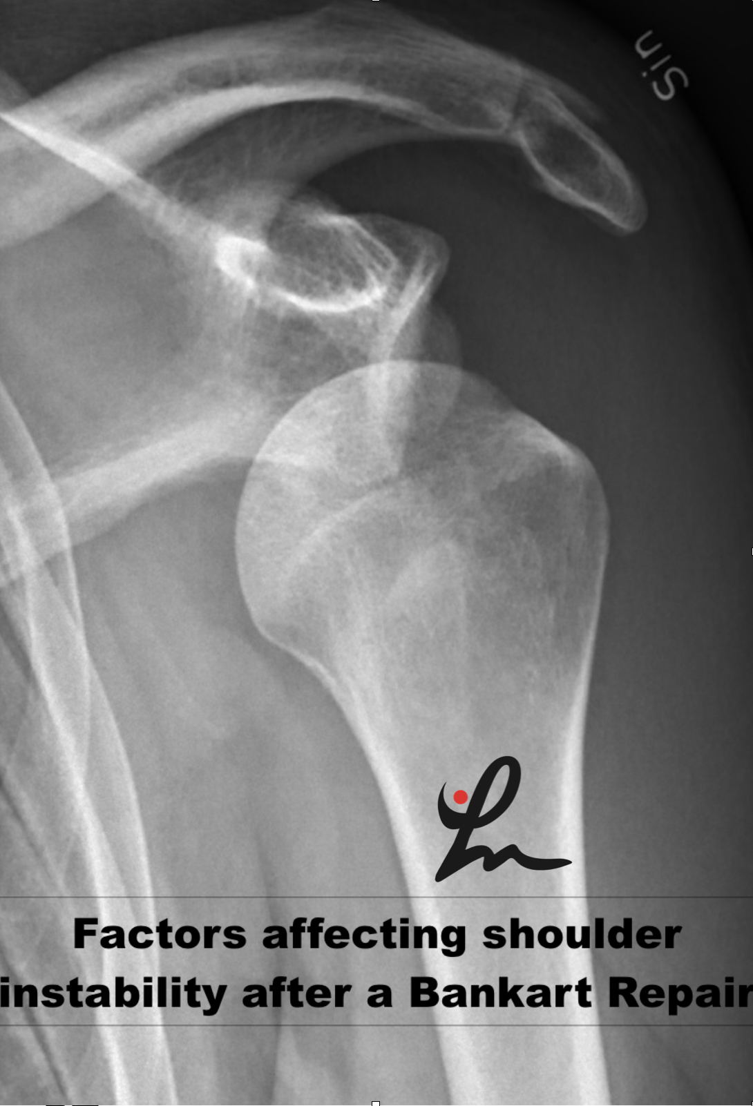
Risk Factors for Recurrent Instability After a Bankart Repair Surgery

Recurrent instability after a Bankart repair surgery is unfortunately very common. This paper looks to highlight the most common risk factors associated with recurrent instability.

I thnk it’s valuable to understand these various risk factors so you can better educate your patients. It may also help clinicians be mindful of the people that may need to have their rehab modified appropriately.

I see a lot of high school and college students that have had a shoulder injury. In this population, they’ll specifically have a dislocation event.

If there is one factor that you should consider in educating a patient about surgery or not, it is their age.

I’ll review this paper from The Bone and Joint Journal and let you know what you need to consider when a patient presents to you with an acute shoulder dislocation.

Age Influences Recurrence Rates

A patient’s age is a huge factor in determining whether or not they will have a recurrent instability episode. And people younger than 25 years of age, I typically recommend a surgery to stabilize the shoulder joint and prevent future issues.

Hovelius et al has shown that patients in their 20’s exhibited a recurrence rate of 60%, whereas patients in their 30’s to 40’s had a recurrence rate of less than 20%. 

Unfortunatley the long term prognosis in these people does not seem promising. They often develop some form of a shoulder arthropathy, as seen in this study by Hovelius in 2016.

That’s not to say that surgery is 100% required. In this study, they showed that ‘after 25 years, half of the primary anterior shoulder dislocations had been treated nonoperatively. And in these patients with an age of 12-25 years, many had not had any recurrences and had become stable over time.

What are the risk factors for recurrent instability or revision surgery following arthroscopic Bankart repair?

This paper ‘sought to determine the rate and risk factors associated with ongoing instability in patients undergoing arthroscopic Bankart repair for instability of the shoulder.’

They looked at 5719 patients with a mean age was 24.9 years, which is pretty much what we see in the clinic.

Nearly 10% of patients (8.1%) in this study had to undergo a 2nd surgery at a mean of 31 months post-operative. So, the 1st 2 years after a surgery is critical, just like in the ACL literature.

Patients between the ages of 10 and 19 had the highest rate of subsequent procedures (11.0%), and comprised over half the patients (53.8%) undergoing a revision procedure or closed reduction.

They also went to conclude that:

  • Younger age,
  • Caucasian race,
  • bilateral instability,
  • and closed reduction prior to the initial repair were independent risk factors for recurrent instability.

They also showed that a 2nd arthroscopic surgery had significantly higher rates of persistent instability than subsequent open revision procedures.

Treatment for shoulder instability

In this post that I wrote for Medbridge Education, an online continuing education company, I discuss my progression for a rotator cuff related issue.

You can use this progression when developing a plan for these patients that have had an instability episode. These progressions are used to treat both non-operative or post-surgery patients.

There are so many different variables to consider when trying to initiate physical thrapy. I tried to outline them below.

This table outlines some of the variables that Kevin Wilk and I came up with in a recent paper in Clinics in Sports Medicine journal in 2013.

Key factors to consider in the unstable shoulder

Conclusions from this paper

This paper should help you to better understand the populations at risk for recurrent instability. I try to use these papers to educate my future patients that are considering a surgey.

Keep in mind, surgery should be saved for only those that truly need it. Physical therapy can often be employed in most patient populations.

Be mindful of the patients that wuld most benefoit from surgery. Confidently educate them that their decision will be the best for them to return to their function.

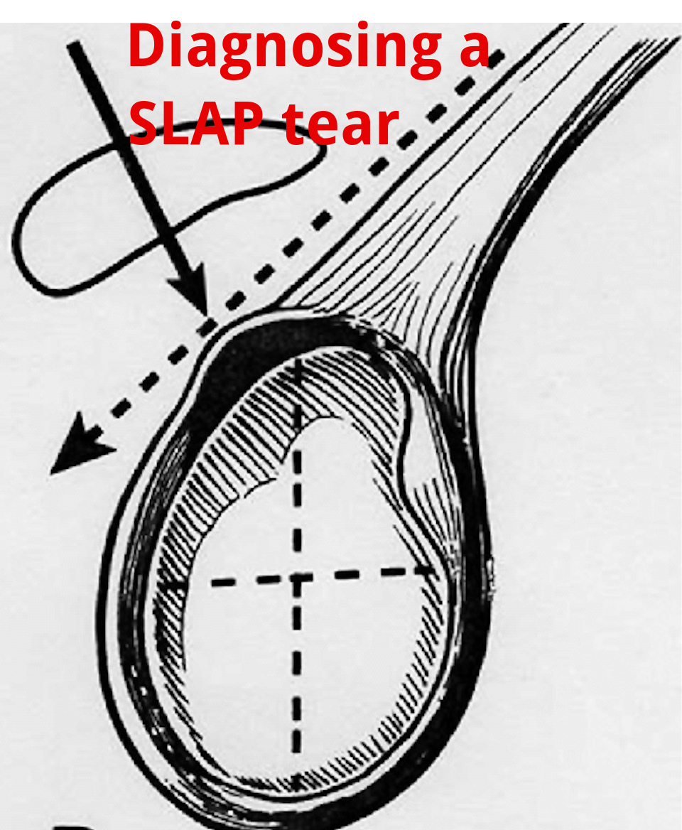
An Update on Diagnosing SLAP tears

Diagnosing a SLAP tear is not easy

We hear a lot about trying to diagnose shoulder pain and to be as specific as possible. It’s often difficult to differentiate SLAP (superior labrum anterior to posterior) tears from other soft tissue injuries of the shoulder.

For a review of the different types of SLAP tears, check out this old blog post that classifies the 10 different types of tears.

from https://commons.wikimedia.org/wiki/File:SLAP-Lesion-front-2.jpg

This paper in IJSPT by Clark et al 2019 attempts to help out the process and recommend a few special tests that MAY aid in diagnosing a SLAP tear.

What do they Recommend to diagnose a slap tear?

They recommend that a combination of at least 3 positive SLAP lesion tests may be clinically useful in diagnosing a shoulder SLAP lesion with greater diagnostic accuracy.

Combo of Tests

The combination of the Biceps Load I/II and O’Brien’s showed the highest sensitivity and specificity.

I have found similar results with this set of special tests so maybe this paper just hits my biases correctly.

In this video at my YouTube channel, I wanted to let you hear my thoughts and small tweaks to the evaluation process.

It’s not easy to diagnose a SLAP tear.

Differential Diagnosis is Critical

Furthermore, does it really matter and will it change the treatment plan much at all? I think it may a little but overall it will remain a pretty similar treatment approach to other similar pathologies like:

  • rotator cuff tendonopathy
  • Biceps strain
  • Latissimus strain
  • Subscapularis strain
  • internal impingement
  • pectoralis major strain

I think one also needs to consider the cervical spine and to make sure the pain is not referred from the neck.

Otherwise, a well thought out program should be implemented that addresses the strain on the shoulder and any strength issues.

I talked about this in an article that I wrote for Medbridge a while back so check out that post here:

You can also check out a snippet of one of my courses at this YouTube video where I discuss rotator cuff and labral issues. Hope it helps too!

Will this Change your Practice?

How ever you look at it, I wanted to use this paper to let you know that there MAY be a cluster of tests that better diagnose a suspected SLAP tear in your next patient’s shoulder.

Check out the paper and comment so we can talk it through. Are these tests similar to what you use in your clinical practice? Will this paper change what you do in your clinical practice?

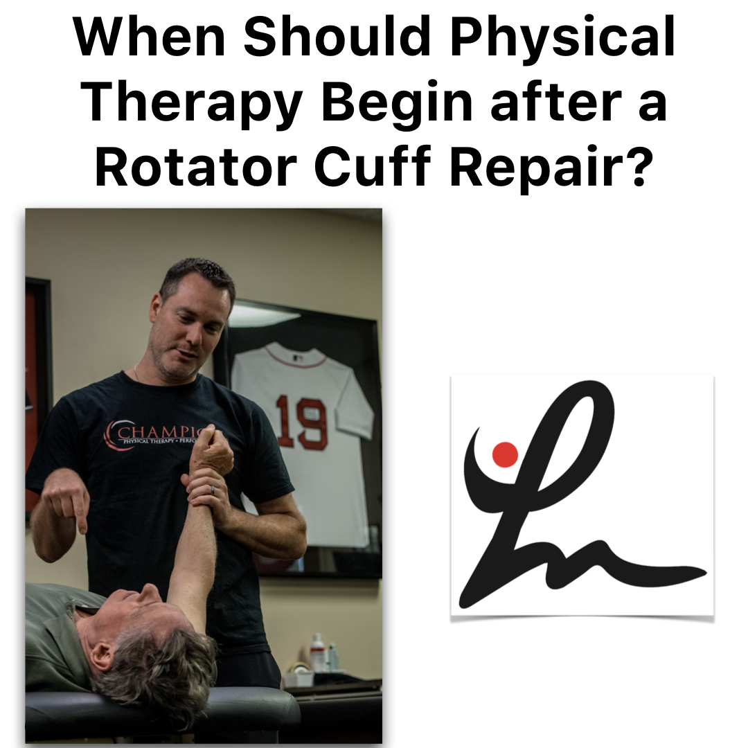
Is Early Physical Therapy Safe After a Rotator Cuff Repair?

Physical therapy is vital after a rotator cuff repair and continues to be common in an outpatient setting. Unfortunately, there’s no true consensus on when to actually begin PT.  Is early physical therapy safe after a rotator cuff repair or should we delay PT to protect the healing tendons?

Let’s dive into this and see what the research is saying…

Why I’m writing this post on rotator cuff rehabilitation

It seems as if we’re all over the place despite the research, which is pretty typical. Some docs prefer early passive range of motion (PROM) while others wait 6-8 weeks (and even up to 12+ weeks for a revision repair) before they allow any form of PT.

For the record, I’m going into this blog post as a firm believer of early PROM. It’s what we’d been doing for years in Birmingham at Champion Sports Medicine.

It’s what I only knew until I moved to Boston in 2014. Now I’ve seen a nearly 180-degree turn in rehab thoughts. Much more conservative!

I recently got in a Twitter discussion (debate) about this same topic with some very respected and prominent PT’s in the field.

It made me think about things so I decided to do a little research to see what the literature says. You can check out the discussion here.

Twitter can be confusing and tough to follow but just trust me, it goes on for a while!

Rotator Cuff Anatomy

The tendon most commonly torn is the supraspinatus tendon. Don’t get me wrong, you can tear the other rotator cuff tendons (infraspinatus, teres minor, subscapularis).

Keep in mind a medium, large or massive rotator cuff tear often will involve the infraspinatus tendon. If it does, then you need to consider modifying your progression appropriately.

But for the sake of this post, I’m going to stick to an isolated supraspinatus tendon for now.

Rotator Cuff Tendon Size and Location

Look at the size of the tendons as they insert on the greater tuberosity. You’ll see it differentiated by antero-posterior and medial-lateral directions.

According to my colleague and friend Jeff Dugas out of Birmingham, he showed in 2002 that the mean dimensions of the supraspinatus insertion were 1.27 cm in the medial-to-lateral direction. For the anterior-to-posterior direction, the supraspinatus dimension was 1.63 cm.

It helps to know this information because you may need to read an operative report and see the size of the tear. The docs will usually mention a 2 cm tear or something like that.

That means that the supraspinatus tendon and a small portion of the infraspinatus tendon were involved (and repaired.)

This is critical information to have when you’re trying to plot the post-op rehab progressions and determine the prognosis. The more tendons involved, then the higher the chance of repair failure.

There are many other factors that influence retear rates but tendon repair size is definitely one to consider.

Rotator Cuff Repair Surgery Types

I’m not going to bore you with the details of a repaired rotator cuff. There are numerous surgical techniques being used by orthopaedic surgeons.

Techniques such as a single row, double row, suture bridge or transosseous repairs are commonly performed. The picture below shows the difference between a single row and a double row repair, for example.

As you can see below, the double row tends to repair more of the tissue back to the humeral insertion point, which in theory has led to better tendon healing. This has been shown in numerous research studies and has become the best technique available.

So you had shoulder surgery…when to start physical therapy?

That seems to be the million dollar question! The research is all over the place. This means that doctors’ opinions are all over the place too, right?

Keep in mind that I’ve written about this in the past. Rotator cuff repair surgery is not always warranted and can be avoided!

Since I joined the group in Birmingham in 2002 (as a PT student), we had our post-op rotator cuff repair patients starting PT post-op day 1. They started PT early regardless of the tear size. This means a small tear of 1 cm in length started PT the same time a massive, 5 cm repair would start PT.

Some may disagree with this start time but it worked…it just worked. At least I think!

Why Early PT after a Rotator Cuff Repair

This is Key!!

There were several reasons why I think it worked:

  • They could chat with a professional.
  • Patients better understand their pain and get reassurance that what they were feeling was normal.
  • Someone could monitor their incisions and answer any and all questions.
  • Begin early, gentle ROM which often helps with pain control, too.

But, that was our ‘protocol’ and it continues to be that way many years later. Most other doctors that I have dealt with outside of Birmingham have taken a far more conservative approach to post-op rehab.

Agree to Disagree

Here in Boston, most docs wait at least 2-3 weeks and even up to 8-12 weeks to begin PT. Talk about eye-opening!

I don’t agree with this premise and wanted to dive a bit deeper into the literature to see if early physical therapy had a detrimental effect on short-term, mid-term and long-term outcomes.

Structure vs Function

The problem that continues to plague the research is the measurement of outcomes. Doctors care about the structural integrity of their rotator cuff repair. They see the research and are concerned with retear rates that hover in the 25-70%+ stratosphere. Of course I’d be concerned with retear rates that high!

Can you imagine if ACL re-tear rates were that high? Well, guess what they still hover in the 6-40% range even with our tremendous rehab skills and return to play testing.

But fortunately, we have other parameters to consider with our patients after a rotator cuff repair. We can look at the pain-free function!

Huh, what a novel idea. Regardless of the integrity of the repair, many patients can still live their lives to the fullest and in most cases without any pain.

Will Ferrell Yes GIF - Find & Share on GIPHY

Start PT Early after a Rotator Cuff Repair? What does the literature say…

I’m going to do my best and unbiased research to figure out if early PT after a rotator cuff repair is safe and effective compared to a delayed protocol. Let’s take a look…

There are a bunch of studies out there that you need to sift through. You can tell when the lead authors are MD’s or PT’s because the docs want to make sure their repair integrity is intact and the PT’s are concerned with restoring ROM, strength, and function.

With that, I’ve done my best to pull out some studies that have helped guide my practice and continue to influence me today.

Literature Review Findings

Age a BIG Factor!

Mind you, Cho et al showed that healing rates after a rotator cuff repair drastically change for older patients compared to younger patients.

The slide below was taken from my rotator cuff lecture that I’ve done in the past and helps to put things in perspective.

In no particular order…

Parsons et al JSES 2010 looked at 43 full thickness RTC repairs who were in a sling for 6 weeks. All were without PT for that time then evaluated for stiffness in PROM. They defined stiffness as 100° flexion/ 30° ER.

Overall, of the 43 surgeries, 23% (n=10) became stiff after that 1st evaluation session. The whole cohort displayed a 56% retear rate overall at 1 year, which to me seems crazy high!

To break it down further there was:

  • 30% retear in stiff group
  • 64% retear in non-stiff group
  • There was no significant difference in ROM or functional scores.

In my opinion, there were some pretty big limitations to the study that should be exposed, like:

  • Single row repair
  • No consideration for Diabetes or smoking
  • MRI without contrast to re-evaluate the repair status
  • What is “ER by the side???”- need to better define what degree of abduction.

So getting stiff may be a good thing but the repairs were done as single row repairs. We know these did not heal as well as they do with double row repairs.

More Literature Reviews

Moving on to a 2014 Level II systematic review and Meta-analysis, the authors said “the results contradicted our hypothesis that immobilization would increase tendon healing compared with an early-motion rehabilitation protocol, as structural outcomes were similar in the two groups 1 year after the arthroscopic repair of rotator cuff tears.

From the paper: “We speculate that rehabilitation is not the sole factor affecting tendon–bone recovery; the effects of other factors, such as older age, fatty degeneration, larger tears, and surgical technique, may outweigh those of the rehabilitation protocol.”

Kim et al AJSM 2012 looked at small to medium sized RTC repairs. They compared immediate PROM (0-120 degrees) to 4 weeks of absolute immobilization. They eventually showed no difference in ROM, pain or tendon healing. So seems like a smaller tear of less than 3 cm may be appropriate for immediate ROM, albeit it was limited to 120 degrees for some reason.

Not sure why they limited to 120 degrees because it seems as if the tendon would shorten as the humerus is placed in further flexion. Maybe they were concerned with subacromial impingement or something but the limitation is a bit confusing to me.

Healing Affected?

Another study by Lee et al AJSM 2012 wanted to compare ROM and healing rates between 2 different rehabilitation protocols after arthroscopic single-row repair (use caution) for full-thickness rotator cuff tear.

They showed pain, ROM, muscle strength, and function all significantly improved after arthroscopic rotator cuff repair, regardless of early postoperative rehabilitation protocols.

They also looked at the repair integrity with postoperative MRI scans, 7 of 30 cases (23.3%) in the immediate ROM group and 3 of 34 cases (8.8%) in the delayed group had re-tears, but the difference was not statistically significant (P = .106).

Well then, only a trend and all had similar functional outcomes regardless of when they started ROM…I’d say that helps the case to start early.

But again, these repairs were done via a single row repair and they allowed manual therapy 2 times per day and unlimited self-passive stretching exercise, which seems a bit aggressive anyway.

Do we even need a sling for 6 weeks?

No Functional Difference Between Three and Six Weeks of Immobilization After Arthroscopic Rotator Cuff Repair: A Prospective Randomized Controlled Non-Inferiority Trial Arthroscopy 2018

This study looked to compare clinical and radiologic results among patients with 3 versus 6 weeks of immobilization after arthroscopic rotator cuff repair in a prospective randomized controlled non-inferiority trial.

They concluded that “3 weeks of postoperative immobilization with sling use was non-inferior to the commonly used regimen involving 6 weeks of immobilization in a brace.” For the structurally concerned people out there, MRI indicated similar degrees of healing between the groups. 

Well then, that throws a wrench in things for the docs!

Ryan Gosling Giggle GIF - Find & Share on GIPHY

Does Early vs Delayed PT Affect Outcomes?

A systematic review by Gallagher et al 2015 looked to determine if there are differences between early and delayed rehabilitation after arthroscopic rotator cuff repair in terms of clinical outcomes and healing.  Six articles matched their criteria and reported significantly increased functional scores within the first 3-6 months with early rehabilitation compared to the delayed group.

To me, this is huge! Put yourself in their position. Imagine feeling better and being able to get back to work a little quicker. That’s my major argument for starting rehab sooner. Earlier pain relief, improved function and a feeling of being normal again.

Furthermore, none of the included studies reported any significant difference in rates of rotator cuff re-tear.

Medium-Large Tears use Caution

However, two studies noted a trend towards increased re-tear with early rehabilitation that did not reach significance. This was more pronounced in studies including medium-large tears. A similar trend that I’ve seen in the literature.

Here’s a nice table from that Gallagher study that summarized their findings for each research paper they included:

I want my patients to feel good as quick as possible and get back to some semblance of a normal life. But of course I want the long-term integrity of the repair to remain intact. And it seems as if this study helps that argument.

Then you look at a group of PT’s from Turkey (Duzgun et al Acta Orthop Traumatol Turc. 2011) that looked to compare the effects of the slow and accelerated protocols on pain and functional activity level after arthroscopic rotator cuff repair. Patients were randomized in two groups: the accelerated protocol group (n=13) and slow protocol group (n=16).

There was no significant difference between the slow and accelerated protocols with regard to pain at rest.

The accelerated protocol was superior to the slow protocol in terms of functional activity level, as determined by DASH at weeks 8, 12, and 16 after surgery.

I’ll take that!

Function over Structure (at times!)

Told you that the PT groups tend to favor function over structure, haha!

Early passive Motion ok- The French Version

This next study out of France that included French Society for Shoulder & Elbow ( 2012) looked to compare the clinical results after two types of postoperative management: immediate passive motion versus immobilization. Patients were randomized to receive postoperative management of immediate passive motion or strict immobilization for 6 weeks.

They concluded that their results suggested that early passive motion should be authorized: the functional results were better with no significant difference in healing. Functional results were statistically better after immediate passive motion and a lower rate of adhesive capsulitis and complex regional pain syndrome.

Seems as if there may be a trend but certainly not an overwhelming conclusion that early ROM is guaranteed to lead to inferior structural results. But, it seems pretty conclusive that ROM, pain, and function are improved after early ROM.

Let’s continue to look at the research

This group from China (Shen et al Arch Orthop Trauma Surg. 2014 Sep) performed a systematic review and meta-analysis to determine whether immobilization after arthroscopic rotator cuff repair improved tendon healing compared with early passive motion. Three RCTs examining 265 patients were included but we need to be cautious because of the limited number of studies included and the heterogeneity of the samples.

They found that there ‘no evidence that immobilization after arthroscopic rotator cuff repair was superior to early-motion rehabilitation in terms of tendon healing or clinical outcome. Patients in the early motion group may recover ROM more rapidly.’

This recent 2017 study in the Journal of Shoulder and Elbow Surgery included 9 meta-analyses in its review. They basically noted, “No clear superiority was noted in clinical outcome scores for early-motion or delayed-motion rehabilitation.”

They also concluded that “Whereas early motion and delayed motion after cuff repair may lead to comparable functional outcomes and retear rates, concern exists that early motion may result in greater retear rates, particularly with larger tear sizes.”

So, it seems as if function and healing the same after a RTC repair but there may be a discrepancy once we start looking at a larger repair size.

Immediate PT after a Revision Rotator Cuff Surgery

This study in AJSM 2018 looked to evaluate the clinical and radiological outcomes after revision rotator cuff repair surgery. They were able to track 31 of 40 patients (77.5%) for the final assessment at a mean follow-up of mean 50.3 months.

Interestingly enough, physical therapy started on the first postoperative day with passive flexion and abduction.

Revision rotator cuff repair improves clinical outcomes and shoulder function at midterm follow-up. The clinical outcome scores were comparable in patients with an intact repair and those with failed RC healing.

And they started PT 1 day after the surgery and got PT 2-3 times per week.

Retear Rates and Long-term function

This study in JBJS 2006 looked to determine the clinical and structural outcomes of re-ruptures in twenty patients after a longer period of follow-up. Nineteen of the twenty patients continued to be either very satisfied or satisfied with the outcome.

At an average of 7.6 years, the clinical outcomes after structural failure of rotator cuff repairs remained significantly improved over the preoperative state in terms of pain, function, strength, and patient satisfaction.

They also found that re-ruptures of the supraspinatus that had been smaller than 400 mm(2) had the potential to heal….wow!

Failure Rates too High!

Another study in JBJS 2013 looked at 18 patients who had undergone arthroscopic repair of massive rotator cuff tears.  At two years of follow-up, 94% had a failed repair. This current study wanted to evaluate the 10-year results for these patients with known structural failures of rotator cuff repairs.

Despite a high rate of progression of radiographic signs associated with large rotator cuff tears (proximal humeral migration or cuff tear arthropathy), most did well.

Clinical improvements and pain relief after arthroscopic rotator cuff repair of large and massive tears are durable at the time of long-term (10 years) follow-up. They went on to say:

“These results demonstrate that healing of large rotator cuff tears is not critical for long-term satisfactory clinical results in older patients.”

So our obsession with healing rates still appears to be overblown, even in older patients with a known cuff tear.

Pendulum Exercises Effects on Muscle Activity

Activation of the Shoulder Musculature During Pendulum Exercises and Light Activities JOSPT 2010

Look at that Paper in JOSPT 2010 (I’ve pulled out the Results Table for you below).

If there’s one exercise that doctors allow after a rotator cuff repair then it’s a pendulum or Codman exercise. How often does our patient do them correctly and make it a completely passive motion? I’d say rarely if seldom, right?

Most often, the patient is just bent over and actively moving their shoulder. They have no body movement or sway. Most of the movement is shoulder based and are not completely relaxing their shoulders.

Furthermore, they were instructed in the doctor’s office that 1st week or 2 after surgery.

They’ve been doing them incorrectly for weeks on end because they have no one to help them (cough cough!)

Look Over Here Steve Carell GIF - Find & Share on GIPHY

EMG of common Rehab ExerciSES

A study in JOSPT 2016 looked at EMG activity in healthy individuals. They wanted to quantify muscular activity during daily tasks and common PT motions. They showed that “of all the tasks assessed, ambulation without a sling and donning and doffing a sling and a shirt consistently showed the highest activity.”

EMG results table is found below.

Pretty helpful to see it listed by muscle and EMG activity and specific movement.

EMG of contralateral movements

A pretty neat EMG paper from 2004 (small n=6 and healthy individuals) were assessed using fine wire and surface EMG during common functional activities of the contralateral extremity while immobilized.

They found high supraspinatus EMG activity of the immobilized shoulder for all fast pulling activities of the contralateral shoulder (25-32%)!

Furthermore, they found high infraspinatus activity (56%) of the immobilized shoulder when the contralateral extremity performed straight forward reaching activities.

So even if they are immobilized and using their non-operative shoulder for daily activities, the rotator cuff is still sustaining a higher amount of activity than anything that we would do in the early phases (PROM, dowel self-ROM, rope and pulleys, properly performed pendulums).

How about revision rotator cuff repairs, you ask?

This study from AJSM in 2018 looked at outcomes after a revision rotator cuff repair. They showed revision rotator cuff repair improved outcomes regardless of tendon integrity (MRI confirmed).

Oh boy, what is going on??

Dig deeper into the study and they started PT the 1st day post-op with passive flexion and abduction. Sounds familiar, no? And this was in revision surgeries.

We used a very similar approach in Birmingham as they did in this study, so I may be a bit biased.

My Closing Thoughts on Physical Therapy after Rotator cuff repair surgery

I think it just shows you that rehab can begin early, will not affect long-term outcomes and that tendon integrity is not correlated to function.

I honestly don’t think our 15-30 minutes of passive motion early on in the rehab process is significantly affecting outcomes and retear rates.

It seems as if the repair technique, contralateral arm daily use, compliance with proper exercises (like pendulums, for example).

Let’s not blame early PT. There are so many more variables that are more likely to affect rotator cuff repair outcomes than anything that we could do in PT.

So I say let’s get people into PT early, educate them, guide them and help them get over this painful surgery.

I’ve been doing this for years and have seen the benefits of early PT. I say the literature agrees with me!

Treating Patients with Low Back Pain

My thoughts on treating low back pain

As many of you may know, I have been practicing physical therapy since 2003. Throughout that time, the clear majority of my patients have had injuries to their extremities. Yet, I somehow was able to ‘avoid’ treating patients with low back pain (LBP). As has been with most of my career, I wanted to share my thoughts on treating low back pain and how I went from completely confused, to utterly intimidated and finally to embracing a common injury.

My early years of LBP rehab and confusion

When I first graduated from PT school, I worked in an outpatient sports medicine practice in North Carolina. I saw a little of everything. There, I was mentored by a very senior PT who specialized in low back pain.

She tried to introduce me to the rotated innominate (didn’t even know what that meant at the time) and how she could fix people with those issues. I looked at the charts of her patients and they had been coming for years.

To me, that seemed like they trusted her and she was helping them. Then I realized quickly that maybe there was more to the story. Maybe she was or wasn’t helping them, but I could never say anything. Something was working so this new grad just went with it.

She would spend hours of her time explaining the pelvis and how we could influence its position. My mind just couldn’t grasp the biomechanics. To this day, I think I’ve blocked out many of the concepts because they just didn’t make sense to me.

They even sent me to an Institute of Physical Art class out in western NC. Again, I couldn’t grasp it which is ironic because I’m very much a biomechanics type guy when it comes to the extremities.

The back was just a mystery to me!

Nothing against the IPA course, but it didn’t jive with me. I just couldn’t apply it to my patients when I didn’t understand it in the 1st place.

Going back to my rotated innominate… I just faked it that I could feel the sacral torsion or the elevated ASIS.

As a new grad, I didn’t want to seem like my palpation skills sucked so I went along with the process. I just never applied it to my patients. I just did exercise with them, because that’s all I knew.

The Evolution of my LBP Fear

When I moved to HealthSouth in Birmingham, AL we had a separate spine department that was run by 2 PT’s. You know what that meant, I didn’t treat any spine patients!! Not sure why they did that, but I wasn’t going to complain.

This trend continued on when HealthSouth had a little accounting issue and Champion Sports Medicine was founded in late 2004.

Again, no spine patients on my schedule for ~ 10 years. Rarely, a patient would be on my schedule for more than a visit or 2 before I’d move them to the “Spine PT.”

Spine Scaries

I just didn’t have an interest in the spine and from what I had seen and heard, I didn’t agree with many of the ‘theories’ out there. I watched other PT’s look for limitations in individual spinal segments from the cervical spine to the sacrum…and even the coccyx (mind blown!).

Any time someone tried to teach me their assessments, I could never feel what they were feeling. No one felt limited or rotated unless I pushed a little harder with one thumb, then all of them were ‘rotated.’ I’m right handed so everyone seemed left rotated because I could push harder into the patient’s anatomy with my right hand.

So again, my faith in spine rehab dwindled and I built my practice around sports medicine and post-op injuries. That’s my niche but I knew that when I moved to Boston to help open Champion PT and Performance that I would have to make my skills better.

My current Approach to low back rehab

My career has been about simplifying my approach to physical therapy. In my earlier days, people were trying to help me (definitely grateful) but conceptually I just couldn’t grasp it.

I knew that there were very small the joints in the spine, but I just couldn’t ‘feel’ the millimeter or 2 of motion that was present. My palpation skills were dismal, but it turns out we may not be able to palpate what we think we’re palpating. (research) (More research) (even more research)

With that, I needed a game plan for my big return to my hometown, Boston.

My Macro Views

In my head, I could conceptualize watching someone move and trying to figure out if a movement dysfunction was the culprit. More of a macro view of the person instead of a micro view, like looking at individual joint segments.

My macro approach often came back to a strength issue or a simple overuse issue that led to back pain. We often blow this off and don’t account for it in our education and treatment.

Don’t Overcomplicate it!

In PT, we try too hard to complicate things.

Let’s think about it, if someone has pushed their tissues beyond their physiological limits (whatever that means), then something has to give.

In my opinion, most people that I see have overuse injuries as a result of overactivity or inactivity. It’s that simple. They’ve either pushed their muscles/joints beyond their capacity and the body is giving a warning.

Or they just don’t do enough to maintain, and the body is pissed off.

Answer in the details

For example, I see many people with non-specific low back pain that are active. Either playing baseball or working out. But, when you dive a bit deeper into their lives, they may have just started a new program that had more volume (weight training volume, more swings of the bat, more deadlifts).

It’s often stories of their (in)activities that are directly contributing to their current state. Never mind if you dive even deeper, then they reveal a stressor in their lives that MAY also be adding to their pain.

I am certainly not a pain science expert, but I can add up 1+1 and realize that life stress + physical stress can play a HUGE role in someone’s experiences of pain.

How I treat Low back pain

So what I do is pretty simple.

I assure my patients with lots of education and encourage early motions. Like any other joint, our body needs to move. Our joints need to move.

The last thing I want to do is discourage someone from moving.

I need to create an environment that is relatively pain-free and creates confidence in their ability to move. Trust me, I’ve been there!

What helped me, you ask?

I had an acute low back spasm a few years ago that was awful!

Advil, foam rolling my low back (what seemed to be my quadratus lumborum), soft tissue work to my low back (again, maybe my QL) and general low back/hip muscles and exercises. No one told me that I had a rotated innominate or that I had too much motion at L5/S1.

I don’t even know what that means.

All I did was try to move each day with a little less pain. I did things like Cat-Cow and dead bugs. I also did clams and bridging. I stretched my hip flexors and tried to squat a bit. I just did anything to promote a safe and pain-free movement.

It built confidence and it built function.

Most people need that after they hurt their back. I know we’re always looking for the reason why the pain occurs. We try to blame the anatomy or the biomechanics because we’re ‘movement experts’.

Keep it Simple

But I say we need to take a step back and realize that it can be even simpler than that. If we sit 8 hours a day, then we’re stagnant and our tissue capacity dwindles. Imagine sitting all day then randomly try to go play 18 holes of golf!

The muscles, tendons, ligaments and anything else in that area are not conditioned for the 90+ strokes it will take to finish the frustrating round.

You wouldn’t try to run a 10K without training for it 1st. Your legs and the cardiovascular system just wouldn’t allow it.

It’s the same thing with the concept of sitting all day then trying to be active. It often won’t work. You need to train the tissue!

Final Thoughts on Low Back Pain

If anything comes out of this blog, I hope it takes some of the scary thoughts that are out there and simplifies them a bit. The human anatomy is so much more complicated than we think. We can’t just blame a rotated innominate or left rotated lumbar spine segment for the dysfunction.

All of the systems seem to play a role but as PT’s we think we can control a couple of them.

I say build tissue capacity by a general strength training program that builds confidence in the client. Let them leave feeling super positive about themselves and I guarantee that your outcomes will be so much better.

No worrying about fake palpation, popping backs and charts that are years deep of short term relief. Hope I didn’t offend anyone but sometimes the truth hurts and I want my readers to hear my simple perspectives.

Now get out there and embrace your next low back pain patient (but don’t worry about the cavitation!)

Why you need “Feel” as a Physical Therapist

I haven’t written a post in a while but wanted get back into the swing of things. In this post, I wanted to talk about having ‘Feel’ as a PT.

When I say feel, I’m talking about being able to read people and adjust the situation based on their response to things. So, what does that mean? Not really sure… but wanted to give a few examples that I have heard recently that I think happen pretty commonly in our profession.

Exercise Progression (or lack of)

This one happens a bunch in our profession and I was guilty of this early on in my career. It’s much easier to have someone come in 2-3 times per week and give them the same exercises, right? But to do this for multiple weeks, if not months, is a travesty!

Listen, I don’t think we need to progress someone’s program every session. Adding a new exercise each visit can be a bit much. know we want to make people feel as if they are moving forward in their rehab but there are other ways we can progress people besides giving them 17 different exercises that keeps them in PT for 2+ hours.

Again, I was guilty!

There are so many variables that we can manipulate for each session, it’s silly! Think about each move that someone does and break it down.

Exercise Variables to Manipulate

We can play with:

  • Tempo
  • Single leg versus double leg
  • Reps/sets scheme
  • Upper body or lower body
  • Time under tension
  • External resistance (bands, chains)
  • Rest periods between exercises
  • Perceived RPE (stole that one from Kiefer!)
  • Volume

So as you can see, even if you don’t have heavy weights as we do here at Champion PT and Performance, then you can still get creative with progressions.

The training facility at Champion PT and Performance in Waltham, MA

Personally, my clients have the same program for 4 weeks and then we write them a new program. That means that they can focus on the aforementioned variables as needed even though they are doing the same exercises for 4 weeks.

The client likes it because they get really good at that movement plus they can see their progression in their weights, which is a huge mental gain!

Besides the obvious weight progressions, there’s a ton that can be manipulated but I don’t see or hear it enough from the patients that come through here. I rarely hear a client tell me that their previous PT experiences involved any type of variable manipulation but maybe that’s why they find us in the 1st place. Who knows…

My advice, let the patient feel as if they are moving forward in their exercise prescription because they are a smarter consumer than you would think. As PT’s we must do better with this stuff and the above bullet points are a good starting point for you.

Running on Empty

On another note, I recently started treating someone for a knee injury she sustained while skiing. Fortunately, she didn’t require surgery but the fracture needed time to heal. I don’t want to reveal too many details of the case for privacy reasons but just know she could’ve easily done more damage to her knee from the mechanism of injury.

She was given a brace and a prescription for PT to begin immediately for ROM and strengthening. She was limited in weight bearing for a period of time (I don’t remember the exact amount) so she had those effects that she had to deal with too.

At the beginning of the 6th week after the injury, her doc said she was fine to begin running even though no new x-rays were done on her knee. Guess they were just going off of time and that she was a healthy female without any co-morbidities.

Back at PT, she was told to start a running program that she thought was a bit early but she was excited to progress to more aggressive exercises.

According to her, she had been doing straight leg raises, clams, bridging and other low-level exercises for the whole duration of the rehab…see above rant!

Upon beginning her running program, she felt immediate pain and had to stop. She said she felt bad because the PT was surprised that the pain was still present but she wanted to work through it a bit. Despite trying to push through it, she still felt pain and had to stop again. She felt a big sense of failure because the pain persisted and she just couldn’t get over this hump.

Think Mode

Let’s think about this scenario for a second. Six weeks after a joint fracture, little strength training after a decent period of immobilization and the patient was expected to run?

As it turns out, the patient was frustrated enough with the scenario that she sought a second opinion and found us. I’ll never put down another PT’s plan of care but it was obvious that the plan was rushed and the patient’s opinions and communications were not fully observed.

She was frustrated and felt defeated but why? Why would someone be expected to run 6 weeks after a fracture without loading the joint and going through a progressive program?

I’m not sure but I wanted to use this case as a teaching moment for other clinicians, especially the younger crowd that may struggle with rehab progressions.

Listen Up!

Listen to the client and have a good understanding of basic soft tissue healing. I told the client that she needed a good 6-8 weeks of strength training before even talking about running. She was relieved that I wanted to take it slow and we now have a very happy client who has completely bought into my system.

Again, listen to your clients. They’ll tell you what’s wrong with themselves if you listen closely!

My Dad’s Knee Replacement

Switching gears, my Dad has his knee replaced a few weeks ago. Despite having treated a gazillion knee patients in my career, my Dad has yet to step foot in our facility. I’ve given him advice from a distance and have tried to keep an eye on things as they came up.

I’m not a home health PT and respect their jobs. I was surprised that no one ever tried to bend his knee during the home visits he had for nearly 6 weeks. Fortunately, he had about 80-90 degrees of flexion but the home health PT kept telling him he didn’t need more than 90 degrees of motion.

You try to get up from a chair with only 90 degrees of knee flexion! I’ll bet it’s much more difficult than 110+ degrees of motion. For the record, I shoot for 120 degrees of knee flexion ROM for all.

After home health, my Dad started outpatient PT and he sounded confident and happy. Yet again, no one felt the need to bend his knee (he did get some patella mob’s) and just showed him basic exercises like straight leg raises and squats.

Obviously an important component to PT but I still think getting more ROM is critical. I’ve talked about how I like to bend the knee after surgery at my YouTube channel that you can access here. As you can see, I prefer the seated position at the edge of the table for its comfort and isolation of the knee joint without hip compensation.

However, no one is bending his knee and he was feeling stiff. He did get a new PT for one of his sessions and they did bend his knee but only 2 times…and each time they cranked on it to the point he had to tell them to stop because of the pain.

Not how I would’ve initiated ROM!

Bad Ass Arya wouldn’t have been so aggressive either!

He’s now 3 days after the PT session and frustrated. He told me he can’t do anything around the house anymore and has considered taking pain medication to help get over the hump. This is the ‘feel’ that I’m talking about.

Why on Earth would any PT think this technique would be beneficial? I’ve heard this way too often in the past and am frustrated by it.

If that were you 🤔

My advice…as always, put yourself in their position and consider the risk/reward. Is this the best we can do and will the patient absolutely benefit from this?

In my Dad’s situation(s) I say he has received mediocre care so far. Fortunately, I have guided his home program and have tried to keep him positive and realistic.

He’s frustrated and vows to never consider his other knee even if he can’t walk. I hope he changes his mind once he gets stronger and more functional.

My lesson in this post- listen to the client and do what’s in their best interest. Have some FEEL and progress people more appropriately.

We can do better!