Category: hip
-
Lumbar Spine Stress Injuries in Baseball Players
—
by
Low back pain can be very debilitating in an athlete, especially in an adolescent baseball player trying to make it through his season. More specifically, I have noticed an increase in lumbar spine stress fractures in baseball players and it is quite disturbing and frustrating! I wanted to dive deep into this rabbit hole, as…
-

The Week in Research Review, etc 11-19-18
—
by
Great ‘Week in Research Review, etc 11-19-18’ that I hope you find helpful to your practice. I’ve always touted the importance of the subjective portion of the exam so I wanted to share a slide from a recent talk I gave to a group in Canandaigua, NY. Obviously, the squat is a fundamental movement and…
-

The Week in Research Review, etc 11-5-18
—
by
in ACL, DPTstudent, emg, exercise, general, hip, knee, Physical Therapy, rehabilitation, Research, rotator cuff, ShoulderThe Week in Research Review, etc 11-5-18 was filled with more informative and eye-opening posts! Lots of visually stimulating posts to help clarify what exactly is going on in the hip joint with PROM. Another post that shows the suction effect from an intact hip labrum… amongst other great posts. Just some great stuff..hope you…
-

The Week in Research Review, etc 10-15-18
—
by
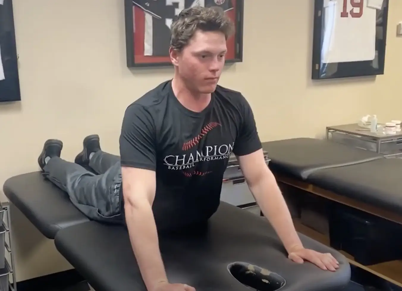
in ACL, baseball, DPTstudent, elbow, exercise, hip, knee, Physical Therapy, rehabilitation, Research, rotator cuff, ShoulderThis week I posted a lot of research and thoughts on shoulder and knee rehab, particularly after an ACL injury. I also shared some others posts that really complimented my posts so there’s some bonus reading to do too. Hope The Physical Therapy Week in Research Review helps your Monday patients and beyond! Take a read and…
-
The Week in Research Review, etc 10-8-18
—
by
in ACL, baseball, DPTstudent, elbow, general, hip, knee, Physical Therapy, rehabilitation, Research, rotator cuff, ShoulderHey all, the Week in Research Review, etc 10-8-18 has some great articles that really got some good discussion going. I highly recommend reading each post and chiming in. Looking forward to the new comments and discussions! PT Continuity of care Fatigue effects on ACL tears Measuring IR in a baseball pitcher Lever sign to…
-
The Week in Research Review, etc 9-24-18
—
by
Hey everyone, another great week of rehab-related posts that brought a lot of topics together. The week in research review for 9-24-18 involved: Blood Flow Restricted Resistance study RTP following an ACL Prevalence of knee osteoarthritis in pain-free people Training your core Dosing Low load Long Duration Using Boditrak during the deadlift Blood Flow…
-

The Week in Research Review, etc 9-17-18
—
by
in ACL, DPTstudent, elbow, hip, knee, Physical Therapy, rehabilitation, Research, rotator cuff, ShoulderAnother week of some great discussions looking at the week in research review. Check it out below and let you friends know they need to subscribe to my blog! Thanks, everyone! Gluteal Tendinopathy: A Review of Mechanisms, Assessment and Management. Grimaldi et al Sports Med 2015. Great review of gluteal tendonopathy, which I…
-
The Week in Research Review, etc 9-10-18
—
by
Lots of good stuff this past week. We talked: Dr. Andrews knowledge bombs Frozen Shoulder video AC joint Classification Whether we should return our ACL patients at 6 months post-op Eric Cressey quote on failing rehab What I have learned about being successful as an orthopedic surgeon by Dr James Andrews Great read by my…
-

The Week in Research Review, etc 8-12-18
—
by
in ACL, baseball, DPTstudent, elbow, general, hip, knee, Physical Therapy, rehabilitation, Research, rotator cuff, ShoulderThis week’s articles discuss a wide variety of research topics. We discussed: Risk Factors for ACL tears Injury after a concussion EMG of the hip to minimize TFL activity We made of our posture and applied it to daily tasks Rhythmic Stabilization drills for the shoulder Hope you enjoy and make sure to share with…