The Week in Research Review, etc 10-8-18

Hey all, the Week in Research Review, etc 10-8-18 has some great articles that really got some good discussion going. I highly recommend reading each post and chiming in. Looking forward to the new comments and discussions!

  1. PT Continuity of care
  2. Fatigue effects on ACL tears
  3. Measuring IR in a baseball pitcher
  4. Lever sign to diagnose an ACL tear
  5. Immediate or delayed ROM after a rotator cuff repair


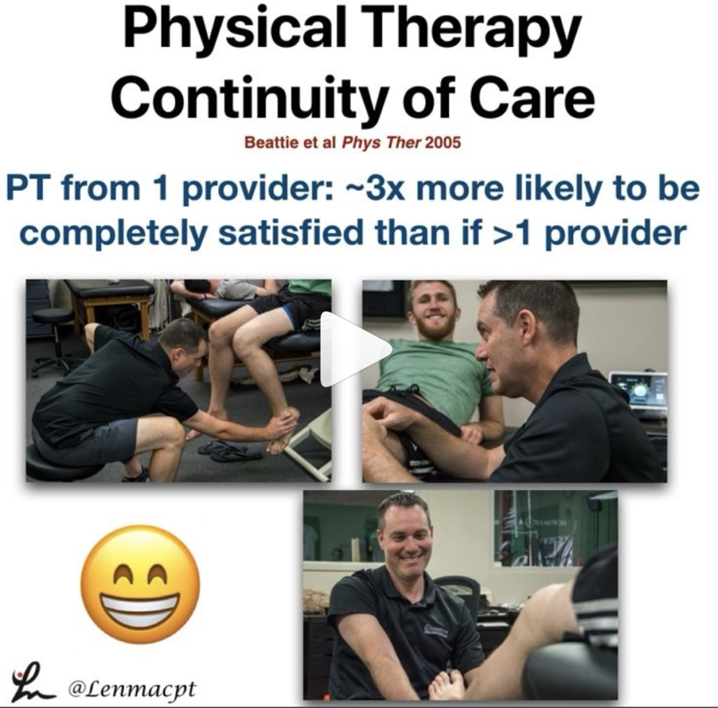
Longitudinal continuity of care is associated with high patient satisfaction with physical therapy. Beattie et al Phys Ther 2005.

I saw a FB post the other day and it reminded me of a study that I had seen about continuity of care and physical therapy.⠀

This study looked to provide ‘preliminary information regarding the association between longitudinal continuity and reports of patient satisfaction with physical therapy outpatient care.’

What they showed was “Subjects who received their entire course of outpatient physical therapy from only 1 provider were approximately 3x more likely to report complete satisfaction with care than those who received care from more than 1 provider.”

All too often, I hear my current clients talk about their past PT sessions and often complain about seeing a tech/aide or a different PT for each session.

I always thought that was such a wrong concept for the client. Throughout my career, I have strived to connect with each client in an attempt to help them overcome an injury.

We did this at @championsportsm in Birmingham and we do it now in Boston at @championptp.

It is such a game changer for the client when they have complete faith in their treatment, can connect with their PT and their PT can connect with them.

Just my little soapbox rant on continuity of care. Are you able to maintain a good continuity of care with your patients or are you constantly sharing and/or just doing evals?

Tag a friend or colleague who may benefit from this post…thanks!⠀


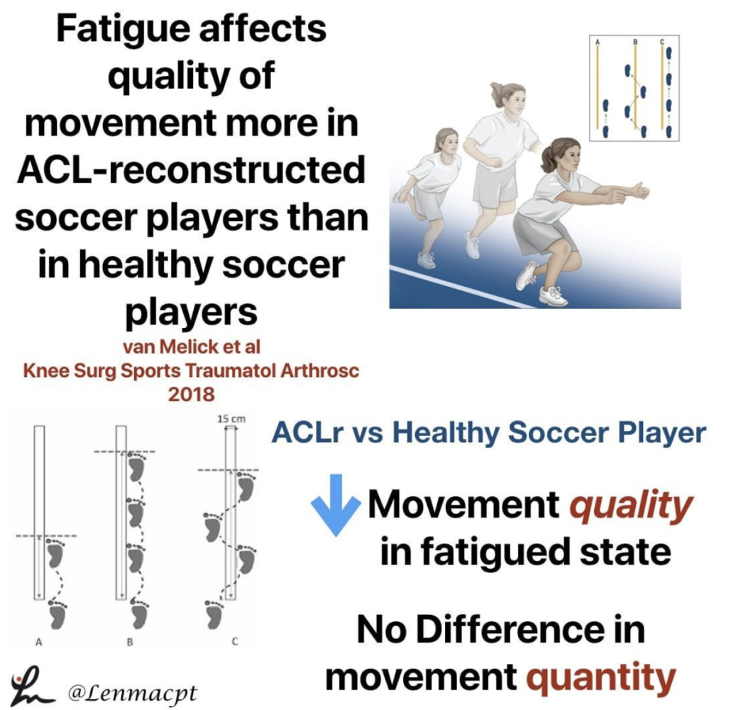
Fatigue affects quality of movement more in ACL-reconstructed soccer players than in healthy soccer players. van Melick et al Knee Surgery, Sports Traumatology, Arthroscopy 2018.

This study looked at the influence of neuromuscular fatigue on both movement quantity and quality in fully-rehabilitated soccer players after ACLR and to compare them with healthy soccer players.

They showed ACL-reconstructed soccer players had a significantly decreased performance when comparing the non-fatigued with the fatigued state.

For movement quantity, they used a single-leg vertical jump, a single-leg hop for distance, and a single-leg side hop.

For movement quality, they used a double-leg countermovement jump with frontal and sagittal plane video analyses. The Borg Rating of Perceived Exertion (RPE) scale was used to measure fatigue after a soccer-specific field training session. In addition to soccer-specific drills, exercises focussing on speed, stability, and coordination were included in this session.

Seems like a pretty neat study that may help to show us that the fatigued state influences quality of movements and not the quantity of movements. I know Tim Hewett has said that there’s no evidence that fatigue influences ACL tears but maybe this study is the 1st step.

Do you agree with this study? Anecdotally it makes sense but there’s little evidence to support the notions.⠀


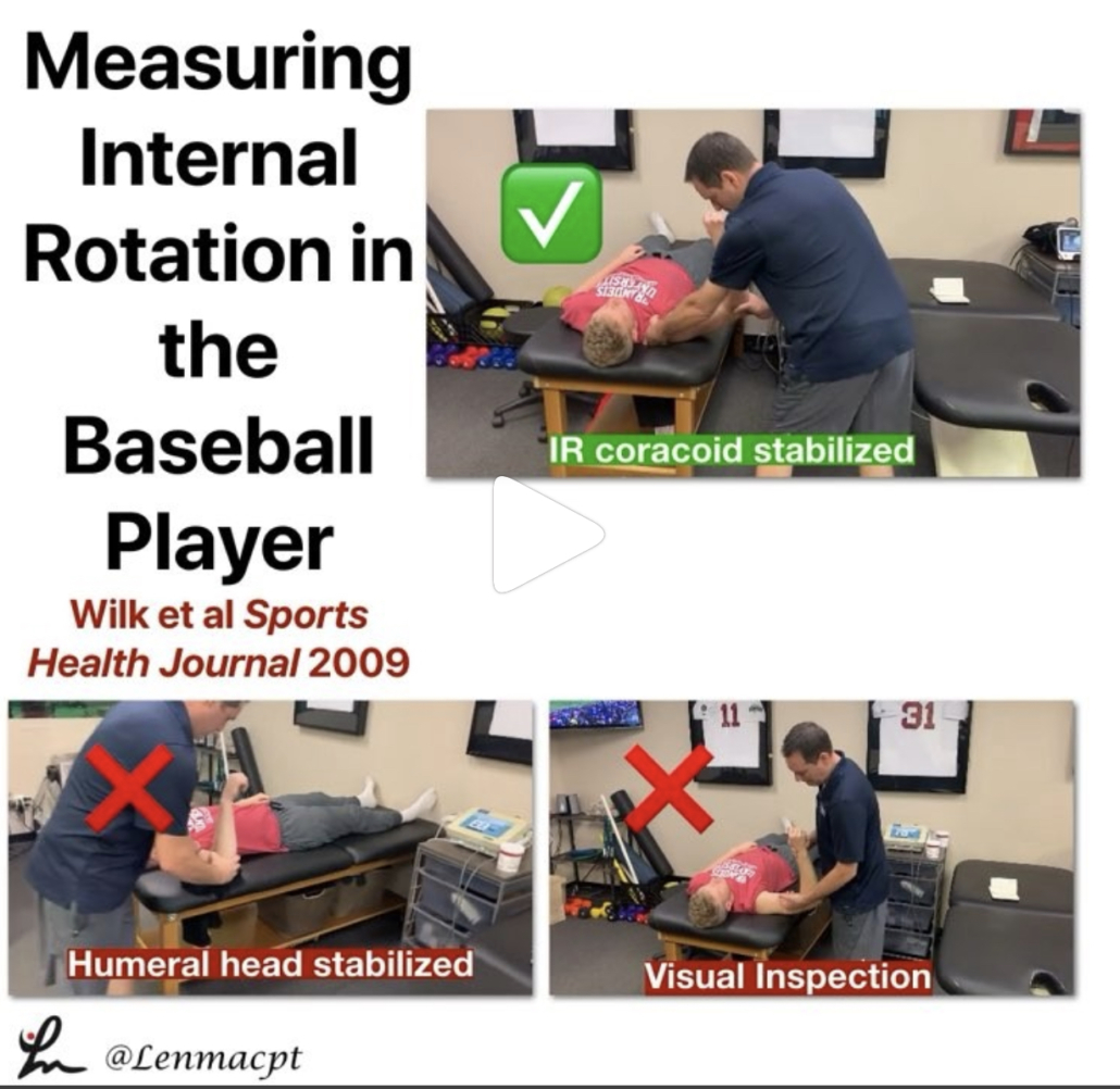
Measuring internal rotation in the baseball player

If you treat baseball pitchers, then you should have a good understanding of how to measure internal rotation of the shoulder joint.

Measuring internal rotation of the shoulder is one part of the equation when obtaining total rotational range of motion (TROM). Total rotational range motion is the sum of external rotation plus internal rotation. I use this equation weekly, if not daily when assessing my baseball players’ shoulders.

In a study in 2009 Sports Health Journal titled “Glenohumeral internal rotation measurements differ depending on stabilization techniques”, we looked at 3 different ways to measure IR. We determined that the scapula stabilized method had the best intra-rater reliability.

We also felt this was the best method to measure pure internal rotation of the glenohumeral joint.

Is this how you measure IR in your baseball pitchers? Do you consider TROM when making treatment recommendations?

Let’s talk it out and discuss the concept of TROM and how to measure it.


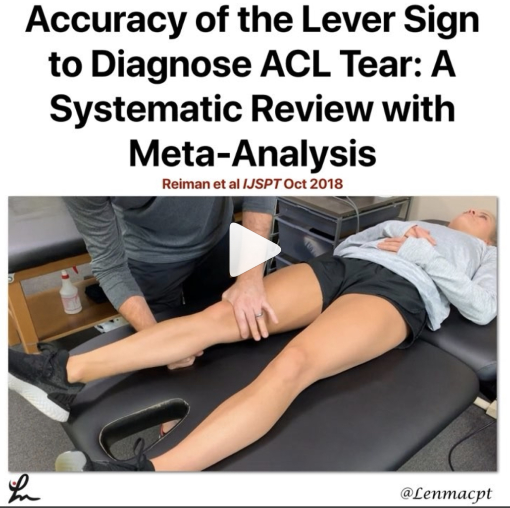
Accuracy of the Lever Sign to Diagnose Anterior Cruciate Ligament Tear: A Systematic Review with Meta-Analysis. Reiman et al IJSPT Oct 2018

This study was a systematic review with meta-analysis that hoped to summarize the diagnostic accuracy of the Lever sign for use during assessment of the knee for an ACL tear.

They showed that based on limited evidence, the Lever sign can moderately change posttest probability to rule in an ACL tear.

I’m a bit surprised by the limited studies because I’ve had a more difficult time getting consistent results compared to the Lachmans test (definitely my go-to!).

For those not familiar with the Lever test, it was 1st published by Dr Lelli in Knee Surg Sports Traumatol Arthrosc. 2016.

From the review, ‘The test requires the evaluator to place their fist under the calf muscle to create a “fulcrum” extending the knee while applying a moderate downward force to the distal part of the femur.

In an intact knee, the ACL completes a lever mechanism, making the heel rise in response to the force applied to the femur. In an ACL-deficient knee, the heel does not rise indicating a positive Lever sign.’ I have personally struggled to get consistent accuracy using the test. My results have been inconsistent with MRI results.

I’ve also struggled to do the test on a plinth that has padding and often have patients lie on a firm surface like the floor (which is very weird) in order to get a better test result.

Some people are freaked out by the method of the test. The clinician has to apply force to the knee in order to create the fulcrum. Many have not liked that force applied to the knee.

In general, this is not my go-to for a suspicious ACL tear. I have tried and still ty to use it but my results have been less than stellar.

Have you used this test for an ACL tear? Do you like it to supplement your Lachmans?


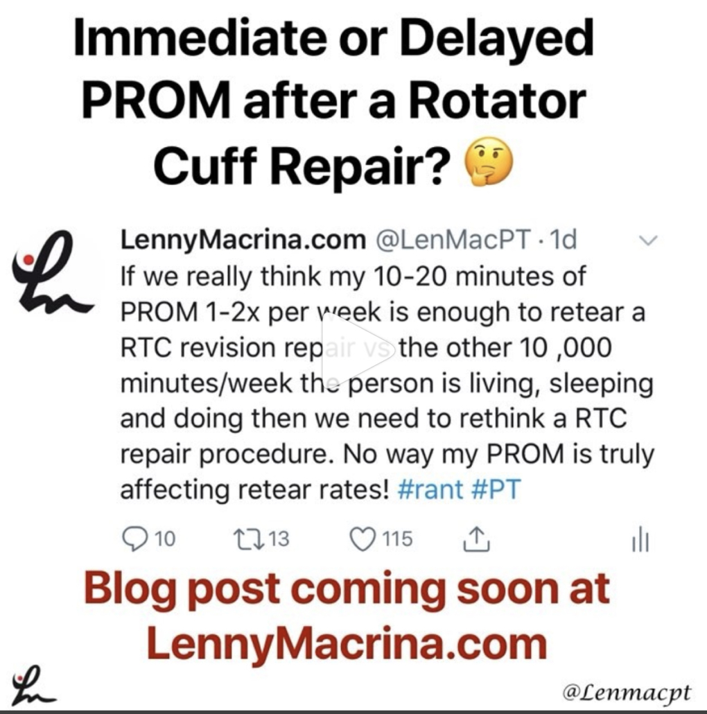
Should we delay PROM after a rotator cuff repair?

It seems as if we’re all over the place, which usually says the research is not cut and dry. There are so many factors that are considered when trying to figure out the best time to initiate motion.

I’m not talking active ROM or strengthening…I”m talking about passive ROM by a rehab specialist like a #PT#OTor #ATC. Obviously, the docs weigh in heavily with this decision. I feel as if patients are restricted for the wrong reasons and could potentially begin PT earlier than we often see.

This is going to be a beast of a blog post and may alter my thinking, we’ll see.

As of now, I fully embrace immediate PROM for most post-op rotator cuff repairs, including Large and Massive repairs.

For revisions, we may need to think it through but I still feel as if most benefit from early PROM. We did it for years and with very good results during my time in Birmingham but feel as if maybe the pendulum is swinging in the conservative direction (for the wrong reasons).

What do you guys do? Do you have any input with your docs and can influence their rehab decisions? Let’s talk it out now and get prepped for my blog release in the coming days, weeks, months…whenever I can make it the best!⠀
 

The Week in Research Review, etc 10-1-18

Another week of some great discussions and learning opportunities. The Week in Research Review included:

  • Risk Factors for Patellofemoral pain
  • Shoulder ROM and elbow injuries
  • Rotator Cuff Exercises
  • Eccentric or Concentric exercise for Tendinopathy
  • Hamstrings Protect the ACL
  • Stretching the Shoulder in the Overhead Athlete

Share with your friends and have them subscribe to the weekly newsletter!


Risk factors for patellofemoral pain: a systematic review and meta-analysis Neal et al BJSM 2018.

This systematic review and meta-analysis of 18 studies involved 4818 participants, of whom 483 developed patellofemoral pain syndrome (PFPS).

First off, PFPS is a wastebasket term that basically tells the client that they have knee pain…that’s it.

My 1st job is to educate the client about this fancy term because they often come in confused and wanting more information.

I use a good subjective exam to have the patient help me narrow in on a potential cause so I can answer the question ‘why’.

My clinical exam will attempt to diagnose the particular culprit…whether it’s mechanical, overuse or something else.

Back to the study…it showed that in patients with PFPS, quadriceps weakness in military recruits and higher hip strength in adolescents were risk factors for PFP.

Not surprised by the quadriceps weakness but kinda surprised by the hip weakness!

The same authors showed this in JOSPT 2012 Lankhorst et al that weaker knee extension strength, expressed by peak torque, appears to be a risk factor for PFPS.

Not sure what to do with the hip strength as a risk factor in adolescents but maybe it becomes a biomechanical issue if the hips are stronger than the quadriceps, relatively.

Do you guys see this out there as well? The key, as usual, is to strengthen the quadriceps!

I would also say activity modification that is causing the quad weakness (overuse) and a progressive return to their activity.

Chime in and let’s talk this out…thanks!⠀


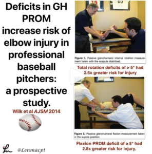
Deficits in glenohumeral passive range of motion increase risk of elbow injury in professional baseball pitchers: a prospective study. @wilk_Kevin, Macrina et al AJSM 2014.

In this paper, we looked to determine whether decreased ROM of the throwing shoulder is correlated with the onset of elbow injuries in professional baseball pitchers.

This one took years to get all of the data collected through multiple spring training trips to the @raysbaseball facilities.

In the end, we were able to show that: ⚾️pitchers with deficits of >5° in total rotation in their throwing shoulders had a 2.6x greater risk for injury.

⚾️Pitchers with deficit of ≥ 5° in flexion of the throwing shoulder had a 2.8x greater risk for injury.

These findings have guided our evaluation and treatment strategies at @championptp.

We hypothesize that loss of flexion may be a result of some soft tissue limitation of the lats, teres, pecs and other muscles.

We focus much of our attention on these muscle groups during our arm care to help regain the flexion and may even help gain back some of the ER in those that are tighter than normal…whatever that means.

After soft tissue work, we look to work on dynamic stability and strength in the newly gained ROM.
Do you use these similar concepts with your baseball pitchers too? Tag a friend who may be interested in this study…thanks!⠀


Rotator Cuff Exercises

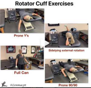
In this post, I wanted to discuss my go-to exercises for the shoulder when someone presents with an injury or pain.

Of course, my exam TRIES to determine the tissue involved but most of our clinical exam tests cannot pinpoint the exact pain generator and pathological tissue.

With that, I have certain exercises that I think, through the available EMG data, are the best to help regain strength and confidence prior to beginning their return to sport (or life) activities.

Numerous studies have looked at the EMG during these specific motions and have determined that the supraspinatus and infraspinatus have higher relative levels compared to other positions, say the full can vs empty can debate, for example.

Take a look at these classic studies to help guide your programs:⠀

❇️Blackburn et al JAT 1990

❇️Townsend et al AJSM 1991

❇️Reinold et al JOSPT 2002

❇️Reinold et al JAT 2007

❇️Kelly et al AJSM 1996

❇️Worrell et al Med Sci Sports Exerc 1992

❇️Jobe et al 1982

❇️Decker et al AJSM 2003

These papers have provided the foundation for today’s shoulder programs and are some that I discuss during my Biomechanics lectures that I give when teaching my course.

Are you familiar with these papers and do you keep them in mind when building your shoulder programs for your clients?

Tag a colleague or friend that may want to see this post…thanks!⠀


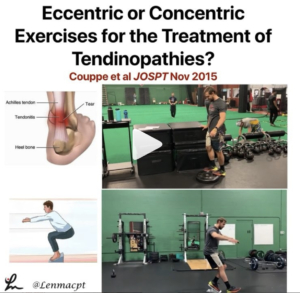
Eccentric or Concentric Exercises for the Treatment of Tendinopathies? Couppe et al JOSPT Nov 2015

Interesting clinical commentary from a few years ago talking about tendinopathy treatments.

Most PT’s and ATC’s generally talk about eccentric loading of tendons to help treat suspected tendon pain.

In this review, they discuss the potential mechanisms that may aid in helping people suffering from tendon pain.

I found this statement very interesting:

👉🏼”There is little evidence for isolating the eccentric component of a loading-based regime.

👉🏼The basic mechanisms that are likely to influence tendon adaptations appear to be related mainly to tendon load/strain magnitude and duration, and there is no theoretical basis for greater tendon loads in eccentric exercises at a given force (body weight or external load).” 🤯

As always, it makes me think that as specific as we think we are with some of our exercises, maybe just putting any strain through the muscle-tendon unit is good enough.

Have you guys read this review? What do you think? is this similar to what you see in your practice?

Tag a friend who may want to read or comment on this post…thanks!⠀


𝐇𝐚𝐦𝐬𝐭𝐫𝐢𝐧𝐠𝐬 & 𝐓𝐡𝐞 𝐀𝐂𝐋

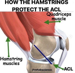
Great post by @rehabscience talking about the influence of the hamstrings on the #ACL. A big focus of my rehab for my patients that have had an ACL reconstruction involves building hamstring strength.

Check out his original post below!

💥𝐇𝐚𝐦𝐬𝐭𝐫𝐢𝐧𝐠𝐬 & 𝐓𝐡𝐞 𝐀𝐂𝐋💥
———–
📌The anterior cruciate ligament (ACL) is an extremely important ligament in terms of overall knee integrity and stability. Specifically, the ACL connects the femur (thigh bone) to the tibia (shin bone) and runs at an oblique angle from the posterior aspect of the femur to the anterior aspect of the tibia. Due to this arrangement, the ACL is responsible for preventing anterior translation of the tibia or posterior translation of the femur.

🔎Now, many of us are aware of the importance of the quadriceps to knee health, but, often times, the hamstrings get neglected. The hamstrings run along the posterior (backside) of the thigh and insert onto the posterior surfaces of the tibia and fibula (shin bones).

When contracting, the hamstrings work to bend the knee, but also pull the tibia posteriorly. In this way, the hamstrings can serve as a dynamic protector of the ACL by limiting excessive anterior displacement of the tibia and strain on the ligament.

✅If you are looking to reduce your risk of ACL injury or recovering from an ACL reconstruction, don’t forget to include hamstring work in your strength training program as this group has an instrumental role in protecting the ACL.

⬅️Swipe left to see several exercises from myself, @jasonbombard@zerenpt and @strengthcoachtherapy that can be incorporated to increase hamstring strength.


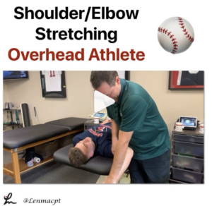
⚾️Stretching the Overhead Athlete ⚾️

In this post, I wanted to give a glimpse into the stretching routine I use on some of my OH athletes before and after a workout, bullpen or a game.

I like to stretch the shoulder into external rotation to make sure the athlete can maintain that important ROM, especially to keep that layback or late cocking position.

I also like to work on horizontal adduction with the lateral border of the scapula stabilized. It’s important that the athlete feels the stretch in the back of the shoulder and nowhere close to the front of the shoulder.

This is the lone reason why I have gone away from the sleeper stretch and focus on horizontal adduction.

I also stretch out the forearm flexors by extending the elbow/wrist and all of the fingers, including the thumb (don’t forget about the thumb!)

I also like to stretch the shoulder joint into flexion by pinning down the scapula and hope I’m somewhere on the lats and/or subscapularis to be able to stretch these muscles out and improve that overhead position.

Remember, in 2014 we showed a loss of flexion increased the risk of medial elbow injuries by almost 3x.
I like to repeat the process a few times until I feel like we maximized the amount of new ROM.⠀
.⠀
At the same time, we’re chatting about the session, how it went, what’s to come, how their fantasy football team is doing, etc.

It’s my way to connect with each client before and after they have a session with me. I feel this is very important and often overlooked by other PT’s.

Do you have any other stretches you like to do? Tag a friend who may want to check out this video…thanks!⠀


The Week in Research Review, etc 9-24-18

Hey everyone, another great week of rehab-related posts that brought a lot of topics together. The week in research review for 9-24-18 involved:

  • Blood Flow Restricted Resistance study
  • RTP following an ACL
  • Prevalence of knee osteoarthritis in pain-free people
  • Training your core
  • Dosing Low load Long Duration
  • Using Boditrak during the deadlift


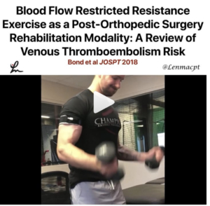
Blood Flow Restricted Resistance Exercise as a Post-Orthopedic Surgery Rehabilitation Modality: A Review of Venous Thromboembolism Risk JOSPT Bond et al 2018.

This paper is more of a review of the literature (so be cautious) but raises some important questions and thoughts about #BFR usage in our typical orthopaedic setting.

They talk about important decisions and concepts to be made including precautions, contraindications, and dosage.

They also admit that there are no universally agreed upon standards indicating which post-surgical orthopedic patients may perform BFR safely.

They also list close to 40 different precautions or contraindications including:⠀
❇️Age >40 yr⠀

❇️Creatine Supplement Use ⠀

❇️Diabetes⠀

❇️General/Local Infection ⠀

❇️Hypertension⠀

❇️Immobility >48 hr in the Past Month⠀

❇️Open or Unhealed Soft Tissue Injuries ⠀

❇️Amongst many others

Seems like a pretty strict list but curious to hear what others are using to determine if their client is appropriate for BFR resistance training.

💪🏼Thanks @kieferlammi for the swole session!

Let’s discuss below… tag a colleague who may be interested in discussing…thanks!


Return to Play after ACL

I posted this the other day on #Twitter because I keep hearing people talk about the failure rates after an ACL.

I feel like 1 major reason why people are failing within the 1st 1-2 years after returning is that they get back on the field with residual weakness.

When the patient’s insurance runs out, they workout on their own or often seek out a personal trainer to help them.

I feel we as PT’s can do a much better job at showing our value to our patients by keeping them under our care an progressing them back to their sport.

If we don’t have the facilities to do this, then we must work with others in our region to help our clients get the best care possible.

At @championptp, we often get referrals from area clinicians asking to take over their client’s care and advance them back to their sport. I definitely respect that clinician for recognizing their care may not be the best for the client at that time in the rehab process.

Do you utilize clinicians in your area in these situations? Do you think we could improve our ACL outcomes if we did this more?

Let’s discuss this below and make sure we have a plan in place when that 2-3 month rehab phase approaches. Tag a friend who may benefit from this post…thanks!⠀⠀


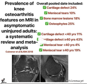
Prevalence of knee osteoarthritis features on MRI in asymptomatic uninjured adults: a systematic review and meta-analysis Culvenor BJSM 2018

This paper ‘performed a systematic review with meta-analysis to provide summary estimates of the prevalence of MRI features of osteoarthritis in asymptomatic uninjured knees.

They basically looked to determine the normal changes in the knee that may be diagnosed on an MRI in people less than and greater than 40 years of age.

The information may help clinicians educate their patients prior to getting an MRI.

Overall pooled date included:⠀
Cartilage defects was 24%⠀
Meniscal tears was 10%⠀
Bone marrow lesions 18% ⠀
Osteophytes 25%⠀

Cartilage defect <40 years 11%⠀
Cartilage defect ≥40 years 43%⠀
Meniscal tear <40 years 4% (seems low to me)⠀
Meniscal tear ≥40 years 19%⠀

Interesting stuff that you need to store in our mental database for future clients.

What do you think of this data? Will it help you in your decisions with your clients?

Tag a friend or colleague who may benefit from this information…thanks!⠀


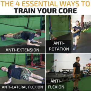
4 WAYS YOU SHOULD BE TRAINING YOUR CORE

Great post by our strength coach @kieferlammi discussing the 4 ways to train your ‘core’. Simple yet a great view of the concepts needed to best address a client’s weaknesses. Give him a follow and see his original post below. @championptp

There are a million different exercises to train your abs/core/trunk/whateveryouwanttocallit. Regardless of which you choose, in my mind there are 4 staple ways that I think belong in every training program:

⠀⠀⠀⠀⠀⠀⠀⠀⠀⠀
1️⃣Anti-Extension – These consist mainly of plank variations, Rollouts, Fallouts, etc.

2️⃣Anti-Rotation – The anti-rotation press or “Pallof Press” is the most popular of this category and can be done from a variety of stances with a variety of tools.

3️⃣Anti-Lateral Flexion – Side planks and unilateral load carries are king here, but this would include anything resisting side bend.

4️⃣Anti-Flexion – Loaded carries and Deadlifts are the top two in my mind, but anything where you have to work to avoid rounding forward, will do.

What are your staples? Anything I’m missing? Disagree? Comment below!


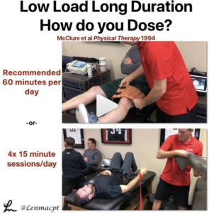
LLLD DOSING

What dosing should you prescribe your patient with a stiff joint when using low load long duration stretching? 🤔

That’s the million dollar question and very little is known.

The one paper that I’m aware of is more of a concepts paper but has been the guidance for my LLLD dosing.

McClure et al talk about 60 minutes per day of total end range of motion time or TERT.

Basically, I tell my clients to apply some form of over-pressure 4x per day for 15 minutes each session. I’ll sometimes do 3x 20 minutes/day if they don’t have the time at work or school.

It allows them to moderately stress the tissue but not take up too much of their time during the day.

The key to this form of stretching (we think) is to elongate the tightened collagen that is limiting the posterior capsule of the knee… see Zhou et al 2018 MSSE ‘Range of Extension Correlates with Posterior Capsule Length after Knee Remobilization.’

Check out this paper and consider this dosing as the basis for your future clients. Do you have any dosing schedules that you use or any research that specifically talk about this?

Let’s talk it out and hopefully help you dose your patients who are tight after knee or elbow surgery…thanks!⠀


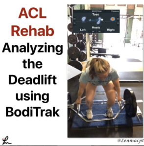
Using Boditrak during the deadlift

This video was recently taken of my #ACL patient who is ~ 12 weeks s/p L ACL reconstruction with a patella tendon autograft and a medial meniscus repair.

I took this video (it’s a mirror image so don’t get confused) to analyze her weight distribution between the involved leg (Left) and the uninvolved leg (right).

As you can see, she spends a lot of time on the front part of her foot throughout much of the deadlift and has her weight shifted to the uninvolved side during her initial pull.

As she ascends up and reaches the max pull position, she is able to redistribute her weight more evenly between each side but continues to keep her weight more toward her toes on that involved side.

To me, I would like to see her weight distribution more equal side to side but also more towards the mid-portion of her foot during the pulling phases.

She self-admits that she is shifting her weight and can’t help it. Through the naked eye, you may be able to see the shift but not necessarily see the anterior/posterior weight distribution (toes/heels).

I like to use the @boditraksports to pick up little nuances and help give feedback to the client.

Did you notice anything else with this video? What would you critique (be nice!!)?

Tag a friend or colleague who may want to see this video and help @lms651 get back to her fencing competitions…thanks!


The Week in Research Review, etc 9-10-18

Lots of good stuff this past week. We talked:

  • Dr. Andrews knowledge bombs
  • Frozen Shoulder video
  • AC joint Classification
  • Whether we should return our ACL patients at 6 months post-op
  • Eric Cressey quote on failing rehab


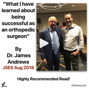
What I have learned about being successful as an orthopedic surgeon by Dr James Andrews

Great read by my friend, mentor and colleague who I was fortunate to work with from 2002-2014, before moving back home to Boston to help open @ChampionPTP with @mikereinold.

I learned so much from my interactions with him and how he handled each and every case. His approach has been the standard by which I carry myself as a PT

In this paper, he talks about: ⠀

✔️Availability⠀

✔️Communication⠀

✔️Compassion⠀

✔️Gentleness⠀

✔️A true love of caring for my patients

He also talked about being successful with a ‘purpose driven life’ and discusses 16 key recommendations to a successful orthopaedic career.

Take a look at this article and implement as much as you can tomorrow and every day thereafter.

Happy reading…share with a friend or colleague in the comments section below!⠀


Frozen Shoulder or adhesive capsulitis can be debilitating and frustrating for the patient.

This video shows why! Look at all of that red and inflamed tissue of the shoulder capsule. Those neovascular changes are a classic sign of frozen shoulder and the main reason why anti-inflammatories are probably effective in the early stages of the disease.

The pain associated with this presentation is often the main limiting factor, combined with the eventual capsular scarring/contracture that develops soon after.

This scarring leads to a loss of joint arthrokinematics, which leads to loss of mobility, functional loss and atrophy. This spiral effect can last months if not years for some.

Regaining mobility, strength and normal function is not guaranteed but PT can help speed up the process a bit by educating, guiding and mobilizing the patient.

if I see someone in an early stage of frozen shoulder, I usually recommend a cortisone shot followed by more PT to help maintain the patient’s shoulder ROM/strength.

What do you recommend? Any tests you use to help diagnose adhesive capsulitis?

Tag a colleague who may benefit from this post…thanks!⠀


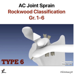
With football season here, we’re going to hear a lot of talk about #shouldersaparation or AC joint sprains

The different Grades (Rockwood Classification) of AC Joint sprains are:⠀

1️⃣ AC ligament sprain, AC joint intact, CC ligaments intact⠀

2️⃣AC Joint disruption, Slight vertical separation of ACJ, CC ligament sprain, CC distance wide⠀

3️⃣AC ligament disruption, AC joint dislocated, CC ligaments torn⠀

4️⃣AC ligament disruption, AC joint dislocated, Clavicle displaced posteriorly into Trapezius, CC ligaments completely torn⠀

5️⃣AC ligament disruption, AC joint dislocated, CC ligaments completely torn,⠀
CC distance 100 to 300 % > than normal side.⠀

6️⃣AC ligament disruption, AC joint dislocated, CC ligaments completely torn, Clavicle in subcoracoid position.

I saw this video that @drroddymcgee put out on #Twitter and loved the visual effects to help simplify the typical tissues involved with each type of AC Joint sprain. You can slo find them at @thesportsdocs00 on Twitter too.

Hope it helps to put the AC joint sprains in a better view for you. Share with a colleague who may have an interest…thanks!


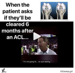
Return to play after an ACL is a complex decision that involves many variables. The research is telling us a 6-month return to play is too quick and should be delayed up to 9 months.

During that time, the focus of the rehab needs to be on gaining strength, power, and confidence in the patient’s lower body, particularly the quadriceps.

✅ 51% reduction for each month return to sport was delayed until 9 months after surgery…research by Grindem et al BJSM 2016.

Hewett et al have advocated for a possible 2-year return to play wait time to account for graft healing and time for the full strength, power and confidence to return.

I have adjusted my practice to educate my clients for a 9-month return and map out a timeframe from the beginning so they buy into that thinking. Often times, doctors will place a 6 month time for RTP and I have to overcome that thinking and ‘convince’ my clients that it’s too soon.

I think that much time is needed to get the quadriceps muscle back completely and to gain the full confidence in the limb through dynamic activities.

Do you agree? What do you recommend and do you agree with Coach Saban’s answer on RTP at 6 months? 😜

Tag a friend who would benefit from seeing this post or at least seeing Coach Saban…thanks! #RTR #notreally #neutral#switzerland 


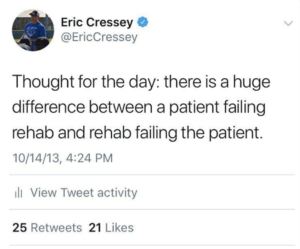
Failing Rehab

As a PT, this really hit home with me and thought @ericcressey nailed it! See his original post below. @cresseysportsperformance 👇🏻

As with any professional offering – training, accounting, contracting, landscaping, or a host of other services – you’ll see good and bad rehabilitation scenarios. Make sure you do your homework about not only a rehab specialist’s experience and credentials, but also the business model in which he/she operates. 😵#cspfamily #sportsmedicine#rehab #physicaltherapy #physicaltherapist#athletictrainer #athletictraining#sportsperformance #rehabilitation


The Week in Research Review, etc 8-19-18

We posted a lot of information this week to review so hopefully you were able to keep up with it all. If not, here’s a bunch of it from the week. Check it out and comment as you want. Lots of good information on:

  1. Advanced Rhythmic Stabilization Drills
  2. Our ACL rehab paper from 2012
  3. PT usage for Frozen Shoulder
  4. Comparison of posterior shoulder stretching techniques
  5. Increased Sweating after an ACL surgery? Seems pretty common in the comments section
  6. @theprehabguys show how to do a posterior shoulder stretch


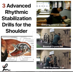
Advanced stabilization drills

These advanced stabilization drills are a great way to progress the patient once they’ve shown improved neuromuscular control with the basic drills from the other day.

I use these for most of my clients that need improved dynamic stability, especially those hypermobile athletes that play baseball, swim, or do gymnastics.

You can modify the speed and amount of force that I give during the drill based on how they are responding that day and how they have progressed overall. You can also increase the time of the drill to bring in an endurance component, as well.

The goal is to use these to prep the shoulder for higher level activities such as plyometrics, throwing, etc.

I want the shoulder joint to be as strong as possible. And most importantly, I want the client to perceive that their shoulder is stronger so that they are more confident!

Do you use these rhythmic stabilization drills with your patients?

Tag a friend who may benefit from these drills and try them on your patients the next time they’re ready to progress their dynamic stability drills!


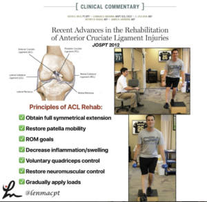
Recent advances in the rehabilitation of anterior cruciate ligament injuries. @wilk_kevin et al JOSPT 2012 @drlylecain @dugasmd1

We wrote this paper in JOSPT to help clinicians better understand the rehab involved after their patient undergoes an ACL reconstruction.

The paper discussed the principles of ACL rehab, including:
✅obtaining full symmetrical extension
✅restoring patella mobility
✅ROM goals
✅Decrease inflammation/swelling
✅voluntary quadriceps control
✅restore neuromuscular control
✅Gradually apply loads

We also discuss special rehab implications for:
✅The Female Athlete
✅Concomitant injuries (MCL, meniscus, LCL, cartilage lesions)
✅Implications of Graft type

Our Accelerated ACL-PTG protocol is included to help guide the rehab process and give the rehab specialist some goals to achieve.

Again, a protocol is just a guide and by no means meant to place aggressive limitations on a patient.

Hope this paper helps you to better understand ACL rehab. Tag a friend or colleague who may benefit from this post.


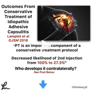
Outcomes From Conservative Treatment of Shoulder Idiopathic Adhesive Capsulitis and Factors Associated With Developing Contralateral Disease Lamplot et al 2018 OJSM 2018

A minimum 2-year follow-up of patients diagnosed with idiopathic adhesive capsulitis.

They were treated with a single intra-articular glenohumeral injection of local anesthetic and corticosteroid as well as 4 weeks of supervised PT.

Physical therapy reduced the use of a second injection as part of treatment. Contralateral disease was more likely in patients with diabetes and those younger than 50 years.

PT is an important component of a conservative treatment protocol, as PT decreased the likelihood of receiving a second injection from 100% to 27.3%.

In my opinion, a cortisone injection + PT is the best treatment ‘cocktail’ for someone with frozen shoulder, particularly in the freezing phase.

Do you guys agree? Let’s discuss this very debilitating pathology. Tag a friend who may want to discuss this further.


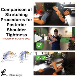
A Randomized Controlled Comparison of Stretching Procedures for Posterior Shoulder Tightness McClure et al JOSPT 2007

This study looked to compare changes in shoulder internal rotation range of motion (ROM), for 2 stretching exercises, the “cross-body stretch” and the “sleeper stretch,” in individuals with posterior shoulder tightness.

From their results, “The improvements in IR ROM for the subjects in the ✅cross-body stretch group (mean ± SD, 20.0° ± 12.9°) were significantly greater than for the subjects in the control group (5.9° ± 9.4°, P = .009). The gains in the ❌sleeper stretch group (12.4° ± 10.4°) were not significant compared to those of the control group (P = .586) and those of the cross-body stretch group (P = .148).”

Just be aware of the large standard deviations and low number of subjects…plus the subjects were all asymptomatic.
With that, I still believe clinically that the cross-body stretch is one of the better stretches for the shoulder. I have gotten away from the sleeper stretch because I don’t believe the risk/reward presents favorably.

✅✅I personally prefer the supine horizontal adduction stretch with the scapula stabilized. I feel like this best isolates the posterior soft tissue (mainly muscle, in my opinion) and that’s my target tissue.

Do you use the sleeper stretch with your patients or do you prefer the cross-body stretch?

Comment below and let’s talk about it. Also, tag a friend who may benefit from this post…thanks!


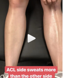
Sweating Leg after an #ACL

Had this in my story and posted to Twitter and got some pretty good discussion going. Is this some altered autonomic nervous system response after #ACLsurgery? I’ve seen it a bunch, where the area inferior to the incision sweats significantly more than the contralateral leg. Anyone else see this phenomenon and have an opinion? Have had people say that maybe it was compartment syndrome or CRPS but it happens with many many ACL patients so it seems very normal. What do you think? #ACL #ACLrehab


Horizontal Adduction Stretching

Perfect timing by @theprehabguys with their horizontal adduction post from the other day. Blends perfectly with the McClure 2007 et al study I posted the other day that showed horizontal adduction is the best way to obtain internal rotation mobility at the shoulder. I often tell people to wedge their lateral scapula against a wall to help stabilize but this way may also benefit them. Check out their post below!⠀
👇🏻⠀
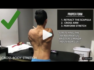
Are You Performing The Arm Across Body Stretch Correctly⁉️⠀
[How to Stretch Your Posterior Cuff]

A tight posterior cuff is associated with a handful of shoulder dysfunctions like subacrominal impingement syndrome, posterior impingement, anterior instability, etc. And thus, ⬇️ tone/increasing extensibility of the posterior cuff is part of the treatment protocol for many with shoulder pain. The cross-body stretch is a fantastic way to target the posterior cuff but far too often it is done INCORRECTLY.

‼️In order to effectively stretch the posterior cuff, you need to keep your SCAPULA STABILIZED ie your scapula CANNOT MOVE!‼️

❌If you pull your arm across your body and your scapula comes with it into horizontal abduction, the only stretch your getting is of your mid-scapular muscles like your rhomboids or traps. Furthermore, in this position there’s more of a distraction force on the glenohumeral joint than a true stretch of the posterior cuff – aka not as specific as it can be.

✅First pull your shoulder blades back. This will keep your scapula in a retracted position. Only WHILE MAINTAINING THE POSITION OF YOUR SHOULDER BLADES BACK can you effectively target the posterior cuff. Pay attention to WHERE you feel the stretch, as you should feel a “deep stretch” in the back of your shoulder in the highlighted area on the video. If you feel a stretch or anything else not in the back of the shoulder, you’re either doing the stretch incorrectly or abutting other structures in your shoulder due to pathology (ie don’t do the stretch anymore and seek out a physio if you’re in pain).

Try it out and let us know how it feels! Tag a friend who NEEDS this stretch! #shoulderstretch #posteriorcuff#posteriorcapsule


The Week in Research Review, etc 8-12-18

This week’s articles discuss a wide variety of research topics. We discussed:

  1. Risk Factors for ACL tears
  2. Injury after a concussion
  3. EMG of the hip to minimize TFL activity
  4. We made of our posture and applied it to daily tasks
  5. Rhythmic Stabilization drills for the shoulder

Hope you enjoy and make sure to share with your friends and colleagues!


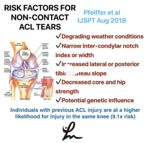
Factors Associated with Non-Contact Anterior Cruciate Ligament Injury: A Systematic Review. Pfeiffer et al IJSPT Aug 2018

This study looks to examine the existing literature for risk factors associated with non-contact anterior cruciate ligament (ACL) injury in both sexes.

A total of 79 full-text articles were reviewed, 55 of which met criteria for inclusion.
✔️Degrading weather conditions⠀
✔️Narrow inter-condylar notch index or width⠀
✔️Increased lateral or posterior tibial plateau slope⠀
✔️Decreased core and hip strength⠀
✔️Potential genetic influence

Individuals with previous ACL injury are at a higher likelihood for injury in the same knee (9.1x risk).

Take a look at this study because they do a good job of breaking down the literature and pointing out some of the common risk factors, both extrinsic and intrinsic.
✅Anatomic factors⠀
✅Neuromuscular Factors⠀
✅Physiologic Factors⠀
✅Biomechanical Factors⠀
✅Genetic Factors

Check out this paper and tag a friend or colleague that may benefit from reading this paper.


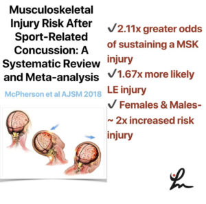
Musculoskeletal Injury Risk After Sport-Related Concussion: A Systematic Review and Meta-analysis – McPherson et al AJSM 2018

This systematic review & meta-analysis looked to determine the odds that athletes will sustain a musculoskeletal injury after a concussion. It was hypothesized that a concussion would increase the risk for MSK injury.

Eight studies met inclusion criteria for meta-analysis reporting 860 male and 163 female athletes with concussion compared with 3719 male and 507 female control athletes without a concussion.

✅Athletes with concussion had 2.11 times greater odds of sustaining a MSK injury compared with control groups and 1.67 times more likely to experience a lower extremity injury.

✅Female athletes with concussion had 2 times greater odds to sustain injury compared with non-concussed female control athletes.

✅Male athletes with concussion similarly demonstrated an increased odds of 2.1x risk for injury compared with non-concussed male control athletes.

Of course, they say further studies are needed to determine the physiological reasoning behind the increased risk for injury after a concussion.

Kinda makes sense, right? Anecdotally I feel like we see this a lot but good to be able to see it in the literature and educate our clients prior to a return to their sport.


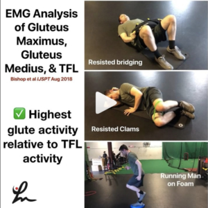
Electromyographic Analysis of Gluteus Maximus, Gluteus Medius, and Tensor Fascia Latae During Therapeutic Exercises With and Without Elastic Resistance. Bishop et al IJSPT 2018

This study looked to determine the gluteal-to-tensor fascia latae muscle activation (GTA index) and to compare electromyographic muscle activation of the GMax, GMed, and TFL while performing 13 commonly prescribed exercises designed to target the GMax and GMed.

Clams with and without resistance, running man gluteus maximus exercise on the stability trainer, and bridge with resistance, generated the highest GTA index respectively.

It seems as if these 3 exercises may give you the biggest bang for your buck when trying to maximize gluteal activity and minimize TFL activity.

Knowing previous research, it seems as if most single-leg-stance exercises are going to give the patient the best gluteal activity per EMG.

Do you guys use any of these exercises? Maybe you need to use them more for your patients looking to improve their hip strength.

Tag a friend who may need bigger 🍑 and may benefit from this research by the esteemed Barton Bishop and his research team! TThanks, carlokoo for modeling!⠀


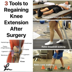
Loss of knee extension after a surgery can greatly impact a patient’s function in the long term.

Although opinions vary greatly in the orthopaedic world, I am a firm believer that regaining symmetrical knee extension is critical for a surgery to be successful.

Shelbourne et al showed that the number one factor in determining long-term satisfaction after a knee surgery was symmetrical knee hyperextension.

Per Benner et al 2016, obtaining full hyperextension that is anatomically normal for most patients does not affect objective stability, ACL graft tear/failure rates, or subjective scores after ACL reconstruction with patellar tendon autograft.

So why do some docs and PT’s still think you only need 0 degrees of knee extension? 🤔⠀

I’m not sure but I’ll tell you it is safe to get hyperextension and here are 3 tools that I use to help regain that last bit of motion.

1. Low load long duration in supine: in my opinion a better position to regain extension and more comfortable than hanging a patient’s leg off of a table. You try doing that and see how you like it!

2. Retro treadmill walking: walking at ~ 1.0 mph on the treadmill has been a great way to get that last bit of motion back and is something they can do at their gym when they’re not with you in PT.

3. Retro cone walking: Again, the goal is to accentuate the end range into hyperextension and it will also challenge their dynamic balance.

So, do you use these techniques with your patients? If not, tag a friend or colleague that may benefit from seeing this post!


I love when we can make fun of ourselves & this post nails it! Give @beardthebestyoucanbe a follow!

There is no such thing as good or bad form or posture. There is no right or wrong “exercise”. What does “functional exercise” even mean, & what is this “core” thing everyone keeps talking about whilst planking or crunching?! My newsfeed is blowing up with these so-called “fitness coaches” and “rehab experts” putting all type of crap out there. For 1 of 2 reasons, maybe both⠀
1. Pure ignorance⠀
2. Trying to grow their following by giving people what they think they want⠀
_⠀
The only bad posture is the posture we spend too much time in (shout-out Andreo Spina). Life is not perfect posture & neither is sports⠀
As human beings, our movement capacity should be vast. If we only stay in a small handful of postures we aren’t fulfilling our movement potential and we’re creating more dysfunctions and weaknesses because of it. Your body will devolve rapidly⠀
If you always hammer down the same posture, even if it’s “good posture”, you’re only creating strength and neurological control in that specific posture. So when your body slips out of that particular shape, weakness and injury is the outcome – and that’s not a good sign⠀

There is no such thing as a functional exercise, just functional (or non-functional) joints that can fulfill movement potential. If your joints can’t successfully control a range of motion that is necessary for your daily life, you should probably make that a priority for your training practice⠀

So in conclusion, is it that you need to practice good form (and never lose it, ever)…or practice multiple forms⠀

How do we practice in those ways? Well, it’s going to take a philosophical change first and foremost. Understanding our biology and neurology is important⠀

If you do not know how to train in such ways, find those who do.


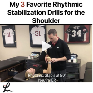
I use Rhythmic Stabilization drills daily in my practice for most of my upper body patients.

I think they are critical to use to help the patient regain neuromuscular control, a sense of their joint and it’s a great way for the PT to get a sense of how the patient is doing overall.

The hands-on strength training (trying not to trigger dependency by the patient, so relax!) is a great way to gauge a patient’s progress.

Various tools can be used to progress and regress these exercises. I hope these few basic RS drills can give you a basic understanding of what we do @championptp

I hope to get more of these videos out in the future.

Share with your friends and colleagues so they can see these valuable drills for the shoulder joint!


The Week in Research Review, etc 8-5-18

The Week in Research Review, etc 8-5-18 we discuss a wide variety of topics including:

  • Long-term disability if weak during adolescence
  • Using heat during rehabilitation
  • OKC vs CKC exercises after an ACL
  • Live look at an Achilles rupture (with sound too!)
  • A fun look at the different types of PT’s
  • Congrats to all of the newly licensed PT’s!

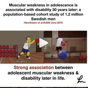
Muscular weakness in adolescence is associated with disability 30 years later: a population-based cohort study of 1.2 million Swedish men. Henriksson et al BJSM June 2018.

Conclusion: There was a strong association between muscular weakness and disability. A combination of muscular weakness and low aerobic fitness was an especially important risk factor for disability. This adds weight to call for muscular strength and fitness-enhancing exercise for adolescents in all BMI categories.

This study out of Sweden looked at the associations of muscular strength in adolescence with later disability pension.

A total of 1 212 503 adolescent males aged 16–19 years, recruited from the Swedish military conscription register between 1969 and 1994.

Moral of the story: exercise as an adolescent may help to reduce issues later in life, including the potential for disability.

Taking it 1 step further… why is physical education being cut out of school requirements when studies like this show the potential negative effects of inactivity?!


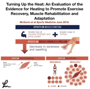
Turning Up the Heat: An Evaluation of the Evidence for Heating to Promote Exercise Recovery, Muscle Rehabilitation, and Adaptation McGorm et al Sports Medicine June 2018.

Key Points: Animal and human trials have shown that various forms of heating can be used in conjunction with exercise or stress to enhance recovery, adaptation and limit muscle atrophy.

Heating muscle activates protective mechanisms, reduces oxidative stress and inflammation, and stimulates genes and proteins involved in muscle hypertrophy.

Further studies highlighting the differences between various heating modalities will help inform athletes and coaches on the best heating practices for specific situations.

This article has a ton of great information that I highly recommend any PT, strength coach, athletic trainer or massage therapist.

It is a review of the literature and there are still many questions to be answered so, as always, take with a grain of salt.

I am a fan of heating before treatment…I do it daily with 99% of the clients that I see and they love it…so that says something.

What do you think? Do you like to heat your clients up before treatment or before a workout? Tag a friend that may benefit from this post! Thanks, guys!


The Effect of Open- Versus Closed-Kinetic-Chain Exercises on Anterior Tibial Laxity, Strength, and Function Following Anterior Cruciate Ligament Reconstruction: A Systematic Review and Meta-analysis Perriman et al JOSPT July 2018 Level 1a

FINDINGS: There was no significant difference in anterior tibial laxity, strength, patient-reported function, or physical function with the early or late introduction of open-kinetic-chain exercises in those who have had anterior cruciate ligament reconstruction, when compared to closed-kinetic-chain exercises, at all follow-up time points.

They wanted to determine whether OKC quadriceps exercises result in differences in anterior laxity, when compared to CKC exercises, at any time point following ACLr.

Also, they wanted to determine whether there are differences in strength, function, quality of life, and adverse events with OKC quadriceps exercises when compared to CKC exercises at any time point.

Overall, calculated effect sizes showed a slight increased laxity in the OKC groups, particularly for the hamstrings graft. The⠀
pooled difference was not statistically significant (P>.05)

When considering all graft types, there was low- to moderate-quality evidence from 3 studies suggesting that there were no between-group differences in laxity at any time point when OKC exercises were introduced earlier than 6 weeks post ACLR, compared to CKC exercises.

There seemed to be a trend that showed early OKC knee extension was safer after a PTG than a hamstring autograft but protocols varied so data was inconsistent.

Of interest to me, they said “The early introduction of OKC quadriceps exercises did not appear to offer additional significant benefits in function and strength for the average patient post ACLR; therefore, this early introduction is questionable, especially in patients with a hamstring graft.


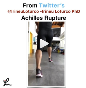
From Twitter’s @IrineuLoturco showing the moment this athlete ruptured their achilles tendon. Pretty impressive and you can see the eccentric loading of the tendon that caused the rupture. See his original post below…

A very impressive recording of the exact moment when an elite sprinter had an acute and complete rupture of the Achilles tendon. Pay attention to the “boom”.


[REPOST] If there was an ESPY for a post by a PT then @theperformancedoc would definitely get it for these videos! Great job and keep pumping out great content. Give him a follow if you haven’t already!

👇🏻
💥Different Types of Physical Therapists In the Real World💥 SWIPE 👉🏽 (Turn on 🔊) Sometimes we have trouble “turning it off” when we are outside of the clinic. Which one are you?! Tag, Comment, & Share with a Physical Therapist‼️

▪️
#ThePerformanceDoc #RehabWithTheDoc
#TeamMovement


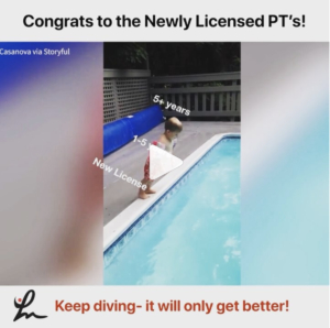
Congrats to all of the newly licensed PT’s out there…Welcome to the profession!

My advice to you:

Stay humble and put the patient first, always

Keep learning and try to avoid complacency

The road will seem rough but it does get a little easier. Get experience…as much as you can. Each interaction with a patient is a snapshot to help guide your future interactions.

Put yourself in their shoes… give them the best experience as you would expect to receive if you were sitting on the plinth being asked questions.

Follow people on social media that help you to learn and keep an open mind. Don’t get pulled into 1 system. Take a little from each and package it nicely.

The research is often biased. Opinions come and go. Stay somewhere in the middle…remember the bell curve, always!

Each “system” has huge overlap despite their ‘trademarked’ proprietary information…they all involve motion and strengthening. That’s the key to PT- keep people moving and keep them stronger…or at least keep them positive and hopeful.

I recently wrote a blog post discussing the evolution of a PT. Take a look…the link is in my bio on Instagram.

These words are the basis of my practice. Take what you think is important and apply it to your practice. Good luck, now work on your dives!


A great week of content that I hope you found valuable and willing to share with your friends and colleagues! Thanks for reading!

The Week in Research Review, etc 7-22-18

The Week in Research Review, etc 7-22-18

I’m trying out this new concept of publishing my social media posts into a nice package for a weekly delivery to my subscribers.

  1. Knee Case Study
  2. Contralateral ACL Strengthening
  3. Shoulder Static Stabilizers
  4. Weighted Ball Research
  5. Glute Activation


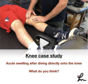
This kid came to me the other day with L knee swelling after sliding headfirst into 2nd base during a baseball game.⠀

Continued to play in the game and even pitched the next day, all without pain or loss of motion.⠀

As you can see from the video, he has a bunch of fluid in his knee, medial ecchymosis (bruising) but full pain-free ROM.

Ligamentous tests appear negative and he has absolutely no pain or stiffness with anything.

I took this video to show what appears to be a bursal sac disruption from the impact of his knee into the ground as he was sliding.

The mechanism fits the presentation and clinical exam.

I advised him to monitor his swelling, wear a knee sleeve and continue his activities per his tolerance.

He is going to touch base with me next week to make sure the fluid is dissipating (and not worsening) and he remains asymptomatic.

What do you think? Am I missing anything? What’s your diagnosis? Tag a friend who may be interested in this case.

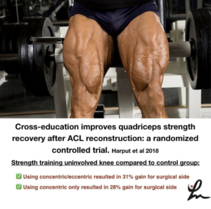
Cross-education improves quadriceps strength recovery after ACL reconstruction: a randomized controlled trial. Harput et al Knee Surg Sports Traumatol Arthrosc. 2018

This study looked at a group of ACL reconstructed patients that were divided into 3 groups.

All 3 groups performed the same standardized ACL rehab, but one group was the control group that performed the standardized rehab only.

The other 2 groups did either 3x per week extra concentric knee extensions on their uninjured leg for 2 months (beginning at 1-month post-op through 3-months post-op) or additional eccentric knee extensions on their uninjured leg 3x per week for 2 months between months 1-3 post-op.
💪🏼
They found that the quads strength for the concentric group was 28% greater compared to the control group. 💪🏼
The eccentric group was 31% greater when compared to the control group.

Conclusion: Concentric and eccentric quadriceps strengthening of healthy limbs in early phases of ACL rehabilitation improved post-surgical quadriceps strength recovery of the reconstructed limb.

Pretty crazy stuff and one more reason to work on bilateral strengthening with most of our patients, especially when they’re post-op ACL reconstruction.

Do you work on bilateral strengthening? if not, why? If you do, what other studies have you seen that show similar results?
Tag a friend who may benefit from this study or let’s discuss in the comments section!

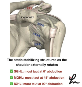
This picture shows a simplified view of the static stabilizers of the shoulder joint. I highly recommend reading a classic paper by Wilk et al 1997 JOSPT that talks about this and cites a paper from Bowen et al Clin Sports Med 1991 @wilk_kevin

When one is picturing these stabilizers, the superior glenohumeral ligament (SGHL) is most taut when the shoulder is externally rotated at 0 degrees of abduction.

As we progress to 45 degrees of GH abduction, we stress the middle glenohumeral ligament (MGHL) as we externally rotate the humerus.

Finally, at 90 degrees of GH abduction, we stress the inferior glenohumeral ligament (IGHL) as we externally rotate. More specifically, the anterior band of the IGHL.

As we internally rotate at 90 degrees of abduction, we stress the posterior band of the IGHL.

These concepts have rehab implications and should be kept in mind when we’re rehabbing people after an injury or surgery.

For example, if someone has an anterior Bankart lesion (front labral repair), then we need to progress them slowly into external rotation, especially at 45 and 90 degrees of abduction.

Another example would be a rotator cuff repair, like the supraspinatus. We would want to progress them slowly at lower degrees of abduction 0-45 degrees but maybe we can progress them a bit quicker at 90 degrees of abduction.

Hope these concepts make sense because they are very important to understand for many patients with shoulder injuries.

Does this make sense? Have you heard this info before? Tag a friend who may benefit from this post!

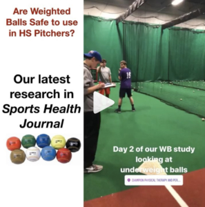
Effect of a 6-Week Weighted Baseball Throwing Program on Pitch Velocity, Pitching Arm Biomechanics, Passive Range of Motion, and Injury Rates. Reinold et al Sports Health Jul-Aug 2018. @mikereinold

Our 1st of potentially 3 research articles looking at the effects of weighted balls on youth baseball pitchers.

High school baseball pitchers performed a 6-week weighted ball training program.

Players gradually ramped up over the 6 weeks to include kneeling, rocker, and run-and-gun throws with balls ranging from 2oz to 32 oz.

🤔After 6 weeks, the weighted ball group did increase velocity by 3.3%, 8% showed no change, and 12% demonstrated a decrease in pitch velocity. Also of note, 67% of the control group also showed an increase in pitch velocity.⠀

The weighted ball group had a 24% injury rate although half of the injuries occurred during the study, and the other half occurred the next season. There were no injuries observed in the control group during the study period or in the following season.

The weighted ball group showed almost a 5-degree increase in passive shoulder external rotation, also known biomechanically as the late cocking position or layback position.

There were no statistically significant differences between pre- and post-testing valgus stress or angular velocity in either group.

✅Our conclusion: Although weighted-ball training may increase pitch velocity, caution is warranted because of the notable increase in injuries and physical changes observed in this cohort.

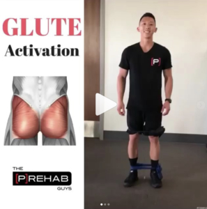
Some great Glute 🍑thoughts buy the @theprehabguys. Check out their videos and content for some great ideas that you can add to your practice!⠀
👇🏼⠀
___________________________________________________________________⠀
Episode 705: “Hip Prep for Glute Activation”⠀
.⠀
Tag a friend looking for a glute🍑 killer!⠀
Hip prep is a series of 6 exercises I’ve adopted from my girlfriend @smenzz and her clinic @eliteorthosport. I use it with my patients to prime the glutes and lower body in general before getting into more dynamic and plyometric activities. I will make the statement right now: if done RIGHT, it’s an absolute glute killer & I promise you that you will feel your glutes!⠀
.⠀
I like these 6 exercises in particular for a variety of reasons.⠀
✅They challenge the glutes in all 3 planes of motion.⠀
✅They hit all types of muscle contractions: isometric, concentric, and eccentric⠀
✅They are performed upright in a functional position⠀
✅There is a variety of double leg, single leg, and split stance variations⠀
✅They train proper lower extremity alignment in a variety of hip and trunk flexed/neutral/extended positions⠀
.⠀
The 6 exercises are:⠀
1️⃣3 way clams: 5 per leg per position⠀
2️⃣Side steps: Alternating steps to the left and right starting with 1 step all the way to 5 steps⠀
3️⃣Monster Walks: 10 steps forward, 10 steps backwards⠀
4️⃣W’s: 10 steps to the left, 10 steps to the right⠀
5️⃣Squats: 10 squats⠀
6️⃣Single leg fire hydrants: 30s per side⠀
.⠀
💡Understand that you first need to teach these exercises in isolation first, before throwing someone all 6 at once⠀
.⠀
Have fun!⠀


Hope this helps you keep up to date and fulfill my goal of this website…simplify the literature and bring great content to you so you can apply it 1st thing Monday morning! Happy Reading! 👊🏼

Follow me on Social Media here:

Image result for instagram logo vectorImage result for twitter iconImage result for facebook logo

Testing the elbow for a UCL sprain in baseball players

I have assessed hundreds if not thousands of elbows for various injuries. One of the most common ailments that I see in my practice is a UCL (ulnar collateral ligament) sprain, aka ‘Tommy John ligament’, especially in a baseball player. This post discusses the typical presentation of a UCL sprain, testing the elbow for a UCL sprain and how I rule in/out with a few simple tests.

Factors contributing to UCL sprains

Injuries to the elbow UCL continue to grow due to many factors. Some think it may be due to:

  • current training regimens,
  • sports specialization,
  • overuse/fatigue,
  • weighted ball training or
  • increased velocity.

(Seems like a great outline for future blog posts!)

Whatever the thought, the clinician needs to be able to differentially diagnose the issue and come up with a game plan that meets the goals and desires of the athlete. No algorithm is going to fit perfectly into each individual’s current/future baseball plans.

That’s why I carefully consider each factor and review it with my baseball players so we can come up with a good game plan in case they get diagnosed with the dreaded UCL sprain!

The Typical UCL Story

Most of the pitchers that come to me with elbow pain have a similar story… and it’s probably not what you would think. Often times, it’s not a dramatic blowout where the ball goes flying into the stands while the pitcher is writhing in pain. Most commonly, it’s a slow onset of elbow soreness, loss of velocity and/or location, or tingling into their fingertips.

The epidemic of Tommy John injuries has freaked out most pitchers. They automatically think they’ve blown their elbow out if they even feel a slight hint of pain or soreness.

It’s up to the clinician to determine what structures may be involved and to have a firm plan in place that allows for a gradual return to throwing or to refer out to a surgeon that you trust.

Unfortunately, it’s not always clear-cut. We need to be able to sift through the stories and recognize that there are many potential causes for elbow pain in a baseball player.

Differential Diagnosis

There are many structures that could become injured in a baseball pitcher. Let’s quickly run through the most common injuries and give my thoughts on each:

  • Flexor-pronator strain– often accompanies a UCL sprain because static stability compromised; painful resisted wrist flexion and sometimes extension. Palpable tenderness in muscle belly not close to the UCL insertion (sublime tubercle.) Often pain-free UCL special tests.
  • Loose bodies: pain-free UCL special tests, pain with late follow-through phase of throwing; significant posterior elbow pain with bounce home test that replicates their symptoms
  • Ulnar neuritis: tingling in the 4th-5th fingers (ring and pinkie fingers); often accompanies a UCL sprain due to increased medial elbow laxity; rarely an isolated event in my opinion.
  • Little Leaguer’s elbow: younger pitcher/player less than 14 years of age; may have painful UCL tests; pain at similar location but often closer to epicondyle; MRI to confirm; rest for at least 3 months; don’t mess with these (would’ve been a UCL if a couple years older and has a higher propensity for a UCL issue later in life.)
  • Thoracic Outlet Syndrome: pain-free UCL tests; loss of control (some call it the yips); vague heaviness and weakness with loss of velocity and location; TOS sees us more than we see it
  • Cervical spine: negative UCL tests; need to consider myotomes and dermatomes; not as common in younger-aged baseball players but may be seen in older pitchers

My go-to UCL tests

There are many tests out there but after many years of playing around with lots them, I have narrowed it down to 4 tests that I feel are the best to help diagnose a UCL sprain. Check out the video below.

 UCL Treatment Options

The treatment options will vary case by case and highly dependent on many variables. The clinician and client need to consider:

  • How much rest, if any has occurred- should try AT LEAST 4-6 weeks of no throwing and rehab to restore ROM (GH flexion, external rotation, and horizontal adduction) then attempt an ITP if pain-free on the clinical exam.
  • Time of year- If it’s the end of the baseball season, may shut it down to give the athlete plenty of time to rest. If clearly needs surgery, plan surgery and rehab, including throwing programs, to last 12-18 months before return to competition.
  • Previous injury history- previous history of elbow issues, especially Little Leaguer’s elbow sets off many bells and whistles; had tingling into fingers a previous time but it went away with some rest; Rest may help but usually leans toward surgery.
  • Tommy John Surgery- reconstruct the ligament; need at least 9-12 months if a pitcher…the longer the better it seems
  • Internal brace UCL repair- a new procedure that repairs the ligament then braces it with a collage-dipped fiber sewn into the joint; currently a quicker rehab but no long-term outcomes and not for all.

To Tommy John or not…that is the question

Wrapping it up, I’d just like to add that there are probably many more scenarios that could play out. My goal was to give you some sort of framework and guidelines for someone presenting with a medial elbow issue and looking for answers.

The cluster of tests seems to be pretty straightforward and simple to understand, hopefully! There are a bunch of possibilities but Tommy John issues should be diagnosed with a good history and careful examination…good luck!