Tag Archive for: rotator cuff

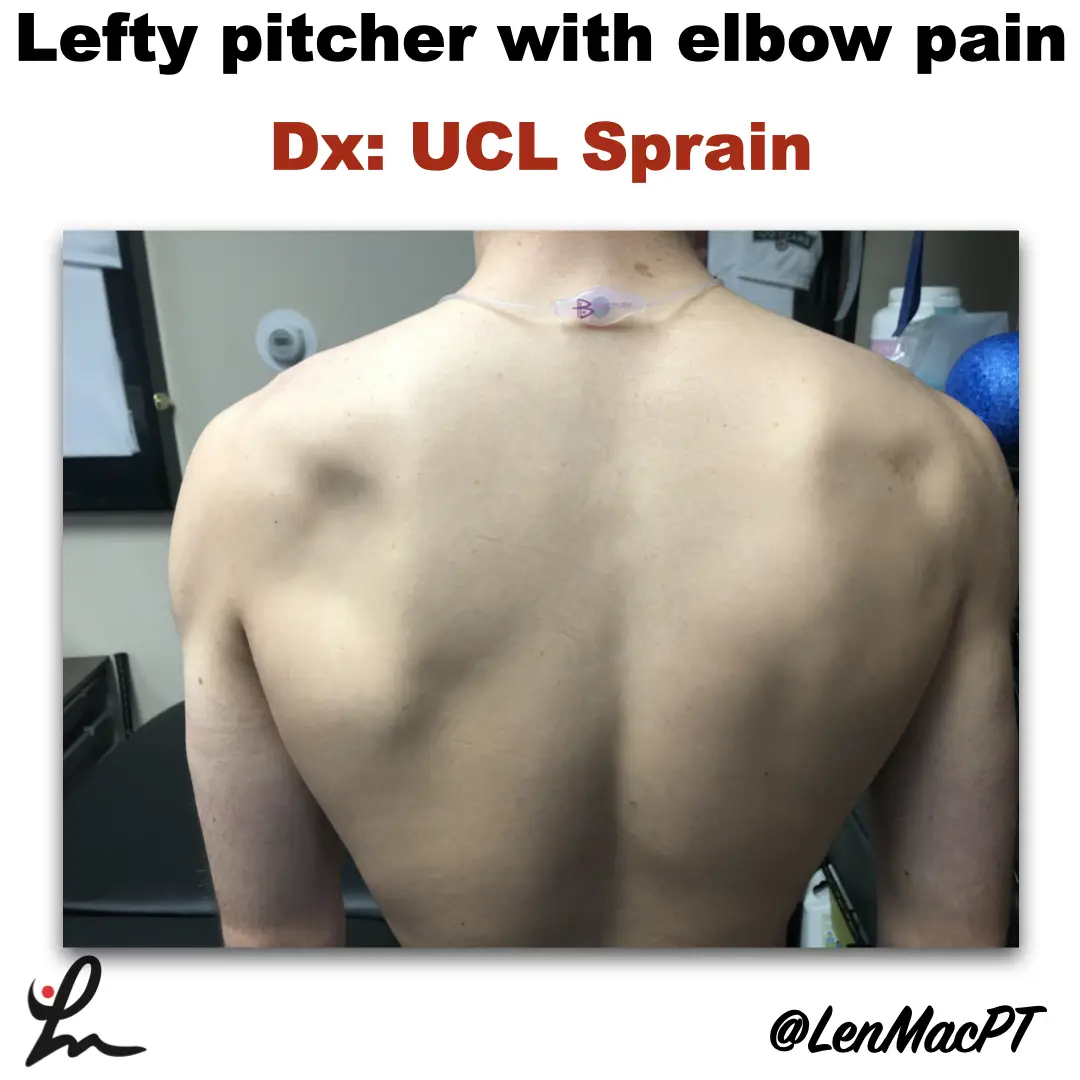
Infraspinatus atrophy in a baseball pitcher with a UCL sprain

Infraspinatus atrophy due to a spinoglenoid notch cyst

This was an incidental finding of mine in a collegiate baseball pitcher. He presented with a UCL sprain during his senior year and was struggling.

He came in looking to salvage his senior year and attempt to pitch at some point. UCL surgery would’ve been warranted if he was anything but a senior.

UCL Evaluation

We evaluated his elbow and he presented with typical findings of painful valgus stress tests. I’ve discussed my UCL exam previously in a post that you can find here.

UCL Tommy John Assessment

His shoulder PROM was slightly limited compared to what we normally see in our overhead throwers, which may have contributed to his elbow issues.

He only had 120 degrees of external rotation compared to 110 degrees on the right side. If you’re wondering how I measure external rotation, then this video below may be a good one to review.

External rotation passive range of motion (PROM)

We use these concepts for our research studies that we have published on weighted ball usage the past few years.

For those that will ask about GIRD, he had 60 degrees of IR on the L side and 50 degrees on the R side. I’m not a big GIRD guy like has been written by others. I still think GIRD is pretty normal and our research has shown that too.

Remember, we showed that differences in total rotational range of motion greater than 5 degrees was significant. This study was published in AJSM back in 2011.

Furthermore, we showed a connection between the loss of passive flexion and elbow injury.

This result was also shown by Camp et al in a separate study, as well.

I had him remove his shirt and noticed a large defect in the infraspinatus area that showed obvious atrophy (see picture below).

Infraspinatus atrophy due to a spinoglenoid notch cyst

The cyst was compressing the suprascapular nerve as it coursed along the spine of the scapula.

His external rotation strength was significantly weaker on his throwing side compared to his right side. This would make complete sense considering the amount of atrophy present.

Often times surgery is warranted to decompress the nerve. There’s often an underlying labral tear that has resulted in a cyst to form. The surgery would remove the cyst and repair the labrum to prevent future issues.

Quick Literature Review on Surgical Outcomes

Reports of improved outcomes are in the literature, here and here and here.

Obviously, this may have contributed to his elbow issues as the posterior cuff muscles are tremendous stabilizers of the arm, especially during the deceleration phase of throwing.

Treatment for infraspinatus muscle atrophy

Our goal was to calm the elbow down through active rest and focus on strengthening of his forearm and shoulder musculature.

We used neuromuscular electrical stimulation (NMES) to the infraspinatus. We felt as if the electrical stimulation would allow for a better muscle contraction than what he could do actively. This is a similar concept to using NMES to the quads after a knee surgery.

NMES to the infraspinatus to help with muscle recruitment

We have previously shown in AJSM that NMES to the infraspinatus was beneficial to patients after a rotator cuff repair. It helped improve their muscle contraction by 22% in the group with the NMES applied during testing.

Outcomes and final thoughts

Believe it or not, we were able to salvage his senior year despite all of the issues that presented.

He made numerous pitching outings and was pretty successful for his team.

The lesson here: Don’t take anything for granted, observe and test.

Make sure you look distal and proximal. It could’ve been very easy to just treat the elbow with some exercises and massage. Don’t get me wrong, we did a bunch of strengthening exercises to his forearm.

His secondary finding was most likely a leading cause for his elbow dysfunction.

This isn’t the 1st time that I’ve run across an issue like this. I’ve found numerous scapula dysfunctions like this involving the infraspinatus. But also a winging issue due to a long thoracic nerve palsy.

You must look at the whole picture… shirts off for a guy and a sports bra for a female to make sure we’re not missing anything obvious!

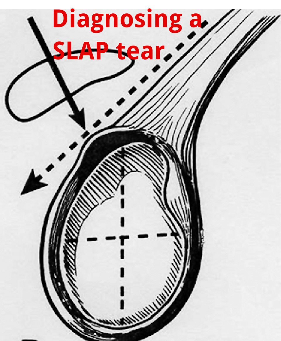
An Update on Diagnosing SLAP tears

Diagnosing a SLAP tear is not easy

We hear a lot about trying to diagnose shoulder pain and to be as specific as possible. It’s often difficult to differentiate SLAP (superior labrum anterior to posterior) tears from other soft tissue injuries of the shoulder.

For a review of the different types of SLAP tears, check out this old blog post that classifies the 10 different types of tears.

from https://commons.wikimedia.org/wiki/File:SLAP-Lesion-front-2.jpg

This paper in IJSPT by Clark et al 2019 attempts to help out the process and recommend a few special tests that MAY aid in diagnosing a SLAP tear.

What do they Recommend to diagnose a slap tear?

They recommend that a combination of at least 3 positive SLAP lesion tests may be clinically useful in diagnosing a shoulder SLAP lesion with greater diagnostic accuracy.

Combo of Tests

The combination of the Biceps Load I/II and O’Brien’s showed the highest sensitivity and specificity.

I have found similar results with this set of special tests so maybe this paper just hits my biases correctly.

In this video at my YouTube channel, I wanted to let you hear my thoughts and small tweaks to the evaluation process.

It’s not easy to diagnose a SLAP tear.

Differential Diagnosis is Critical

Furthermore, does it really matter and will it change the treatment plan much at all? I think it may a little but overall it will remain a pretty similar treatment approach to other similar pathologies like:

  • rotator cuff tendonopathy
  • Biceps strain
  • Latissimus strain
  • Subscapularis strain
  • internal impingement
  • pectoralis major strain

I think one also needs to consider the cervical spine and to make sure the pain is not referred from the neck.

Otherwise, a well thought out program should be implemented that addresses the strain on the shoulder and any strength issues.

I talked about this in an article that I wrote for Medbridge a while back so check out that post here:

You can also check out a snippet of one of my courses at this YouTube video where I discuss rotator cuff and labral issues. Hope it helps too!

Will this Change your Practice?

How ever you look at it, I wanted to use this paper to let you know that there MAY be a cluster of tests that better diagnose a suspected SLAP tear in your next patient’s shoulder.

Check out the paper and comment so we can talk it through. Are these tests similar to what you use in your clinical practice? Will this paper change what you do in your clinical practice?

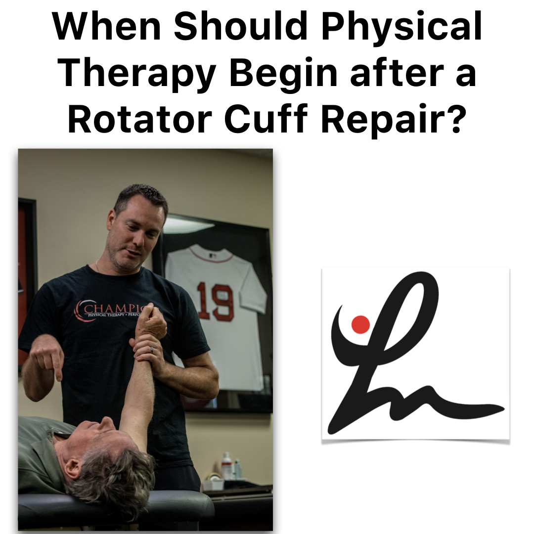
Is Early Physical Therapy Safe After a Rotator Cuff Repair?

Physical therapy is vital after a rotator cuff repair and continues to be common in an outpatient setting. Unfortunately, there’s no true consensus on when to actually begin PT.  Is early physical therapy safe after a rotator cuff repair or should we delay PT to protect the healing tendons?

Let’s dive into this and see what the research is saying…

Why I’m writing this post on rotator cuff rehabilitation

It seems as if we’re all over the place despite the research, which is pretty typical. Some docs prefer early passive range of motion (PROM) while others wait 6-8 weeks (and even up to 12+ weeks for a revision repair) before they allow any form of PT.

For the record, I’m going into this blog post as a firm believer of early PROM. It’s what we’d been doing for years in Birmingham at Champion Sports Medicine.

It’s what I only knew until I moved to Boston in 2014. Now I’ve seen a nearly 180-degree turn in rehab thoughts. Much more conservative!

I recently got in a Twitter discussion (debate) about this same topic with some very respected and prominent PT’s in the field.

It made me think about things so I decided to do a little research to see what the literature says. You can check out the discussion here.

Twitter can be confusing and tough to follow but just trust me, it goes on for a while!

Rotator Cuff Anatomy

The tendon most commonly torn is the supraspinatus tendon. Don’t get me wrong, you can tear the other rotator cuff tendons (infraspinatus, teres minor, subscapularis).

Keep in mind a medium, large or massive rotator cuff tear often will involve the infraspinatus tendon. If it does, then you need to consider modifying your progression appropriately.

But for the sake of this post, I’m going to stick to an isolated supraspinatus tendon for now.

Rotator Cuff Tendon Size and Location

Look at the size of the tendons as they insert on the greater tuberosity. You’ll see it differentiated by antero-posterior and medial-lateral directions.

According to my colleague and friend Jeff Dugas out of Birmingham, he showed in 2002 that the mean dimensions of the supraspinatus insertion were 1.27 cm in the medial-to-lateral direction. For the anterior-to-posterior direction, the supraspinatus dimension was 1.63 cm.

It helps to know this information because you may need to read an operative report and see the size of the tear. The docs will usually mention a 2 cm tear or something like that.

That means that the supraspinatus tendon and a small portion of the infraspinatus tendon were involved (and repaired.)

This is critical information to have when you’re trying to plot the post-op rehab progressions and determine the prognosis. The more tendons involved, then the higher the chance of repair failure.

There are many other factors that influence retear rates but tendon repair size is definitely one to consider.

Rotator Cuff Repair Surgery Types

I’m not going to bore you with the details of a repaired rotator cuff. There are numerous surgical techniques being used by orthopaedic surgeons.

Techniques such as a single row, double row, suture bridge or transosseous repairs are commonly performed. The picture below shows the difference between a single row and a double row repair, for example.

As you can see below, the double row tends to repair more of the tissue back to the humeral insertion point, which in theory has led to better tendon healing. This has been shown in numerous research studies and has become the best technique available.

So you had shoulder surgery…when to start physical therapy?

That seems to be the million dollar question! The research is all over the place. This means that doctors’ opinions are all over the place too, right?

Keep in mind that I’ve written about this in the past. Rotator cuff repair surgery is not always warranted and can be avoided!

Since I joined the group in Birmingham in 2002 (as a PT student), we had our post-op rotator cuff repair patients starting PT post-op day 1. They started PT early regardless of the tear size. This means a small tear of 1 cm in length started PT the same time a massive, 5 cm repair would start PT.

Some may disagree with this start time but it worked…it just worked. At least I think!

Why Early PT after a Rotator Cuff Repair

This is Key!!

There were several reasons why I think it worked:

  • They could chat with a professional.
  • Patients better understand their pain and get reassurance that what they were feeling was normal.
  • Someone could monitor their incisions and answer any and all questions.
  • Begin early, gentle ROM which often helps with pain control, too.

But, that was our ‘protocol’ and it continues to be that way many years later. Most other doctors that I have dealt with outside of Birmingham have taken a far more conservative approach to post-op rehab.

Agree to Disagree

Here in Boston, most docs wait at least 2-3 weeks and even up to 8-12 weeks to begin PT. Talk about eye-opening!

I don’t agree with this premise and wanted to dive a bit deeper into the literature to see if early physical therapy had a detrimental effect on short-term, mid-term and long-term outcomes.

Structure vs Function

The problem that continues to plague the research is the measurement of outcomes. Doctors care about the structural integrity of their rotator cuff repair. They see the research and are concerned with retear rates that hover in the 25-70%+ stratosphere. Of course I’d be concerned with retear rates that high!

Can you imagine if ACL re-tear rates were that high? Well, guess what they still hover in the 6-40% range even with our tremendous rehab skills and return to play testing.

But fortunately, we have other parameters to consider with our patients after a rotator cuff repair. We can look at the pain-free function!

Huh, what a novel idea. Regardless of the integrity of the repair, many patients can still live their lives to the fullest and in most cases without any pain.

Will Ferrell Yes GIF - Find & Share on GIPHY

Start PT Early after a Rotator Cuff Repair? What does the literature say…

I’m going to do my best and unbiased research to figure out if early PT after a rotator cuff repair is safe and effective compared to a delayed protocol. Let’s take a look…

There are a bunch of studies out there that you need to sift through. You can tell when the lead authors are MD’s or PT’s because the docs want to make sure their repair integrity is intact and the PT’s are concerned with restoring ROM, strength, and function.

With that, I’ve done my best to pull out some studies that have helped guide my practice and continue to influence me today.

Literature Review Findings

Age a BIG Factor!

Mind you, Cho et al showed that healing rates after a rotator cuff repair drastically change for older patients compared to younger patients.

The slide below was taken from my rotator cuff lecture that I’ve done in the past and helps to put things in perspective.

In no particular order…

Parsons et al JSES 2010 looked at 43 full thickness RTC repairs who were in a sling for 6 weeks. All were without PT for that time then evaluated for stiffness in PROM. They defined stiffness as 100° flexion/ 30° ER.

Overall, of the 43 surgeries, 23% (n=10) became stiff after that 1st evaluation session. The whole cohort displayed a 56% retear rate overall at 1 year, which to me seems crazy high!

To break it down further there was:

  • 30% retear in stiff group
  • 64% retear in non-stiff group
  • There was no significant difference in ROM or functional scores.

In my opinion, there were some pretty big limitations to the study that should be exposed, like:

  • Single row repair
  • No consideration for Diabetes or smoking
  • MRI without contrast to re-evaluate the repair status
  • What is “ER by the side???”- need to better define what degree of abduction.

So getting stiff may be a good thing but the repairs were done as single row repairs. We know these did not heal as well as they do with double row repairs.

More Literature Reviews

Moving on to a 2014 Level II systematic review and Meta-analysis, the authors said “the results contradicted our hypothesis that immobilization would increase tendon healing compared with an early-motion rehabilitation protocol, as structural outcomes were similar in the two groups 1 year after the arthroscopic repair of rotator cuff tears.

From the paper: “We speculate that rehabilitation is not the sole factor affecting tendon–bone recovery; the effects of other factors, such as older age, fatty degeneration, larger tears, and surgical technique, may outweigh those of the rehabilitation protocol.”

Kim et al AJSM 2012 looked at small to medium sized RTC repairs. They compared immediate PROM (0-120 degrees) to 4 weeks of absolute immobilization. They eventually showed no difference in ROM, pain or tendon healing. So seems like a smaller tear of less than 3 cm may be appropriate for immediate ROM, albeit it was limited to 120 degrees for some reason.

Not sure why they limited to 120 degrees because it seems as if the tendon would shorten as the humerus is placed in further flexion. Maybe they were concerned with subacromial impingement or something but the limitation is a bit confusing to me.

Healing Affected?

Another study by Lee et al AJSM 2012 wanted to compare ROM and healing rates between 2 different rehabilitation protocols after arthroscopic single-row repair (use caution) for full-thickness rotator cuff tear.

They showed pain, ROM, muscle strength, and function all significantly improved after arthroscopic rotator cuff repair, regardless of early postoperative rehabilitation protocols.

They also looked at the repair integrity with postoperative MRI scans, 7 of 30 cases (23.3%) in the immediate ROM group and 3 of 34 cases (8.8%) in the delayed group had re-tears, but the difference was not statistically significant (P = .106).

Well then, only a trend and all had similar functional outcomes regardless of when they started ROM…I’d say that helps the case to start early.

But again, these repairs were done via a single row repair and they allowed manual therapy 2 times per day and unlimited self-passive stretching exercise, which seems a bit aggressive anyway.

Do we even need a sling for 6 weeks?

No Functional Difference Between Three and Six Weeks of Immobilization After Arthroscopic Rotator Cuff Repair: A Prospective Randomized Controlled Non-Inferiority Trial Arthroscopy 2018

This study looked to compare clinical and radiologic results among patients with 3 versus 6 weeks of immobilization after arthroscopic rotator cuff repair in a prospective randomized controlled non-inferiority trial.

They concluded that “3 weeks of postoperative immobilization with sling use was non-inferior to the commonly used regimen involving 6 weeks of immobilization in a brace.” For the structurally concerned people out there, MRI indicated similar degrees of healing between the groups. 

Well then, that throws a wrench in things for the docs!

Ryan Gosling Giggle GIF - Find & Share on GIPHY

Does Early vs Delayed PT Affect Outcomes?

A systematic review by Gallagher et al 2015 looked to determine if there are differences between early and delayed rehabilitation after arthroscopic rotator cuff repair in terms of clinical outcomes and healing.  Six articles matched their criteria and reported significantly increased functional scores within the first 3-6 months with early rehabilitation compared to the delayed group.

To me, this is huge! Put yourself in their position. Imagine feeling better and being able to get back to work a little quicker. That’s my major argument for starting rehab sooner. Earlier pain relief, improved function and a feeling of being normal again.

Furthermore, none of the included studies reported any significant difference in rates of rotator cuff re-tear.

Medium-Large Tears use Caution

However, two studies noted a trend towards increased re-tear with early rehabilitation that did not reach significance. This was more pronounced in studies including medium-large tears. A similar trend that I’ve seen in the literature.

Here’s a nice table from that Gallagher study that summarized their findings for each research paper they included:

I want my patients to feel good as quick as possible and get back to some semblance of a normal life. But of course I want the long-term integrity of the repair to remain intact. And it seems as if this study helps that argument.

Then you look at a group of PT’s from Turkey (Duzgun et al Acta Orthop Traumatol Turc. 2011) that looked to compare the effects of the slow and accelerated protocols on pain and functional activity level after arthroscopic rotator cuff repair. Patients were randomized in two groups: the accelerated protocol group (n=13) and slow protocol group (n=16).

There was no significant difference between the slow and accelerated protocols with regard to pain at rest.

The accelerated protocol was superior to the slow protocol in terms of functional activity level, as determined by DASH at weeks 8, 12, and 16 after surgery.

I’ll take that!

Function over Structure (at times!)

Told you that the PT groups tend to favor function over structure, haha!

Early passive Motion ok- The French Version

This next study out of France that included French Society for Shoulder & Elbow ( 2012) looked to compare the clinical results after two types of postoperative management: immediate passive motion versus immobilization. Patients were randomized to receive postoperative management of immediate passive motion or strict immobilization for 6 weeks.

They concluded that their results suggested that early passive motion should be authorized: the functional results were better with no significant difference in healing. Functional results were statistically better after immediate passive motion and a lower rate of adhesive capsulitis and complex regional pain syndrome.

Seems as if there may be a trend but certainly not an overwhelming conclusion that early ROM is guaranteed to lead to inferior structural results. But, it seems pretty conclusive that ROM, pain, and function are improved after early ROM.

Let’s continue to look at the research

This group from China (Shen et al Arch Orthop Trauma Surg. 2014 Sep) performed a systematic review and meta-analysis to determine whether immobilization after arthroscopic rotator cuff repair improved tendon healing compared with early passive motion. Three RCTs examining 265 patients were included but we need to be cautious because of the limited number of studies included and the heterogeneity of the samples.

They found that there ‘no evidence that immobilization after arthroscopic rotator cuff repair was superior to early-motion rehabilitation in terms of tendon healing or clinical outcome. Patients in the early motion group may recover ROM more rapidly.’

This recent 2017 study in the Journal of Shoulder and Elbow Surgery included 9 meta-analyses in its review. They basically noted, “No clear superiority was noted in clinical outcome scores for early-motion or delayed-motion rehabilitation.”

They also concluded that “Whereas early motion and delayed motion after cuff repair may lead to comparable functional outcomes and retear rates, concern exists that early motion may result in greater retear rates, particularly with larger tear sizes.”

So, it seems as if function and healing the same after a RTC repair but there may be a discrepancy once we start looking at a larger repair size.

Immediate PT after a Revision Rotator Cuff Surgery

This study in AJSM 2018 looked to evaluate the clinical and radiological outcomes after revision rotator cuff repair surgery. They were able to track 31 of 40 patients (77.5%) for the final assessment at a mean follow-up of mean 50.3 months.

Interestingly enough, physical therapy started on the first postoperative day with passive flexion and abduction.

Revision rotator cuff repair improves clinical outcomes and shoulder function at midterm follow-up. The clinical outcome scores were comparable in patients with an intact repair and those with failed RC healing.

And they started PT 1 day after the surgery and got PT 2-3 times per week.

Retear Rates and Long-term function

This study in JBJS 2006 looked to determine the clinical and structural outcomes of re-ruptures in twenty patients after a longer period of follow-up. Nineteen of the twenty patients continued to be either very satisfied or satisfied with the outcome.

At an average of 7.6 years, the clinical outcomes after structural failure of rotator cuff repairs remained significantly improved over the preoperative state in terms of pain, function, strength, and patient satisfaction.

They also found that re-ruptures of the supraspinatus that had been smaller than 400 mm(2) had the potential to heal….wow!

Failure Rates too High!

Another study in JBJS 2013 looked at 18 patients who had undergone arthroscopic repair of massive rotator cuff tears.  At two years of follow-up, 94% had a failed repair. This current study wanted to evaluate the 10-year results for these patients with known structural failures of rotator cuff repairs.

Despite a high rate of progression of radiographic signs associated with large rotator cuff tears (proximal humeral migration or cuff tear arthropathy), most did well.

Clinical improvements and pain relief after arthroscopic rotator cuff repair of large and massive tears are durable at the time of long-term (10 years) follow-up. They went on to say:

“These results demonstrate that healing of large rotator cuff tears is not critical for long-term satisfactory clinical results in older patients.”

So our obsession with healing rates still appears to be overblown, even in older patients with a known cuff tear.

Pendulum Exercises Effects on Muscle Activity

Activation of the Shoulder Musculature During Pendulum Exercises and Light Activities JOSPT 2010

Look at that Paper in JOSPT 2010 (I’ve pulled out the Results Table for you below).

If there’s one exercise that doctors allow after a rotator cuff repair then it’s a pendulum or Codman exercise. How often does our patient do them correctly and make it a completely passive motion? I’d say rarely if seldom, right?

Most often, the patient is just bent over and actively moving their shoulder. They have no body movement or sway. Most of the movement is shoulder based and are not completely relaxing their shoulders.

Furthermore, they were instructed in the doctor’s office that 1st week or 2 after surgery.

They’ve been doing them incorrectly for weeks on end because they have no one to help them (cough cough!)

Look Over Here Steve Carell GIF - Find & Share on GIPHY

EMG of common Rehab ExerciSES

A study in JOSPT 2016 looked at EMG activity in healthy individuals. They wanted to quantify muscular activity during daily tasks and common PT motions. They showed that “of all the tasks assessed, ambulation without a sling and donning and doffing a sling and a shirt consistently showed the highest activity.”

EMG results table is found below.

Pretty helpful to see it listed by muscle and EMG activity and specific movement.

EMG of contralateral movements

A pretty neat EMG paper from 2004 (small n=6 and healthy individuals) were assessed using fine wire and surface EMG during common functional activities of the contralateral extremity while immobilized.

They found high supraspinatus EMG activity of the immobilized shoulder for all fast pulling activities of the contralateral shoulder (25-32%)!

Furthermore, they found high infraspinatus activity (56%) of the immobilized shoulder when the contralateral extremity performed straight forward reaching activities.

So even if they are immobilized and using their non-operative shoulder for daily activities, the rotator cuff is still sustaining a higher amount of activity than anything that we would do in the early phases (PROM, dowel self-ROM, rope and pulleys, properly performed pendulums).

How about revision rotator cuff repairs, you ask?

This study from AJSM in 2018 looked at outcomes after a revision rotator cuff repair. They showed revision rotator cuff repair improved outcomes regardless of tendon integrity (MRI confirmed).

Oh boy, what is going on??

Dig deeper into the study and they started PT the 1st day post-op with passive flexion and abduction. Sounds familiar, no? And this was in revision surgeries.

We used a very similar approach in Birmingham as they did in this study, so I may be a bit biased.

My Closing Thoughts on Physical Therapy after Rotator cuff repair surgery

I think it just shows you that rehab can begin early, will not affect long-term outcomes and that tendon integrity is not correlated to function.

I honestly don’t think our 15-30 minutes of passive motion early on in the rehab process is significantly affecting outcomes and retear rates.

It seems as if the repair technique, contralateral arm daily use, compliance with proper exercises (like pendulums, for example).

Let’s not blame early PT. There are so many more variables that are more likely to affect rotator cuff repair outcomes than anything that we could do in PT.

So I say let’s get people into PT early, educate them, guide them and help them get over this painful surgery.

I’ve been doing this for years and have seen the benefits of early PT. I say the literature agrees with me!

The Week in Research Review, etc 11-19-18

Great ‘Week in Research Review, etc 11-19-18’ that I hope you find helpful to your practice.

I’ve always touted the importance of the subjective portion of the exam so I wanted to share a slide from a recent talk I gave to a group in Canandaigua, NY. Obviously, the squat is a fundamental movement and I wanted to give some basic positions that I use to help assess. So excited that I’ve launched a brand new Medbridge course that helps the rehab specialist better eval and treat the baseball pitcher. On my YouTube channel, I discussed my thoughts on setting the scapula with various upper and lower body exercises. And finally, my co-worker Kiefer Lammi discusses the landmine with exercise.


Importance of the Subjective Exam

Assessing the Squat

My New Baseball Medbridge Course

Set the Scapula with Shoulder Exercises?

6 Ways to use the Landmine by @kieferlammi


💥Subjective the most important aspect of the Evaluation💥

This slide, taken from this past weekend’s course in Canandaigua, NY is always a favorite of mine.

I try to keep a slide like this in all of my lectures because I have found that this portion of the examination can give the rehab specialist a huge look into what is going on with the person in front of them.

Don’t get me wrong, I still consider the biomechanical aspect of what may be causing their symptoms.

It often comes down to a tissue capacity issue but it’s up to me to determine the appropriate course of treatment.

These questions will help build confidence in your client and guide the early stages of rehab.

Do you have any specific questions that you like to ask your clients during their 1st few sessions? Remember, these questions are just not for the evaluation. You should be asking these questions periodically to gauge progress and help guide the next phases of rehab, too!


🔅Assessing the Squat 🔅

Squatting is a fundamental movement that all of us have to do on a daily basis.

Utilizing several different positions can help the rehab specialist better assess the squat and develop a treatment plan that enables their client the ability to improve their squat pattern.

In the above videos, I have utilized 3 different squat patterns and will outline them by the degree of difficulty.

✅The Overhead Squat- by far the most challenging version which challenges the shoulders, thoracic spine, lumbar spine, pelvis, knee and ankles.

A movement limitation at any of these joints will most likely cause the squat pattern to break down. Using overhead resistance would further challenge the system and potentially cause the squat to further breakdown.

✅Arms Crossed Chest Squat- alters the challenge by taking most of the shoulder and thoracic spine out of the equation and isolates the motions to the lumbar spine, hips, knees and ankles.

I often use this position as my fundamental motion because most people don’t have to squat with any weights over their head. This position, in my opinion, should be the most informational and utilized.

✅Counter-weight Squat

This position changes the center of mass by moving some of the weight distribution more anteriorly (front) and making the squat motion slightly easier. I use this position as a regression, for some, which allows them to squat with less stress and potential difficulty.

There are many other variations to the squat that you can make but I wanted to highlight a few of the major changes that you cause successfully. Assessing the squat is essential and can give the rehab specialist a nice picture of the function of multiple joints during a common movement.


My BRAND NEW course on Medbridge’s platform

…that helps the sports and ortho rehab specialist (PT, OT, ATC) better understand the anatomy and biomechanics involved in the baseball pitching motion.

Advanced Rehab for the Baseball Pitcher to Improve ROM & Strength@medbridge_education

The goal of this course was to allow the clinician to be able to evaluate and treat the baseball pitcher using evidence-based guidelines that I use on a daily basis.

Numerous research studies discuss the adaptive changes that occur with the pitching motion followed by numerous videos to help guide the treatment process.

If you’re already a Medbridge subscriber, then you have immediate access today.

If you’re not a Medbridge member, then you can use my promo code “Lenny2018” to save up to 40% off a yearly membership.

This gets you unlimited CEU’s for 1 year and potential access to their online HEP and a lot more!

Students can also get 1 year of unlimited courses (no CEU’s) by using promo code LennySTUDENT2018 and pay only $100.

Check out my other shoulder courses as well by using the Medbridge platform…along with many other great speakers!

Hope you enjoy and good luck!


💥Should you Set the Scapula with your Shoulder Exercise?💥

In this video excerpt from my YouTube channel, I wanted to discuss my opinion on setting the scapula during common exercises.

I think there’s an obvious role for setting the scapula during a heavier lower body lift like a deadlift.

But for a classic upper body exercise like the Full Can (Scaption Raises) or prone T (horizontal abduction), prone Y (Prone full can), etc then I definitely want the scapula to freely move along the rib cage.

I did a quick literature search and didn’t see anything obvious that helped to guide my thoughts so most of this is anecdotal. Check out the video and comment below.

Do you coach your clients to set their scapulae before a rotator cuff workout? If so, why? If not, do you think we should reconsider?


6 WAYS TO USE THE LANDMINE!⁣

Great post from our own @kieferlammi at @championptp on various ways to use the landmine in your client’s workout routine.

If you don’t have one, then I’d highly recommend you try to obtain one because they are highly versatile and can be used in many stages of rehab. See Kiefer’s original post below 👏🏼

_____________

6 WAYS TO USE THE LANDMINE!⁣

The landmine attachment is a super versatile tool for loading that is traditionally known for being used for angled pressing variations. While that’s probably my most programmed use for it, it also provides benefit to a ton of other movements by placing the load and direction of force at a bit of an angle, which can help to promote a particular path of movement, like sitting back more in a squat or lunge. Here are 6 of my favorite ways to use the landmine:⁣

1️⃣1-Leg RDL⁣

2️⃣Split Stance Row⁣

3️⃣Reverse Lunge⁣

4️⃣Deadlift⁣

5️⃣Squat⁣

6️⃣Russian Twist⁣⠀


Save 25% off our OnLine Knee Seminar Course…all this week!

Expires Sunday, November 25th at midnight ET

If you want to learn more about how I treat ACL’s or the knee in general, then you can check out our all online knee seminar at www.onlinekneeseminar.com and let me know what you think.

We cover the anatomy, rehab prescription, ACL, meniscal injuries knee replacements and patellofemoral issues. Furthermore, the course covers both the non-operative and post-operative treatment.t

This is an awesome course if you’re interested in learning more about rehabilitating the knee joint. And if you’re a PT, there’s a good chance you can get CEU’s as well.

The Week in Research Review, etc 11-12-18

This week in research review for 11-12-18 we focused a bit more on assessment and also dabbled in some basic treatment strategies for the back and shoulder. Check out the topics below and like them or comment on Instagram to keep the conversation going…thanks all!


  • A quick fix for a sore low back?
  • Knee Fat Pad Testing and Diagnosis
  • How to Assess the Elbow for a Tommy John (UCL) Sprain
  • Lumbopelvic control on shoulder and elbow kinetics in elite baseball pitchers
  • Full Can or Empty Can? – by @mikereinold


Looking for a quick fix for a sore low back?

I’m speaking from personal experiences when I post a few of the common exercises that have helped me tremendously in the past.

I’m not saying that this is all you have to do but I do think that new onset of low back soreness, you know that tightness that you feel on either side of your spine, can be somewhat alleviated with some foam rolling and active range of motion.

I would definitely include more focal strengthening of the core like deadbugs and bird dogs, squats, deadlifts (when they’re ready), etc.

But for the purpose of this post, I think some foam rolling and motion to the area can take the edge off of someone’s soreness and get them feeling a little better. That’s my goal for many and hopefully those small gins can add up to big gains in the long run!

Do you utilize these techniques as well? If you don’t, then I suggest that you try! They’ve helped me numerous times and continue to help me when my soreness gets a bit out of control.

Tag a friend who may want to check out this post…thanks!

Thanks @corrine_evelyn for the demos!


Knee Fat Pad Testing and Diagnosis

Here’s an excerpt from a previous blog post where I talked about anterior knee pain fat pad irritation. Link in bio!

Keep in mind, my differential diagnosis is all over the place at times. With knee pain you need to consider:

Meniscus (see my previous blog post)⠀

ITB

Osteochondral lesion

Patella tendonitis

Pes anserine bursitis

MPFL sprain

Hamstring strain

Plica syndrome

MCL/LCL

Tumor

Infrapatellar fat pad irritation can be functionally debilitating. I believe it presents itself pretty often in the clinic, more than most PT’s realize.

Use this test to see if it truly is a fat pad issue.


How to Assess the Elbow for a Tommy John (UCL) Sprain

In this excerpt from my YouTube channel, I discuss the tests that I use to help identify an elbow sprain, typically seen in the baseball players that I treat.

In the full video, I discuss:

✅Joint Palpation

✅Seated Milking Sign

✅Prone Valgus Test (maybe a new one for you!)

✅Supine end range External Rotation with Valgus Extension Overload (VEO)

I also wrote a blog post about this topic so hopefully you’ll go to my site and read a bit more about this.

If you treat baseball players of all ages, then you should know how to diagnose a UCL sprain.


The influence of lumbopelvic control on shoulder and elbow kinetics in elite baseball pitchers

Laudner et al JSES 2018.

This study looked at 43 asymptomatic, #NCAA Division I and professional minor league baseball pitchers. They measured the bilateral amount of anterior-posterior lumbopelvic tilt during a single-leg stance trunk stability test.

The Level Belt Pro (Perfect Practice, Columbus, OH, USA) was used to assess anterior-posterior lumbopelvic control. The LevelBelt Pro consists of an iPod–based digital level secured to a belt using hook-and-loop fasteners.

This test has been used and studied previously by Chaudhari et al (JSCR 2011) and he showed that pitchers with less lumbopelvic control produced more walks and hits per inning than those with more control.

Also, pitchers with less lumbopelvic control have been shown to have an increased likelihood of spending more days on the disabled list than those with more control (Chaudhari et al AJSM 2014).

“The results of our study show that as lumbopelvic control of the drive leg decreases, shoulder horizontal abduction torque and elbow valgus torque increase.”

Have you tried this simple test? I will say that having the ability to detect millimeters of motion is clinically difficult.

It is good to see such a simple test utilized clinically can help aid in determining the need for more core/hip exercises for our pitchers. In all, I think it’s a safe bet to incorporate these exercises in all pitchers’ programs.


Full Can or Empty Can?

– by @mikereinold 

Great Post by @mikereinold on which motion is BEST to isolate the supraspinatus during arm elevation. I know you can’t isolate the supraspinatus but numerous studies have (Kelly et al 1996, Reinold et al 2004) shown that the full can (or thumb up position) is better than the empty can position.

Check it out below! 👇🏼

Full Can or Empty Can? – by @mikereinold⠀⠀
-⠀⠀
🧠 WANT TO LEARN MORE FROM ME? Head to my website MikeReinold.com, link in bio.⠀⠀
-⠀⠀
I’m still surprised after all these years that I still see the empty can exercise kicking around. I analyzed these two movements many years ago in an article in JOSPT and showed that the full can exercise (thumbs up 👍) had similar EMG of the supraspinatus with lower levels of deltoid EMG, while the empty can (thumbs down 👎) had higher levels of deltoid EMG.

Why does this matter?

Well, think about it. If you are performing this exercise you probably are trying to strengthen the rotator cuff. And if you are weak and performing an exercise with more deltoid, the ratio of cuff to deltoid will be lower and you’ll have more potential for superior humeral head migration.

Plus, let’s be honest, the empty can just hurts… It’s also a provocative test, and I don’t like to use provocative tests as exercises. 😂😂😂⠀


The Week in Research Review, etc 11-5-18

The Week in Research Review, etc 11-5-18 was filled with more informative and eye-opening posts! Lots of visually stimulating posts to help clarify what exactly is going on in the hip joint with PROM. Another post that shows the suction effect from an intact hip labrum… amongst other great posts.  Just some great stuff..hope you enjoy!


  1. Manual Forearm Resistance Drills
  2. ACL Graft Healing Times to Maturation
  3. Hip Capsule Stress with PROM External Rotation
  4. Muscle Activation Affected by Hip Thrust Variation
  5. Hip Thrust Form by Bret Contreras
  6. Hip Joint Suction Affected by labral Status


Manual Resistance Forearm Exercises

In this post, I wanted to show you guys some of the manual resistance drills we use @championptp on our shoulder and elbow clients, especially our baseball players. We love to use these drills because we can control so many variables with each athlete and tailor it for their specific needs.

We can control the speed and tempo, the direction of forces (eccentric, concentric), and the magnitude of the forces. Plus it’s a great way to interact with our clients. It’s also a great way to feel how well they’re progressing in their programs instead of just giving them dumbbells.

I have found these manual resistance drills to be very helpful with my overhead athletes and hope you give them a try on your clients soon! Let me know what you think or tag a friend below who may like to use these drills too.

In my course that I teach around the US, I try to include these concepts so you can practice and be able to utilize these drills for your clients…thanks!


ACL Graft Harvesting and Healing times

In this post, I wanted to show some research studies on graft healing times and why we need to respect tissue biology.

The systematic review from AJSM 2011 looked at ‘The ‘‘Ligamentization’’ Process in Anterior Cruciate Ligament Reconstruction.’

They essentially looked at 4 different biopsy studies on BPTB and Hamstring autograft reconstructions. They concluded that maturation of the graft, as determined by mainly vascularity and cellularity, was not complete until 12 months at the earliest. The healing time even extended to 24+ months as well.

The ligamentization endpoint is defined as the time point from which no further changes are witnessed in the remodeled grafts. The surgical procedure is quite involved, as you can see in the video that I took from @drlylecain on #YouTube.

As I’m rehabbing my clients, my decision making and post-op progressions often take into account:

✔️Healing biology

✔️Graft harvesting

✔️Graft Type

✔️Bone bruise presence (often!)

✔️Other concomitant issues (meniscus, articular cartilage).

So, respect the tissue and allow natural healing to occur before you add more exercises or are concerned that they’re not making the gains you’d expect.⠀


Hip Capsular Closure: A Biomechanical Analysis of Failure Torque

Chahla et al AJSM 2016

Interesting look at tissue failure, albeit in a cadaver graft, that should help to guide the physical therapist or ATC early in the rehab process after a hip scope.

The purpose of this study was to determine the failure torques of 1-, 2-, and 3-suture constructs for hip capsular closure to resist external rotation and extension.

The 3-suture construct withstood a significantly higher torque (91.7 Nm) than the 1-suture construct (67.4 Nm) but no significant difference was found between the 2- and 3- suture construct.

The hip external rotation degree in which the capsule failed was:

✅1-suture construct: 34 degrees

✅2-suture construct: 44.3 degrees

✅3-sutures: 30.3 degrees (yes, smaller than 2-suture construct)

I think as a #PT, we need to keep this study in mind and respect the healing tissues after a hip scope.

Love when we can get this information and put it into practice, similar to RTC repairs, ACL, etc.

Obviously, this was on a cadaver where there’s no guarding, pain or muscle contraction. We still need to know that there MAY be enough tension on the capsule to create potential issues (like tissue failure).

If you treat patients after hip scopes, then I recommend you read this cadaveric study.


Barbell Hip Thrust Variations Affect Muscle Activation

COLLAZO GARCIA et al JSCR 2018

This study looked at the EMG activity of various lower body muscles while performing the hip thrust in various positions.

Their results showed that by varying the foot position into more external rotation, you can recruit the glute max and medius more than by the traditional hip thrust.⠀ …”the activity of the gluteus maximus increases significantly reaching up to 90% MVIC with only 40% of 1RM” with this hip ER variation.

Also, ‘when the distance between the feet is increased, the activity of knee flexors increases. Therefore, this is a very recommendable option to increase hamstring: quadriceps co-activation ratio.’

I like this study because it helps guide our rehab if we’re targeting a specific muscle group a bit more because of an injury or surgery.

It’s one of my go exercises for anyone with a lower body injury, especially after an ACL reconstruction. But I do use this exercise for most of my clients rehabbing from any injury, including the upper body.

It’s a great way to recruit the gluteus maximus and medius, which we know are hugely? (is that a word?) important to help produce and dissipate forces during athletic movements.

The exercise was widely researched by @bretcontreras1 and should be a staple in your rehab programs.

Check it out and add this to your go-to exercise list…thanks!


Hip Thrust Form

[REPOST] and a great one from @bretcontreras1 talking hip thrust form, which is perfectly coinciding with my post earlier today on variations to the hip thrust and how they affect muscle activation. Check out his original post below…highly recommended!

Teaching optimal hip thrust form is complicated. While the occasional lifter prefers and functions better staying fairly neutral in the head, neck, and spine, the vast majority of lifters do best maintaining a forward head position, which leads to ribs down and a posterior pelvic tilt.

It’s not just the forward eye gaze; the whole head has to maintain its forward position. You’re not hinging around the bench; the body mass above the bench stays relatively put, while the body mass below the bench is where the movement occurs.

The astute science geeks out there will rightfully point out that posterior pelvic tilt is associated with some lumbar flexion, and that lumbar flexion under load can be problematic. However, lumbar flexion is only dangerous when the discs are simultaneously subjected to compressive forces. With this style of hip thrust, the glutes are driving hip extension and posterior pelvic tilt, and erector spinae activation is greatly diminished. Core activation is what creates the bulk of the compressive forces, so with the erectors more “silenced,” the discs aren’t as compressed. This makes the exercise very safe. In fact, it’s safer than the “neutral” technique because as you rep to failure or go a bit too heavy, you will inevitably arch the chest and hyperextend the spine, which can lead to lower back pain. ⁣

We have 200 members at Glute Lab hip thrusting day in and day out, and there have been zero injuries to date. Considering how heavy we go, this is astounding.⠀
⁣⠀
#gluteguy #glutelab #thethrustisamust


Hip Joint Suction and Stability

[REPOST] From @chicagosportsdoc and a very cool look at the suction within the hip joint that contributes to its stability. As the video progresses, they have simulated a labral tear that shows how easily the joint can dislocate. Once the labrum is repaired, the suction effect is recreated, and joint stability is re-established.

That’s 2 posts this week on the hip…if you want to see some awesome posts, then follow him. He just got on Instagram but his visual posts really aid in learning the mechanics of the various joints…see below!

An impressive demonstration of the powerful hip suction seal. When the hip labrum is injured, the seal is disrupted which can potentially produce microinstability. A labral reconstruction can restore the suction seal #labrum #sportsmedicine #hip #anatomy#orthopedicsurgery #medicine


The Week in Research Review, etc 10-29-18

This week we started the week off with a couple shoulder posts, specifically the rotator cuff and SLAP tears. As usual, I can’t resist a good ACL paper so included that NM control program that should be in all knee patients’ programs. We ended the week with a recorded knee scope as the surgeon was mobilizing the patella. It was a very informative and fun way to see the patella. We closed the week off with an old school video of myself performing a proprioception drill for the shoulder. I recommend you read these posts and like them on Instagram. Take a look at The Week in Research Review, etc 10-29-18


  1. Topics on the Rotator Cuff including post-op
  2. Classifying SLAP tears
  3. Essential Components of a neuromuscular control program
  4. Live Patellar scope during mobilization
  5. Shoulder Proprioception Drill


Topics on the Rotator Cuff including post-op

A Systematic Summary of Systematic Reviews on the Topic of the Rotator Cuff- Jancuska et al OJSM 2018

Nice summary of systematic reviews for you guys if you treat patients after a rotator cuff surgery. I’ve been doing a pretty good literature on the topic and wanted to share some of the articles that I have found helpful.

Their conclusions:

❇️There is substantial evidence indicating that the most accurate physical examinations for diagnosing RC tears are a positive painful arc and positive ER lag test

❇️Considerable evidence showing that rehabilitation is better than no rehabilitation for non-op management of RC tears, although RC repair was shown to be superior to rehabilitation alone.⠀

❇️No evidence to support the use of injections for nonoperative management of RC tears.

❇️Double Row repair results in better outcomes and fewer re-tears than Single Row repairs, especially for tears >3 cm.

❇️Predictors of re-tears and poor postoperative outcomes:⠀

✔️older age⠀

✔️female sex⠀

✔️smoking⠀

✔️increased tear size⠀

✔️preoperative fatty infiltration⠀

✔️preoperative shoulder stiffness⠀

✔️diabetes⠀

✔️workers’ compensation claim⠀

✔️decreased preoperative muscle strength⠀

✔️concomitant procedures.

Overall, a good review of the literature on rotator cuffs and anything associated.⠀


Classification of SLAP Tears

If you treat patients with shoulder pain, then you may run into different labral tears of the shoulder.

This post hopes to summarize the 10 different types of #SLAP tears that are currently known.

Type 1️⃣: Fraying but intact biceps

Type 2️⃣: Superior Labrum and biceps detached from the glenoid rim

Type 3️⃣: Bucket handle tear of the superior labrum but biceps anchor attached

Type 4️⃣: Bucket handle tear of the superior labrum that extends up into the biceps tendon

Type 5️⃣: BankartTear and also a detached biceps anchor

Type 6️⃣: an unstable flap of the superior labrum with a detached biceps anchor

Type 7️⃣: Anterior superior labral tear that extends to the middle Glenohumeral ligament; Biceps anchor detached

Type 8️⃣: Superior and posterior labral tear along with detached biceps anchor

Type 9️⃣: 360° labral tear

Type 🔟: Superior labral tear along with reverse Bankart tear and a detached biceps anchor.

That’s a lot and some are pretty rare but it helps to be able to communicate effectively with the medical team or to read an operative report.⠀


Neuromuscular training to reduce ACL injuries in female athletes

Critical components of neuromuscular training to reduce ACL injury risk in female athletes: meta-regression analysis. Sugimoto et al BJSM 2016.

This meta-regression analysis looked at the effects of combining key components in neuromuscular training (NMT) that optimize ACL injury reduction in female athletes.

They looked at a total of 14 studies that met the inclusion criteria of the current analyses. A total of 23 544 athletes were included.

They showed that there are 4 Key components

✅14-18 years old better than other age groups

✅2x/week for 30 minutes/session

✅Balance, planks, ‘posterior chain’ and plyometrics

✅Verbal cues like ‘Land softly’ or ‘Don’t let knees cave in’

Furthermore, inclusion of 1 of the 4 components in NMT could reduce ACL injury risk by 17.2–17.7% in female athletes. A great look that really specifics what age groups would best benefit from a NMT program. Do you incorporate any of these key concepts into your programs, even 1-2 of them?

I know I try to with most of my clients, whether or not they’re returning from an ACL or not.


Patella mobility during a knee scope

Great video by @physionetwork looking at the patella during a knee scope. This stuff is just exciting to see (in my opinion) because it gives us a little bit of insight into what is exactly going on during a patella mobilization.

In my opinion, the PF joint is often overlooked when it comes to knee surgery and it can affect joint mechanics, quadriceps activation and patient function. You need to mobilize the patella and normalize the motion…can’t stress this enough!

Check out the post below…good stuff!

Patellar mobilization is important to avoid stiffness after surgery. In this video, you can see from an arthroscopic view that little motion outside the knee, translates into a significant motion inside the knee. Mobilization may help prevent the formation of scar tissue and allow for better biomechanics of the knee joint.

We review the latest and most clinically relevant research in physiotherapy. Click link in bio to learn more and boost your knowledge 🔗

Video by Jorge Chahla, MD, PhD – Orthopaedic Surgeon -Sports Medicine Specialist


Active Reposition Drill after a Passive Motion

Loss of proprioception after a shoulder injury has been documented numerous times in the literature and can affect long-term function.

This drill may help the rehab specialist to test proprioception by measuring the exact active position difference that the patient attains.

You can also use this drill as a treatment reproduce the exact position that you passively brought them into.

Give it a shot and see what you think…you can use this drill for any joint in which you have assessed proprioception loss.


The Week in Research Review, etc 10-22-18

That was a milestone week as my Instagram account finally hit 10k followers, whatever that means! I’ve really been pushing a daily post to help other rehab professionals better simplify the research. One milestone hit but I still want to keep publishing good quality research reviews. The Week in Research Review, etc 10-22-18 included:

  1. Do baseball Pitchers really have a tight posterior capsule?
  2. ACL strain curve during the squat
  3. Does the pec minor length influence shoulder pain?
  4. What does the literature say about the EMG activity of the rotator cuff, particularly of the supraspinatus, with ROM
  5. Classification of Meniscus Tears and Osteoarthritis

Do baseball Pitchers really have a tight posterior capsule?

My guess is emphatically no based on what I see on a daily basis, the general anatomy of the glenohumeral joint and some research studies.

Anatomy
1️⃣When I stretch a baseball pitcher’s shoulder, it is usually very mobile. I find this in both symptomatic and asymptomatic individuals.

If I try to assess their posterior capsule with a joint play technique, I can often translate the humeral head pretty far over the glenoid rim. Sometimes, I can even sublux the humerus!

2️⃣Anatomically, the posterior capsule is relatively thin compared to the anterior and inferior capsule (see the post).

In general, that capsule is thinner probably because of the glenoid position that is not strictly in the frontal plane.
Because of that, it is theorized that the capsule evolved to have less of a role in stabilizing the humerus.

3️⃣There are a couple of research studies that have specifically looked at baseball pitchers to determine their humeral head translation.

Borsa et al AJSM 2005 reported that posterior translation was actually greater than anterior translation in both the dominant and non-dominant shoulders of professional baseball pitchers.

Crawford et al J Ath Train 2006 found no significant differences in posterior glenohumeral laxity and stiffness between the throwing and non-throwing shoulders.

I understand why the theory exists and think it could be plausible but just don’t think it’s truly responsible for what we think.

We just don’t think we can stretch the posterior capsule with any joint mobilization or contract-relax procedure, including a sleeper stretch. I often giggle at all of those MD prescriptions that say ‘#GIRD, posterior capsule tightness’. I just treat what I find on my examination and ignore the script.


ACL strain curve during the squat

As you can see, the strain curve from the Beynnon et al study is very similar to the strain curve during resisted knee extension in a full ROM (90-0).

We argue all of the time about anterior tibial translation during the open chain exercises but often ignore the other side of the story.

The strain on the ligament is barely 4%, which is in line with many functional activities like walking, descending steps, etc. The argument that we’re going to stretch the ligament out just has not been proven in the literature.

I wanted to show the closed chain strain curve so you could compare it to the open chain strain curve. I know the n=8 argument is present but we really don’t have much more data on the ligament in vivo that shows the true effects of open vs closed chain exercises on the ACL.

Again, as @barbhoogie mentioned, you need to monitor the PF joint, especially after a patella tendon autograft but as long as we’re not aggravating that joint, then I begin early 90-0’s and mini squats as tolerated.

Do you agree with this? Do you prevent squats early on during the ACL rehab process? If you don’t, then why do you hold back on full active knee extension exercises?


Does the pec minor length influence shoulder pain

Does the pectoralis minor length influence acromiohumeral distance, shoulder pain-function, and range of movement? Navarro-Ledesma et al Phys Ther Sport Aug 2018.

Their conclusion: Pectoralis minor length is not a distinguishing factor in shoulder⠀
assessment when a chronic condition exists, and it seems not to play a key role in pain perception and ROM.

54 participants with chronic shoulder pain in their dominant arm were recruited, as well as fifty-four participants with a pain-free shoulder.

The resting muscle length is measured between the caudal edge of the 4th rib to the inferomedial aspect of the coracoid process with a sliding caliper.

The acromiohumeral distance (AHD) was defined as the shortest linear distance between the most inferior aspect of the acromion and the adjacent humeral head, measured by ultrasound.

An interesting study that used an asymptomatic control group along with the contralateral shoulder of the symptomatic subject. A pretty clean study that is very interesting. I’m not going to say that the pec minor doesn’t play a role in shoulder pain but maybe its role is not as prominent as we think.

What do you think? Do you find pec minor length has a substantial role in your patients with shoulder pain?⠀


EMG of the rotator cuff during rehab exercises

What does the literature say about the EMG activity of the rotator cuff, particularly of the supraspinatus, with ROM?

Many PT’s and doctors are uncertain when to safely begin physical therapy after a shoulder surgery, particularly after a rotator cuff repair. In my 15+ years as a PT, I’ve seen docs begin PT post-op day 1 or wait as long as 6 weeks (which drives me bonkers!!)

In this snippet that I’ve taken from an upcoming blog post at LennyMacrina.com. I discuss the research that’s helping to guide best practice, in particular, the research that looks at PROM and AAROM and how much EMG activity is actually going on in the supraspinatus with each movement.

As you can see in the video, there’s minimal supraspinatus activity (<20% is considered minimal) for all motions. Keep in mind, many of these studies are done on healthy individuals but who in their right mind would volunteer their newly repaired RTC repair to have fine-wire EMG done on them?

So, I can only draw my conclusions from a limited body of evidence and my own anecdotal evidence (which consists of 12+ years of immediate PROM POD 1). Many still think it’s safe to get a RTC repair patient’s shoulder moving early for many reasons that I will describe in this blog post.

I just wanted to get this early point out there to get another discussion going. I think our patients can do much better after a RTC repair and this is one of the reasons.

Do you agree? Do you advocate for early PROM after a RTC repair, especially a small-medium repair?


Classification of Meniscus Tears and Osteoarthritis

Great post by @physicaltherapyresearch talking about the various types of meniscus tears. Nice visual & description of each type and the incidence of OA. Take a look! 👇🏼
_______________
Meniscus Tears and Osteoarthritis

💡

Prevalence of meniscal tears is estimated as ~24-31% of some populations, increasing with age and ranging from 19% in women aged 50–59 years to 56% among men between 70 and 90 years and is markedly higher in established OA subjects.
💡

Medial meniscus and/or the posterior horn tears make up 66% of cases, with horizontal and complex tears being the most common.
💡

Most subjects with a meniscal tear are asymptomatic.
💡

Regardless of morphologic type, meniscal tears are strongly associated with OA cross-sectionally and predict OA longitudinally and are considered to be part of the spectrum of early or pre-radiographic disease

📝📝📝

TEAR TYPES INFO:

Often enough, meniscal tear types are categorized into varying groups for comparison rather than separately compared to each other.
📝

There is a striking lack of data on the relevance of different morphologic types of meniscal tears in OA.

📝

Horizontal and complex tears are common findings in knees with OA

📝

Posterior radial tears of the medial meniscus are associated with a high degree of cartilage loss and meniscal extrusion, and appear to be a highly relevant event in the progression of OA in the knee. 📝

Lateral meniscus radial tears affect younger individuals and are considered post-traumatic.

📝
Despite their suggested high relevance, radial tears are more commonly misdiagnosed on MRI than any other type of tear.

📝

While medial meniscus posterior root tears are of “radial” morphology, there is growing interest in regarding them as a separate entity.
📝

Longitudinal and bucket handle tears affect younger individuals and are highly associated with ACL injuries, favoring a traumatic etiology.
📝

MRI is important to detect and locate a possible displaced tear.
📝

Further epidemiologic studies should focus on the morphology of specific meniscal tears to better understand their relevance in the genesis and progression of knee OA.
📚📚📚
SOURCE:
Jarraya et al. 2017 Semin Arthritis Rheum


The Week in Research Review, etc 10-15-18

This week I posted a lot of research and thoughts on shoulder and knee rehab, particularly after an ACL injury. I also shared some others posts that really complimented my posts so there’s some bonus reading to do too. Hope The Physical Therapy Week in Research Review helps your Monday patients and beyond! Take a read and share with your friends!


  1. Co-morbidities in the first 2 years after arthroscopic hip surgery: substantial increases in mental health disorders, chronic pain, substance abuse, and cardiometabolic conditions. Rhon et al BJSM 2018.⠀
  2. Range of Extension Correlates with Posterior Capsule Length after Knee Remobilization Zhou et al Med Sci Sports Exerc 2018⠀
  3. Sidelying External Rotation- The 1 exercise in all upper body programs
  4. @dr.jacob.harden talking Infraspinatus release.
  5. Do you account for Bone Bruises after an ACL
  6.  @cbutlersportspton bone bruises and the specifics
  7. When is it safe to initiate full AROM knee extension after an ACL-PTG autograft
  8. @mickhughes.physio on when it MAY be safe to initiate full knee extension from 90-0 after an ACL reconstruction.


Comorbidities after Hip Arthroscopy

Co-morbidities in the first 2 years after arthroscopic hip surgery: substantial increases in mental health disorders, chronic pain, substance abuse and cardiometabolic conditions. Rhon et al BJSM 2018.

This is an interesting study on 1870 mainly US Military personnel between 2004-13 (~33% were not active duty).

Relative to baseline, cases of:

❇️mental health disorders rose 84%

❇️chronic pain diagnoses increased by 166%

❇️substance abuse disorders rose 57%

❇️cardiovascular disorders rose by 71%

❇️metabolic syndrome cases rose by 85.9%

❇️systemic arthropathy rose 132%

❇️sleep disorders rose 111%

The comorbidity with the greatest increase of new cases was that of mental health disorders (26% of the entire cohort). Age and socioeconomic status had significant associations on outcomes as well.

Just an eye-opening study that followed each subject 2 years after their respective surgeries. One giant variable that jumped out at me was that they used mainly military personnel only as the subjects.

We certainly can’t extrapolate on non-military personnel but need to keep this study in mind for others treating a similar cohort. Did the surgery cause these disorders? Absolutely not! No causation can be associated and that is very important!

What do you think about this study and how mainly military personnel and civilians that were tracked ending up developing many chronic disorders? I say it is very troubling! Let’s chat…and remember, this is not a causation study but just a reminder to educate and monitor your patients’ well-being after a surgery.


Posterior Capsule Limits Knee Extension after an ACL

Range of Extension Correlates with Posterior Capsule Length after Knee Remobilization Zhou et al Med Sci Sports Exerc 2018

This study is a confirmation bias for me because it showed that the knee’s posterior capsule limits extension after immobilization (in rats!) This is why I’m a huge proponent of low load long duration stretching of most joints when they begin to get stiff.

It seems as if the prolonged stretching is needed to regain collagen length and return the ROM. I know it’s in rats so calm down…but we need to get the data from somewhere.

Take it with a grain of salt but know that LLLD is going to be the best mode to return ROM (and not just hamstring stretching).⠀
.⠀
Do you agree? Do you treat rats with stiff knees? Then this study was created for you!


The Best Exercise for the Rotator Cuff

❗️Sidelying External Rotation- The 1 exercise in all upper body programs❗️

I really think this exercise should be in everyone’s program, whether going through rehab for a painful shoulder or a high level, healthy powerlifter. The role of the infraspinatus and other rotator cuff muscles is crucial to maintaining humeral head stability.

Sidelying external rotation has been shown to elicit the highest amount of EMG activity for the infraspinatus so I give this exercise to everyone, once there are no precautions for tissue healing. The infraspinatus and subscapularis (front rotator cuff muscle) are force couples that help to stabilize the humerus within the glenoid. Weakness of the infraspinatus may affect this force couple and create an inefficient movement within the joint.

My goal for all of my clients is to create an efficient movement that allows them to work at their highest level. The infraspinatus is a critical muscle of the shoulder complex so MOST of my programs include this exercise.


Myofascial Release of the Infraspinatus

Great post by @dr.jacob.harden talking Infraspinatus release. Perfect timing for my earlier post today looking at my go to exercise for the shoulder joint. Check his post out below!👉🏻 🔴 𝙃𝙊𝙒 𝙏𝙊 𝙍𝙀𝙇𝙀𝘼𝙎𝙀 𝙄𝙉𝙁𝙍𝘼𝙎𝙋𝙄𝙉𝘼𝙏𝙐𝙎

Coming at ya with a little #throwbackthursday since I’m about to jump on a plane across the pond to London. So we’re looking at how to do a pin and stretch for the rotator cuff, specifically the infraspinatus. The infraspinatus is the main external rotator of your shoulder, so it’s that muscle we see everyone working when they swing there 5 pound plates side to side in their warm-ups. (Side note: if you do that, please use a band or do it sidelying. Standing with plates does nothing but work the bicep.👍)

This can also help with some those little hypersensitive areas in the back of the shoulder. If you’re feeling those spots or having shoulder pain or just want to improve your internal rotation a bit, this release can help.

𝗛𝗲𝗿𝗲’𝘀 𝗵𝗼𝘄 𝘁𝗼 𝗱𝗼 𝗶𝘁:

🔹️Ball placement is below the spine of the scapula.

🔹️Internally rotate, flex, and adduct the shoulder

🔹️Work back and forth for a minute or so


Bone Bruises after an ACL

Do you even consider a bone bruise after an ACL when progressing your patients? I know I certainly do and one of the major reasons why I have gone a bit slower with my latter stage progression, especially to impact activities like plyometrics and running.

There are a few studies that have shown the presence of a bone bruise after an ACL injury but we are not 100% certain this eventually leads to joint degradation.

Hanypsiak et al included 44 patients (82%) who underwent unilateral ACLR without multi-ligament involvement. Thirty-six (82%) patients had a bone bruise on index MRI. Potter et al reported all patients in their cohort sustained chondral damage at the time of injury.

Faber et al examined 23 patients with occult osteochondral lesion (bone bruise) who underwent ACLR. They found that at 6-year follow-up, a significant number of patients had evidence of cartilage thinning adjacent to the site of the initial osteochondral lesion (13/23 patients).

So as you can see, bone bruises are more common than most people think. This may be one reason why osteoarthritis rates are much higher in ACL reconstructed knees.

Additional factors, such as cartilage and meniscus injury, associated with ACL rupture may play an important role in subsequent outcomes following surgical reconstruction independent of a bone bruise.

Do you consider a bone bruise when progressing your patients back from a knee injury like an ACL reconstruction?


Types of Bone Bruises after an ACL Injury

@cbutlersportspton bone bruises, which fits perfectly with my post earlier today. He talks about the 3 different types of common bone bruises…check it out below!

❗️What is a Bone Bruise❗️We often hear that one of our Fantasy Football players has a Bone Bruise and may be out for a few weeks.

It sounds like something that an NFL athlete should be able to tough out, right?

Here’s why you may need to put in a backup for a few games.

A bone bruise occurs when several trabeculae in the bone are broken, whereas a fracture occurs when all the trabeculae in one area have broken. Trabecular bone is also known as spongy bone.

—-Three Types of Bone Bruises—-⠀
1️⃣Subperiosteal hematoma: A bruise that occurs due to an impact on the periosteum that leads to pooling of blood in the region.⠀
2️⃣Intraosseous Bruising: The bruise occurs in the bone marrow and is due to high impact stress on the bone.⠀
3️⃣Subchondral Bruise: This bruise is bleeding between cartilage and bone such as in a joint.

—-Symptoms of Bone Bruises—-

•Pain and tenderness in the region of injury

•Swelling in the region of injury

•Skin discoloration in the region of injury

Bone bruises often occur with joint injuries, such as ankle sprains and ACL tears, therefore a bone bruise can also coincide with stiffness and swelling in the joint.⠀


When is it safe to initiate full AROM knee extension after an ACL-PTG autograft?

I posted this video in my the other day and had a ton of people message me about the exercise.

Most people wanted to know how far out of surgery the patient was and when I felt it was safe to begin full, active knee extension after an ACL.

I’ve always been relatively conservative with my rehab (at least I think so) but I wanted to dig a little deeper. I recently saw a post by @mickhughes.physio and he was talking about the Fukuda et al study from 2013.

The study looked at 90-40 knee extensions and ‘ACLR patients can perform 3×10 at a 70% 1RM load through a restricted 45-90deg ROM between weeks 4-12 post-op, and then the same load full ROM from 12 weeks post-op. ‘

It made me dive a bit deeper and I went to my trusty Beynnon et al AJSM studies from the late 90’s. You can see the strain on the ACL decreases as we approach 40 degrees and stays low out to 90 degrees…but is 3-4% strain on the ligament significant?

If you look at the study (yes, it’s only on 8 subjects) you’ll see a similar strain curve for closed chain exercises as well…but we do mini squats immediately after surgery without 2nd guessing!

In 2011, Beynnon et al AJSM showed that an accelerated program that initiated full resisted knee extension (90-0) at 4 weeks showed similar knee laxity throughout the study. The other group initiated full resisted knee extension at 12 weeks. Also, those who underwent accelerated rehabilitation experienced a significant improvement in thigh muscle strength at the 3-month follow-up.

So, what do we do with this data? I have begun to do full, resisted knee extensions with my patients between 4-6 weeks post-op, as long as it’s a patella tendon autograft. For allografts or HS autografts, I tend to delay it a bit longer because of the soft tissue healing that is delayed.

What do you think? When do you initiate full AROM after an ACL? Do you know of a study that definitively says the strain on the ACL graft is detrimental to the healing ligament?


How much Resistance Should we Recommend Open Chain Exercises After an ACL

This is the post from @mickhughes.physio that made me dive a bit deeper into the research on when it MAY be safe to initiate full knee extension from 90-0 after an ACL reconstruction. Check out his post below! ⠀
____________________

So if we can safely perform OKC exercises (knee extensions) as part of ACLR rehab; how heavy can lift?⠀
*⠀
*

This is a question I often get asked. Based on the work by Fukuda et al (2013), ACLR patients can perform 3×10 at a 70% 1RM load through a restricted 45-90deg ROM between weeks 4-12 post-op, and then the same load full ROM from 12 weeks post-op. *⠀
*⠀

From then you can progressively load as per what can be tolerated. Usually the first sign that the knee is unhappy with the load is that the underneath the kneecap will be sore/painful. That’s a sign you need to back the load off a little so the exercise is felt in the quads only. *

If you’re still unsure about OKC exercises (knee extensions) during ACLR rehab read my blog by clicking on the link in my bio ⠀
#ACL #Physio #Knee #Rehab


The Week in Research Review, etc 10-8-18

Hey all, the Week in Research Review, etc 10-8-18 has some great articles that really got some good discussion going. I highly recommend reading each post and chiming in. Looking forward to the new comments and discussions!

  1. PT Continuity of care
  2. Fatigue effects on ACL tears
  3. Measuring IR in a baseball pitcher
  4. Lever sign to diagnose an ACL tear
  5. Immediate or delayed ROM after a rotator cuff repair


Longitudinal continuity of care is associated with high patient satisfaction with physical therapy. Beattie et al Phys Ther 2005.

I saw a FB post the other day and it reminded me of a study that I had seen about continuity of care and physical therapy.⠀

This study looked to provide ‘preliminary information regarding the association between longitudinal continuity and reports of patient satisfaction with physical therapy outpatient care.’

What they showed was “Subjects who received their entire course of outpatient physical therapy from only 1 provider were approximately 3x more likely to report complete satisfaction with care than those who received care from more than 1 provider.”

All too often, I hear my current clients talk about their past PT sessions and often complain about seeing a tech/aide or a different PT for each session.

I always thought that was such a wrong concept for the client. Throughout my career, I have strived to connect with each client in an attempt to help them overcome an injury.

We did this at @championsportsm in Birmingham and we do it now in Boston at @championptp.

It is such a game changer for the client when they have complete faith in their treatment, can connect with their PT and their PT can connect with them.

Just my little soapbox rant on continuity of care. Are you able to maintain a good continuity of care with your patients or are you constantly sharing and/or just doing evals?

Tag a friend or colleague who may benefit from this post…thanks!⠀


Fatigue affects quality of movement more in ACL-reconstructed soccer players than in healthy soccer players. van Melick et al Knee Surgery, Sports Traumatology, Arthroscopy 2018.

This study looked at the influence of neuromuscular fatigue on both movement quantity and quality in fully-rehabilitated soccer players after ACLR and to compare them with healthy soccer players.

They showed ACL-reconstructed soccer players had a significantly decreased performance when comparing the non-fatigued with the fatigued state.

For movement quantity, they used a single-leg vertical jump, a single-leg hop for distance, and a single-leg side hop.

For movement quality, they used a double-leg countermovement jump with frontal and sagittal plane video analyses. The Borg Rating of Perceived Exertion (RPE) scale was used to measure fatigue after a soccer-specific field training session. In addition to soccer-specific drills, exercises focussing on speed, stability, and coordination were included in this session.

Seems like a pretty neat study that may help to show us that the fatigued state influences quality of movements and not the quantity of movements. I know Tim Hewett has said that there’s no evidence that fatigue influences ACL tears but maybe this study is the 1st step.

Do you agree with this study? Anecdotally it makes sense but there’s little evidence to support the notions.⠀


Measuring internal rotation in the baseball player

If you treat baseball pitchers, then you should have a good understanding of how to measure internal rotation of the shoulder joint.

Measuring internal rotation of the shoulder is one part of the equation when obtaining total rotational range of motion (TROM). Total rotational range motion is the sum of external rotation plus internal rotation. I use this equation weekly, if not daily when assessing my baseball players’ shoulders.

In a study in 2009 Sports Health Journal titled “Glenohumeral internal rotation measurements differ depending on stabilization techniques”, we looked at 3 different ways to measure IR. We determined that the scapula stabilized method had the best intra-rater reliability.

We also felt this was the best method to measure pure internal rotation of the glenohumeral joint.

Is this how you measure IR in your baseball pitchers? Do you consider TROM when making treatment recommendations?

Let’s talk it out and discuss the concept of TROM and how to measure it.


Accuracy of the Lever Sign to Diagnose Anterior Cruciate Ligament Tear: A Systematic Review with Meta-Analysis. Reiman et al IJSPT Oct 2018

This study was a systematic review with meta-analysis that hoped to summarize the diagnostic accuracy of the Lever sign for use during assessment of the knee for an ACL tear.

They showed that based on limited evidence, the Lever sign can moderately change posttest probability to rule in an ACL tear.

I’m a bit surprised by the limited studies because I’ve had a more difficult time getting consistent results compared to the Lachmans test (definitely my go-to!).

For those not familiar with the Lever test, it was 1st published by Dr Lelli in Knee Surg Sports Traumatol Arthrosc. 2016.

From the review, ‘The test requires the evaluator to place their fist under the calf muscle to create a “fulcrum” extending the knee while applying a moderate downward force to the distal part of the femur.

In an intact knee, the ACL completes a lever mechanism, making the heel rise in response to the force applied to the femur. In an ACL-deficient knee, the heel does not rise indicating a positive Lever sign.’ I have personally struggled to get consistent accuracy using the test. My results have been inconsistent with MRI results.

I’ve also struggled to do the test on a plinth that has padding and often have patients lie on a firm surface like the floor (which is very weird) in order to get a better test result.

Some people are freaked out by the method of the test. The clinician has to apply force to the knee in order to create the fulcrum. Many have not liked that force applied to the knee.

In general, this is not my go-to for a suspicious ACL tear. I have tried and still ty to use it but my results have been less than stellar.

Have you used this test for an ACL tear? Do you like it to supplement your Lachmans?


Should we delay PROM after a rotator cuff repair?

It seems as if we’re all over the place, which usually says the research is not cut and dry. There are so many factors that are considered when trying to figure out the best time to initiate motion.

I’m not talking active ROM or strengthening…I”m talking about passive ROM by a rehab specialist like a #PT#OTor #ATC. Obviously, the docs weigh in heavily with this decision. I feel as if patients are restricted for the wrong reasons and could potentially begin PT earlier than we often see.

This is going to be a beast of a blog post and may alter my thinking, we’ll see.

As of now, I fully embrace immediate PROM for most post-op rotator cuff repairs, including Large and Massive repairs.

For revisions, we may need to think it through but I still feel as if most benefit from early PROM. We did it for years and with very good results during my time in Birmingham but feel as if maybe the pendulum is swinging in the conservative direction (for the wrong reasons).

What do you guys do? Do you have any input with your docs and can influence their rehab decisions? Let’s talk it out now and get prepped for my blog release in the coming days, weeks, months…whenever I can make it the best!⠀