Tag Archive for: pain

Lumbar Spine Stress Injuries in Baseball Players

Low back pain can be very debilitating in an athlete, especially in an adolescent baseball player trying to make it through his season. More specifically, I have noticed an increase in lumbar spine stress fractures in baseball players and it is quite disturbing and frustrating!

I wanted to dive deep into this rabbit hole, as I always do. Low back pain is way too common in youth athletes. I wanted to know why it’s happening so often and try to figure out a solution for my clients.

It can get complicated so I will try to simplify the process.

My goals:

  1. What is a stress fracture or a stress reaction?
  2. How common is this injury in youth sports compared to other sports and age groups?
  3. What is the typical clinical presentation?
  4. What are some clinical tests that PT’s can use in the clinic?
  5. Demonstrate a typical treatment of a young athlete with a low back stress injury?
  6. What are the expected outcomes?

Pain is Complicated

Low back pain can take on many etiologies. Pain, in and of itself, is extremely complicated and very personal.

It can manifest in different ways for each person. In this post, I want to talk about the athlete with an acute injury. Specifically, the youth and adolescent baseball player.

Many factors can contribute to someone’s pain including stress, anxiety, medical history, their environment, support system, etc.

This topic is a bit more personal because of what do on a daily basis. I see a ton of youth athletes with a variety of injuries. Plus, I was diagnosed with spondylolisthesis (bilateral fracture in the spine and some shifting of the vertebral body) while a junior in college so I’ve lived it myself!

Recently, I’ve started to see a lot more athletes with low back pain as a result of a stress reaction or fracture. It really made me wonder what was truly causing this in the 12-18 year old athlete.

Video example

courtesy of pitching coach Anthony Meo of The Farm Baseball Waltham, MA

As you can see in the video, the pre-injury video shows how long the pitching motion is for this athlete. His throwing arm is wrapped around his body which forces him to keep his weight back.

His arm is trailing his body significantly and he tries to make up for it by hyperextending his back. His timing is way off and his arm is not keeping up with his forwardly moving body.

You can see in the post-injury video that he is much more balanced. His arm also has better timing with his body. He is driving toward home plate with his lower body while maintaining a better upper body sequence

There are many issues going on here but you can see the significant changes that were made after the injury to help him improve his mechanics and hopefully prevent a future injury.

LUMBAR STRESS INJURIES DEFINED

Stress Reaction Defined

Basically, a stress reaction is the early breakdown of the bone without a clinical diagnosis of a true fracture. Often times, a suspected fracture will be diagnosed as a stress reaction. On MRI, there is only edema noted in the area and not a true fracture.

I hear a lot of people talk about fractures in different terms. Simply stated, a fracture is a crack, break, defect in the bone. Whether it’s a hairline fracture or a gross disruption of the bone may determine prognosis but the bone has been compromised.

taken from: Link
taken from: Link

Obviously pain is an early sign but it needs to fit the clinical presentation. Has there been a history of excessive activity or a change in the volume of playing?

Factors to consider in a baseball player with low back pain

I usually have to dive a bit deeper into the player’s life to figure out the root cause. It’s often something very overlooked but is often the main contributor.

These are just some, off the top of my head, that may be involved in the athlete’s predicament and that may nee to be changed.

It is an early indicator that something is wrong and it can progress to a stress fracture if not carefully treated. We’ll talk treatment later on so be patient!

How common is a stress reaction in youth sports?

As with any research, it is quite mixed and will heavily depend on the sport that you are tracking. From what I can see, if a kid has low back pain for greater than a week, then that’s a pretty good sign that a more serious injury may be present.

For example, this retrospective case study from 2017 showed that 30% of pediatric cases with low back presenting to this hospital were diagnosed with spondylolysis.

Specializing in a single sport was not associated with an increased risk of spondylolysis when compared with multisport athletes. Male athletes were 1.5 times more likely to have a spondylolysis than female athletes.

Males with LBP who participated in baseball had the highest risk of being diagnosed with a spondylolysis, followed by soccer and hockey.

Females with LBP who participated in gymnastics had the highest risk of spondylolysis, followed by marching band and softball. I will refer to my good friend Dave Tilley and his website for his professional judgment on this one. Based on what I see at our facility, I’m not surprised.

As I usually tell my clients and their families…I’m going to treat this as a spondy until proven otherwise.

More Research on Spondylolysis Prevalence

According to recent research, it looks as if persistent pain for 1-2 weeks is associated with a positive MRI diagnosis for spondylolysis in 33-40% of the kids scanned. You can read the abstracts here and here if you’re interested in more.

This study out of New York showed that the most common athletic activities associated with spondylolysis were:

  • Soccer (19.3%)
  • Basketball (17.2%)
  • Lacrosse (9.4%).

Of note, 71% of the children in this study were male and the most common level to have the spondy was at L5.

Do keep in mind that false positive results are pretty prevalent in patients getting an MRI for low back pain.

Don’t treat the MRI (or x-ray!)

This systematic review looked at the incidence or prevalence of incidental findings on MRI in asymptomatic pediatric patients. The results are pretty eye-opening too!

  • Degenerative disc disease (prevalence 19.6%)
  • Disc herniation/protrusion (prevalence 2.9%)
  • disc height/narrowed disc space (prevalence 33.7%)
  • Spondylolisthesis/spondylolysis (prevalence 2.3%)
  • annular tear and/or nerve root compression (prevalences ranging from 4.5-51.6%)

Pretty amazing to see the prevalence of false positives read on an MRI in a pediatric population. We need to be able to clinically diagnose these first then use the MRI scan to further determine a course of action.

Baseball specific injury rates

In this study from 2015, the proportion of High School baseball players with a low back stress fracture was 3.3%. So, a typical AAU baseball club of 100 kids will have 3 kids that have a stress fracture.

They also reported that High School girls sustained more stress fractures (63.3%) than did boys (36.7%) but for overall injuries and not just in the low back. This seems consistent with what I see in my practice, as well.

In college, 12.1% of the stress fractures noted in this NCAA study from 2017 were in the low back. Women experienced stress fractures at higher rates than men, more often in the preseason, and predominantly in the foot and lower leg.

Baseball Specific low back pain

This nice review of the literature discusses low back pain in general athletes. They went on to further discuss baseball and “the asymmetric baseball postures and motions lead to asymmetric spondylosis in right and left sides and cause mechanical degeneration in intervertebral discs.”

In this study, “students who played baseball were 3.2x more likely to experience LBP in their lifetime only behind volleyball which had an odds ratio of 3.8. 

The repetitive nature of the baseball specific movements and a preponderance to specialize in one sport can definitely affect these injury rates.

The role of mental stress and low back pain

The role of stress, particularly mental stress on bone development, has been studied too. Our teenage athletes are under a tremendous amount of stress at home and socially. This study from 2017 discussed the emerging concept of mental stress as an important player in bone adaptation and its potential cross-talk with physical stress.

Essentially, mental stress can have a negative impact on normal hormone production. Hormones are needed to maintain normal bone health. A shift in normal hormone regulation can negatively affect a youth’s growth and healing capabilities.

They went on to say that there is “compelling evidence has recently emerged that biochemical and psychoneuroendocrinological maladaptations caused by mental stress are not only also relevant for bone quality, but may furthermore considerably interact with physical stress.”

Clinical Presentation of a Spondylolysis or Spondylolisthesis

Clinically, pain is the guide here. It’s often described as a pretty sharp pain that is localized to the site of the injury.

There is rarely a referred pain down the leg or the buttocks. The pain is often close to the spine at the level of the injury.

In this study, activities involving repetitive hyperextension and/or extension rotation of the lumbar spine were described as painful in 98% of the patients.

Pretty powerful statement. In my examination, I look to recreate these symptoms with these exact movements.

Many use a single-legged position, also known as the Stork Test, to test for spondylosis. I’ve simplified the test to just include regular standing on 2 feet to simulate regular daily movements.

A standing extension test can also be used to assess end range extension and rotation irritability. I find this test (see video below) is a great way to begin to hone in on a diagnosis.

I find that pain in full hyperextension and rotation is a pretty powerful indicator of a stress reaction or fracture. It is similar to the standing position that I described above. However, the prone position takes the postural component out of the picture and may help to better isolate the location of the lesion.

I also like to have the patient get in the prone position and use different levels of active extension to help guide my clinical examination.

Again, people will report pain that is very close to the spine and that replicates their pain.

Any young athlete with low back pain for greater than 1 week should be assessed for some kind of bony issue in their low back.

That’s not to say that we will always find something.

But I just think that in younger athletes, this prevalence is all too common and needs to be ruled out to prevent further low back issues.

Treatment for Spondylosis in Baseball Players

Muscle tenderness too

Patients will usually have tenderness right at the region in which they have pain. I suspect it’s the muscles that stabilize those lumbar segments.

The multifidi and most likely quadratus lumborum are often implicated. Soft tissue work of any kind can have positive effects on muscle tightness. The athlete should perform some self-myofascial release with a foam roller or light pressure with a baseball in the affected muscles.

Manual therapy can also be utilized to feel tissue density and help to pinpoint the exact location of their muscle soreness. By utilizing this, I can help to better educate the athlete for their home exercise programs.

Manual Therapy can be helpful

In this photo below, you can see I am applying direct pressure with my elbow right into the muscles of the low back. The patient will usually give me feedback and tell me if I am in the right area. Muscle soreness is often detected and after a short amount of time, the symptoms should decrease.

In the short term, manual therapy can help with some of the acute symptoms. For long-term changes to occur, activity modification and exercises that involve low back, hip and core strengthening are critical.

Soft tissue work to the muscles of the low back in an athlete with low back pain.
Soft tissue work to the low back in an athlete with low back pain.

My treatments for kids with low back pain are pretty simple. I want to rest them from the repetitive activities, such as swinging a bat or even throwing a baseball.

Brace or no brace?

Also during this time, the athlete will most likely have to wear some form of a back brace to prevent excessive lumbar extension and rotation. One particular brace is called a Boston Brace. It was developed by doctors at Children’s Hospital in Boston, MA.

The research is unclear and can favor both methods of treating with or without bracing. A study in 1986 showed that bracing ‘restricted at least some gross body motion to approximately ⅔ to ½ of no-orthosis values.’ The restrictions we’re most pronounced when wearing a larger Thoracolumbosacral orthosis (TLSO) versus just a corset.

If the goal is to restrict motion, then it seems as if a brace will accomplish that. Most research shows about 40-65% restriction in motion no matter the brace. This paper showed similar findings as well.

Here’s a version that our local docs have been using that is less cumbersome and not as embarrassing to wear as a teenager!

Brace used to help restrict motion during a low back stress fracture

These are just some of my quick and easy exercises to help promote movement in the spine.

Obviously, I want to avoid painful motions like lumbar hyperextension and rotation. I still want to promote some sort of motion to build confidence and promote muscle recruitment in that area.

Specific treatment ideas for low back pain

I like foam rolling or some form of self-myofascial release to the area. This will help the muscles to feel better if done multiple times per day. See the video below

Some would say motion is lotion and I tend to agree! I like to use active range of motion through the cat-cow video below.

Cat-Cow exercise to promote active motion in the low back

Some general guidelines for returning to baseball

From what I’ve seen, most baseball players need at least 3 months of active rest.

I’ve written about my general low back treatment philosophies in a previous blog post.

You can find that post here .

Active Rest is key

I generally want to begin a general strength training program at around 6-8 weeks following the diagnosis. It Is critical to include restricted range of motion exercises to avoid painful hyperextension. These exercises are restricted to avoid excessive extension that may place extra stress on the low back. I prefer to bias flexion type exercises like squats, lunges and step-ups.

I like to closely monitor their form and symptoms. We don’t want any pain at all in that region…a very important concept!

I like to perform these exercises for at least six weeks to get a good base of strength in the lower body and core.

Lose the brace at 12 weeks

At about 12 weeks, they will come out of the brace and we can begin to be a bit more aggressive in our strengthening. Again, we are avoiding hyperextension type movements in the low back. We prefer to stay in a flexed or neutral spine as much as possible.

I also like a supine dead bug exercise to promote a flexed spine with active motion. See the video below.

Throughout this time, we encourage cardiovascular exercises that involve the stationary bike. This seems to be a safe modality that allows the athlete to work up some sweat but also maintain a relatively flexed spine, which is usually pain-free.

Unrestricted activities at 5-6 months

At around 5 to 6 months post-diagnosis, we encourage for pain-free activities that involve strength training for the whole body. There are no limits in range of motion in the athlete is free to work out. We then encourage more functional activities that involve rotation. This will allow them to replicate the motions involved with hitting and throwing.

Return to sports at 6-8 months

On average, it seems as if a patient recovering from a low back stress fracture will need at least 5 to 6 months before I would feel comfortable having them return to their support. These injuries usually happen in a younger population so the risk for re-injury is pretty high if they return to their sport too quickly.

I believe the risk is high because of their underdeveloped muscular system. Their bodies are growing and tremendous stress may be placed upon their joints with excessive activities. A slow progression back to their sport is critical.

Concluding thoughts on stress fracture injuries in youth athletes

Baseball players face numerous challenges during their training.

As you can see, numerous injuries can result and not just to the shoulder and elbow.

Low back pain injuries are all too common but are often preventable. It seems as if the volume of throws and hits may play a huge role. It is up to the coach, physical therapist, or athletic trainer to monitor this workload for each athlete.

There is no exact science but listening to the athlete is critical. A well-balanced training program that addresses some of the obvious weaknesses is key.

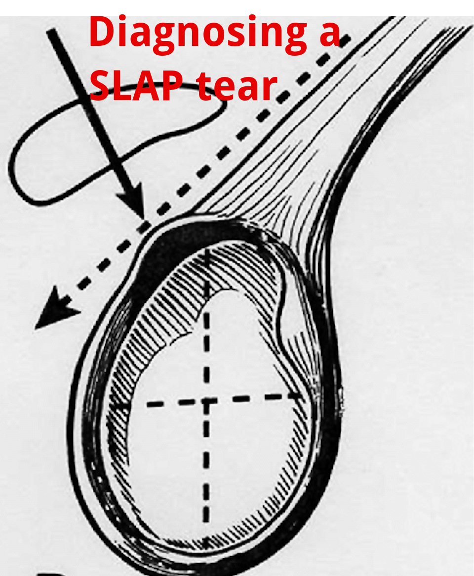
An Update on Diagnosing SLAP tears

Diagnosing a SLAP tear is not easy

We hear a lot about trying to diagnose shoulder pain and to be as specific as possible. It’s often difficult to differentiate SLAP (superior labrum anterior to posterior) tears from other soft tissue injuries of the shoulder.

For a review of the different types of SLAP tears, check out this old blog post that classifies the 10 different types of tears.

from https://commons.wikimedia.org/wiki/File:SLAP-Lesion-front-2.jpg

This paper in IJSPT by Clark et al 2019 attempts to help out the process and recommend a few special tests that MAY aid in diagnosing a SLAP tear.

What do they Recommend to diagnose a slap tear?

They recommend that a combination of at least 3 positive SLAP lesion tests may be clinically useful in diagnosing a shoulder SLAP lesion with greater diagnostic accuracy.

Combo of Tests

The combination of the Biceps Load I/II and O’Brien’s showed the highest sensitivity and specificity.

I have found similar results with this set of special tests so maybe this paper just hits my biases correctly.

In this video at my YouTube channel, I wanted to let you hear my thoughts and small tweaks to the evaluation process.

It’s not easy to diagnose a SLAP tear.

Differential Diagnosis is Critical

Furthermore, does it really matter and will it change the treatment plan much at all? I think it may a little but overall it will remain a pretty similar treatment approach to other similar pathologies like:

  • rotator cuff tendonopathy
  • Biceps strain
  • Latissimus strain
  • Subscapularis strain
  • internal impingement
  • pectoralis major strain

I think one also needs to consider the cervical spine and to make sure the pain is not referred from the neck.

Otherwise, a well thought out program should be implemented that addresses the strain on the shoulder and any strength issues.

I talked about this in an article that I wrote for Medbridge a while back so check out that post here:

You can also check out a snippet of one of my courses at this YouTube video where I discuss rotator cuff and labral issues. Hope it helps too!

Will this Change your Practice?

How ever you look at it, I wanted to use this paper to let you know that there MAY be a cluster of tests that better diagnose a suspected SLAP tear in your next patient’s shoulder.

Check out the paper and comment so we can talk it through. Are these tests similar to what you use in your clinical practice? Will this paper change what you do in your clinical practice?

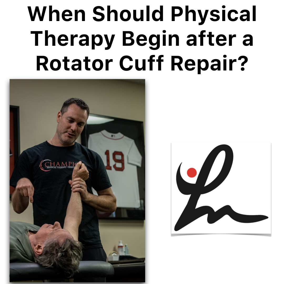
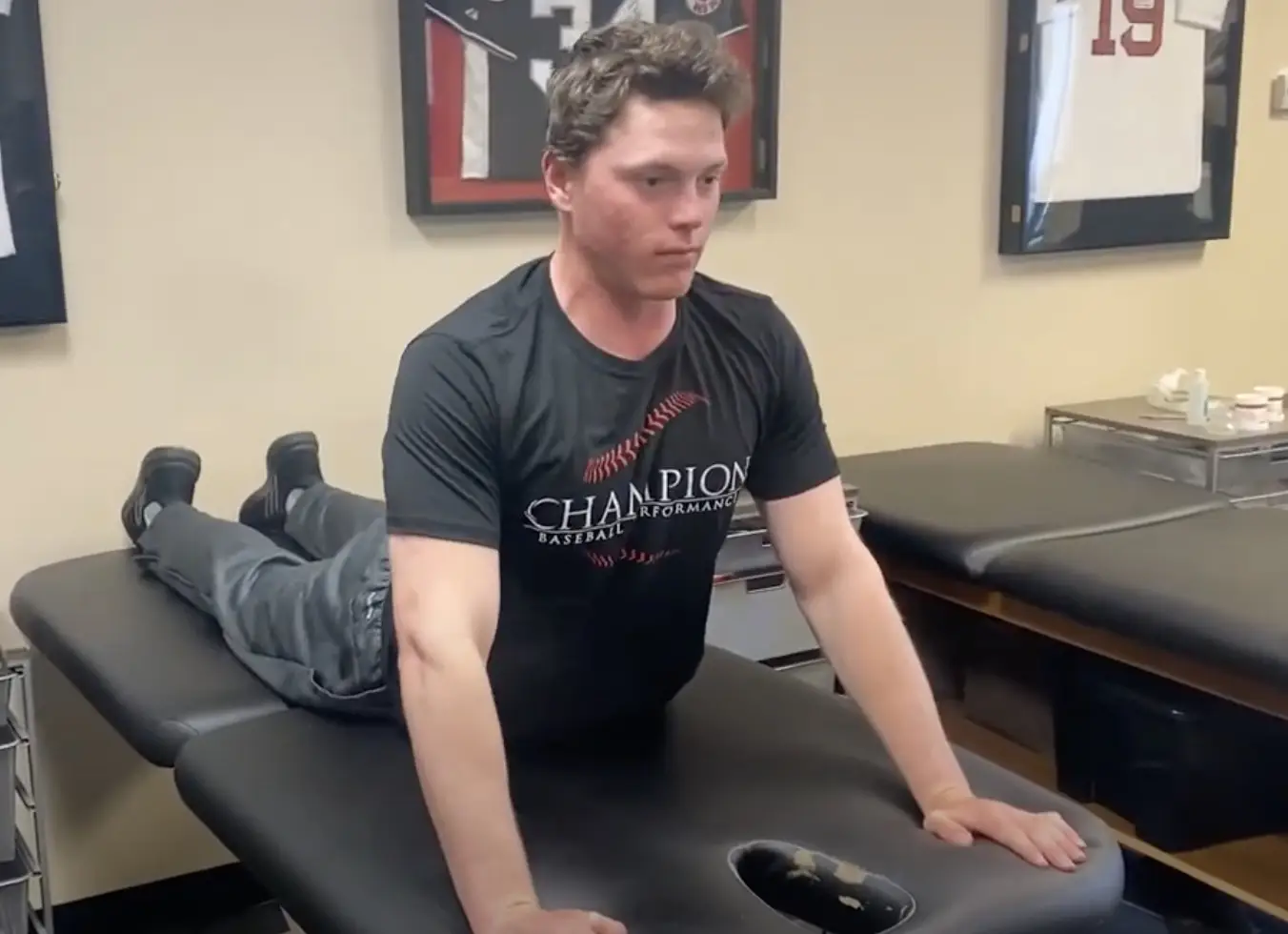
Is Early Physical Therapy Safe After a Rotator Cuff Repair?

Physical therapy is vital after a rotator cuff repair and continues to be common in an outpatient setting. Unfortunately, there’s no true consensus on when to actually begin PT.  Is early physical therapy safe after a rotator cuff repair or should we delay PT to protect the healing tendons?

Let’s dive into this and see what the research is saying…

Why I’m writing this post on rotator cuff rehabilitation

It seems as if we’re all over the place despite the research, which is pretty typical. Some docs prefer early passive range of motion (PROM) while others wait 6-8 weeks (and even up to 12+ weeks for a revision repair) before they allow any form of PT.

For the record, I’m going into this blog post as a firm believer of early PROM. It’s what we’d been doing for years in Birmingham at Champion Sports Medicine.

It’s what I only knew until I moved to Boston in 2014. Now I’ve seen a nearly 180-degree turn in rehab thoughts. Much more conservative!

I recently got in a Twitter discussion (debate) about this same topic with some very respected and prominent PT’s in the field.

It made me think about things so I decided to do a little research to see what the literature says. You can check out the discussion here.

Twitter can be confusing and tough to follow but just trust me, it goes on for a while!

Rotator Cuff Anatomy

The tendon most commonly torn is the supraspinatus tendon. Don’t get me wrong, you can tear the other rotator cuff tendons (infraspinatus, teres minor, subscapularis).

Keep in mind a medium, large or massive rotator cuff tear often will involve the infraspinatus tendon. If it does, then you need to consider modifying your progression appropriately.

But for the sake of this post, I’m going to stick to an isolated supraspinatus tendon for now.

Rotator Cuff Tendon Size and Location

Look at the size of the tendons as they insert on the greater tuberosity. You’ll see it differentiated by antero-posterior and medial-lateral directions.

According to my colleague and friend Jeff Dugas out of Birmingham, he showed in 2002 that the mean dimensions of the supraspinatus insertion were 1.27 cm in the medial-to-lateral direction. For the anterior-to-posterior direction, the supraspinatus dimension was 1.63 cm.

It helps to know this information because you may need to read an operative report and see the size of the tear. The docs will usually mention a 2 cm tear or something like that.

That means that the supraspinatus tendon and a small portion of the infraspinatus tendon were involved (and repaired.)

This is critical information to have when you’re trying to plot the post-op rehab progressions and determine the prognosis. The more tendons involved, then the higher the chance of repair failure.

There are many other factors that influence retear rates but tendon repair size is definitely one to consider.

Rotator Cuff Repair Surgery Types

I’m not going to bore you with the details of a repaired rotator cuff. There are numerous surgical techniques being used by orthopaedic surgeons.

Techniques such as a single row, double row, suture bridge or transosseous repairs are commonly performed. The picture below shows the difference between a single row and a double row repair, for example.

As you can see below, the double row tends to repair more of the tissue back to the humeral insertion point, which in theory has led to better tendon healing. This has been shown in numerous research studies and has become the best technique available.

So you had shoulder surgery…when to start physical therapy?

That seems to be the million dollar question! The research is all over the place. This means that doctors’ opinions are all over the place too, right?

Keep in mind that I’ve written about this in the past. Rotator cuff repair surgery is not always warranted and can be avoided!

Since I joined the group in Birmingham in 2002 (as a PT student), we had our post-op rotator cuff repair patients starting PT post-op day 1. They started PT early regardless of the tear size. This means a small tear of 1 cm in length started PT the same time a massive, 5 cm repair would start PT.

Some may disagree with this start time but it worked…it just worked. At least I think!

Why Early PT after a Rotator Cuff Repair

This is Key!!

There were several reasons why I think it worked:

  • They could chat with a professional.
  • Patients better understand their pain and get reassurance that what they were feeling was normal.
  • Someone could monitor their incisions and answer any and all questions.
  • Begin early, gentle ROM which often helps with pain control, too.

But, that was our ‘protocol’ and it continues to be that way many years later. Most other doctors that I have dealt with outside of Birmingham have taken a far more conservative approach to post-op rehab.

Agree to Disagree

Here in Boston, most docs wait at least 2-3 weeks and even up to 8-12 weeks to begin PT. Talk about eye-opening!

I don’t agree with this premise and wanted to dive a bit deeper into the literature to see if early physical therapy had a detrimental effect on short-term, mid-term and long-term outcomes.

Structure vs Function

The problem that continues to plague the research is the measurement of outcomes. Doctors care about the structural integrity of their rotator cuff repair. They see the research and are concerned with retear rates that hover in the 25-70%+ stratosphere. Of course I’d be concerned with retear rates that high!

Can you imagine if ACL re-tear rates were that high? Well, guess what they still hover in the 6-40% range even with our tremendous rehab skills and return to play testing.

But fortunately, we have other parameters to consider with our patients after a rotator cuff repair. We can look at the pain-free function!

Huh, what a novel idea. Regardless of the integrity of the repair, many patients can still live their lives to the fullest and in most cases without any pain.

Will Ferrell Yes GIF - Find & Share on GIPHY

Start PT Early after a Rotator Cuff Repair? What does the literature say…

I’m going to do my best and unbiased research to figure out if early PT after a rotator cuff repair is safe and effective compared to a delayed protocol. Let’s take a look…

There are a bunch of studies out there that you need to sift through. You can tell when the lead authors are MD’s or PT’s because the docs want to make sure their repair integrity is intact and the PT’s are concerned with restoring ROM, strength, and function.

With that, I’ve done my best to pull out some studies that have helped guide my practice and continue to influence me today.

Literature Review Findings

Age a BIG Factor!

Mind you, Cho et al showed that healing rates after a rotator cuff repair drastically change for older patients compared to younger patients.

The slide below was taken from my rotator cuff lecture that I’ve done in the past and helps to put things in perspective.

In no particular order…

Parsons et al JSES 2010 looked at 43 full thickness RTC repairs who were in a sling for 6 weeks. All were without PT for that time then evaluated for stiffness in PROM. They defined stiffness as 100° flexion/ 30° ER.

Overall, of the 43 surgeries, 23% (n=10) became stiff after that 1st evaluation session. The whole cohort displayed a 56% retear rate overall at 1 year, which to me seems crazy high!

To break it down further there was:

  • 30% retear in stiff group
  • 64% retear in non-stiff group
  • There was no significant difference in ROM or functional scores.

In my opinion, there were some pretty big limitations to the study that should be exposed, like:

  • Single row repair
  • No consideration for Diabetes or smoking
  • MRI without contrast to re-evaluate the repair status
  • What is “ER by the side???”- need to better define what degree of abduction.

So getting stiff may be a good thing but the repairs were done as single row repairs. We know these did not heal as well as they do with double row repairs.

More Literature Reviews

Moving on to a 2014 Level II systematic review and Meta-analysis, the authors said “the results contradicted our hypothesis that immobilization would increase tendon healing compared with an early-motion rehabilitation protocol, as structural outcomes were similar in the two groups 1 year after the arthroscopic repair of rotator cuff tears.

From the paper: “We speculate that rehabilitation is not the sole factor affecting tendon–bone recovery; the effects of other factors, such as older age, fatty degeneration, larger tears, and surgical technique, may outweigh those of the rehabilitation protocol.”

Kim et al AJSM 2012 looked at small to medium sized RTC repairs. They compared immediate PROM (0-120 degrees) to 4 weeks of absolute immobilization. They eventually showed no difference in ROM, pain or tendon healing. So seems like a smaller tear of less than 3 cm may be appropriate for immediate ROM, albeit it was limited to 120 degrees for some reason.

Not sure why they limited to 120 degrees because it seems as if the tendon would shorten as the humerus is placed in further flexion. Maybe they were concerned with subacromial impingement or something but the limitation is a bit confusing to me.

Healing Affected?

Another study by Lee et al AJSM 2012 wanted to compare ROM and healing rates between 2 different rehabilitation protocols after arthroscopic single-row repair (use caution) for full-thickness rotator cuff tear.

They showed pain, ROM, muscle strength, and function all significantly improved after arthroscopic rotator cuff repair, regardless of early postoperative rehabilitation protocols.

They also looked at the repair integrity with postoperative MRI scans, 7 of 30 cases (23.3%) in the immediate ROM group and 3 of 34 cases (8.8%) in the delayed group had re-tears, but the difference was not statistically significant (P = .106).

Well then, only a trend and all had similar functional outcomes regardless of when they started ROM…I’d say that helps the case to start early.

But again, these repairs were done via a single row repair and they allowed manual therapy 2 times per day and unlimited self-passive stretching exercise, which seems a bit aggressive anyway.

Do we even need a sling for 6 weeks?

No Functional Difference Between Three and Six Weeks of Immobilization After Arthroscopic Rotator Cuff Repair: A Prospective Randomized Controlled Non-Inferiority Trial Arthroscopy 2018

This study looked to compare clinical and radiologic results among patients with 3 versus 6 weeks of immobilization after arthroscopic rotator cuff repair in a prospective randomized controlled non-inferiority trial.

They concluded that “3 weeks of postoperative immobilization with sling use was non-inferior to the commonly used regimen involving 6 weeks of immobilization in a brace.” For the structurally concerned people out there, MRI indicated similar degrees of healing between the groups. 

Well then, that throws a wrench in things for the docs!

Ryan Gosling Giggle GIF - Find & Share on GIPHY

Does Early vs Delayed PT Affect Outcomes?

A systematic review by Gallagher et al 2015 looked to determine if there are differences between early and delayed rehabilitation after arthroscopic rotator cuff repair in terms of clinical outcomes and healing.  Six articles matched their criteria and reported significantly increased functional scores within the first 3-6 months with early rehabilitation compared to the delayed group.

To me, this is huge! Put yourself in their position. Imagine feeling better and being able to get back to work a little quicker. That’s my major argument for starting rehab sooner. Earlier pain relief, improved function and a feeling of being normal again.

Furthermore, none of the included studies reported any significant difference in rates of rotator cuff re-tear.

Medium-Large Tears use Caution

However, two studies noted a trend towards increased re-tear with early rehabilitation that did not reach significance. This was more pronounced in studies including medium-large tears. A similar trend that I’ve seen in the literature.

Here’s a nice table from that Gallagher study that summarized their findings for each research paper they included:

I want my patients to feel good as quick as possible and get back to some semblance of a normal life. But of course I want the long-term integrity of the repair to remain intact. And it seems as if this study helps that argument.

Then you look at a group of PT’s from Turkey (Duzgun et al Acta Orthop Traumatol Turc. 2011) that looked to compare the effects of the slow and accelerated protocols on pain and functional activity level after arthroscopic rotator cuff repair. Patients were randomized in two groups: the accelerated protocol group (n=13) and slow protocol group (n=16).

There was no significant difference between the slow and accelerated protocols with regard to pain at rest.

The accelerated protocol was superior to the slow protocol in terms of functional activity level, as determined by DASH at weeks 8, 12, and 16 after surgery.

I’ll take that!

Function over Structure (at times!)

Told you that the PT groups tend to favor function over structure, haha!

Early passive Motion ok- The French Version

This next study out of France that included French Society for Shoulder & Elbow ( 2012) looked to compare the clinical results after two types of postoperative management: immediate passive motion versus immobilization. Patients were randomized to receive postoperative management of immediate passive motion or strict immobilization for 6 weeks.

They concluded that their results suggested that early passive motion should be authorized: the functional results were better with no significant difference in healing. Functional results were statistically better after immediate passive motion and a lower rate of adhesive capsulitis and complex regional pain syndrome.

Seems as if there may be a trend but certainly not an overwhelming conclusion that early ROM is guaranteed to lead to inferior structural results. But, it seems pretty conclusive that ROM, pain, and function are improved after early ROM.

Let’s continue to look at the research

This group from China (Shen et al Arch Orthop Trauma Surg. 2014 Sep) performed a systematic review and meta-analysis to determine whether immobilization after arthroscopic rotator cuff repair improved tendon healing compared with early passive motion. Three RCTs examining 265 patients were included but we need to be cautious because of the limited number of studies included and the heterogeneity of the samples.

They found that there ‘no evidence that immobilization after arthroscopic rotator cuff repair was superior to early-motion rehabilitation in terms of tendon healing or clinical outcome. Patients in the early motion group may recover ROM more rapidly.’

This recent 2017 study in the Journal of Shoulder and Elbow Surgery included 9 meta-analyses in its review. They basically noted, “No clear superiority was noted in clinical outcome scores for early-motion or delayed-motion rehabilitation.”

They also concluded that “Whereas early motion and delayed motion after cuff repair may lead to comparable functional outcomes and retear rates, concern exists that early motion may result in greater retear rates, particularly with larger tear sizes.”

So, it seems as if function and healing the same after a RTC repair but there may be a discrepancy once we start looking at a larger repair size.

Immediate PT after a Revision Rotator Cuff Surgery

This study in AJSM 2018 looked to evaluate the clinical and radiological outcomes after revision rotator cuff repair surgery. They were able to track 31 of 40 patients (77.5%) for the final assessment at a mean follow-up of mean 50.3 months.

Interestingly enough, physical therapy started on the first postoperative day with passive flexion and abduction.

Revision rotator cuff repair improves clinical outcomes and shoulder function at midterm follow-up. The clinical outcome scores were comparable in patients with an intact repair and those with failed RC healing.

And they started PT 1 day after the surgery and got PT 2-3 times per week.

Retear Rates and Long-term function

This study in JBJS 2006 looked to determine the clinical and structural outcomes of re-ruptures in twenty patients after a longer period of follow-up. Nineteen of the twenty patients continued to be either very satisfied or satisfied with the outcome.

At an average of 7.6 years, the clinical outcomes after structural failure of rotator cuff repairs remained significantly improved over the preoperative state in terms of pain, function, strength, and patient satisfaction.

They also found that re-ruptures of the supraspinatus that had been smaller than 400 mm(2) had the potential to heal….wow!

Failure Rates too High!

Another study in JBJS 2013 looked at 18 patients who had undergone arthroscopic repair of massive rotator cuff tears.  At two years of follow-up, 94% had a failed repair. This current study wanted to evaluate the 10-year results for these patients with known structural failures of rotator cuff repairs.

Despite a high rate of progression of radiographic signs associated with large rotator cuff tears (proximal humeral migration or cuff tear arthropathy), most did well.

Clinical improvements and pain relief after arthroscopic rotator cuff repair of large and massive tears are durable at the time of long-term (10 years) follow-up. They went on to say:

“These results demonstrate that healing of large rotator cuff tears is not critical for long-term satisfactory clinical results in older patients.”

So our obsession with healing rates still appears to be overblown, even in older patients with a known cuff tear.

Pendulum Exercises Effects on Muscle Activity

Activation of the Shoulder Musculature During Pendulum Exercises and Light Activities JOSPT 2010

Look at that Paper in JOSPT 2010 (I’ve pulled out the Results Table for you below).

If there’s one exercise that doctors allow after a rotator cuff repair then it’s a pendulum or Codman exercise. How often does our patient do them correctly and make it a completely passive motion? I’d say rarely if seldom, right?

Most often, the patient is just bent over and actively moving their shoulder. They have no body movement or sway. Most of the movement is shoulder based and are not completely relaxing their shoulders.

Furthermore, they were instructed in the doctor’s office that 1st week or 2 after surgery.

They’ve been doing them incorrectly for weeks on end because they have no one to help them (cough cough!)

Look Over Here Steve Carell GIF - Find & Share on GIPHY

EMG of common Rehab ExerciSES

A study in JOSPT 2016 looked at EMG activity in healthy individuals. They wanted to quantify muscular activity during daily tasks and common PT motions. They showed that “of all the tasks assessed, ambulation without a sling and donning and doffing a sling and a shirt consistently showed the highest activity.”

EMG results table is found below.

Pretty helpful to see it listed by muscle and EMG activity and specific movement.

EMG of contralateral movements

A pretty neat EMG paper from 2004 (small n=6 and healthy individuals) were assessed using fine wire and surface EMG during common functional activities of the contralateral extremity while immobilized.

They found high supraspinatus EMG activity of the immobilized shoulder for all fast pulling activities of the contralateral shoulder (25-32%)!

Furthermore, they found high infraspinatus activity (56%) of the immobilized shoulder when the contralateral extremity performed straight forward reaching activities.

So even if they are immobilized and using their non-operative shoulder for daily activities, the rotator cuff is still sustaining a higher amount of activity than anything that we would do in the early phases (PROM, dowel self-ROM, rope and pulleys, properly performed pendulums).

How about revision rotator cuff repairs, you ask?

This study from AJSM in 2018 looked at outcomes after a revision rotator cuff repair. They showed revision rotator cuff repair improved outcomes regardless of tendon integrity (MRI confirmed).

Oh boy, what is going on??

Dig deeper into the study and they started PT the 1st day post-op with passive flexion and abduction. Sounds familiar, no? And this was in revision surgeries.

We used a very similar approach in Birmingham as they did in this study, so I may be a bit biased.

My Closing Thoughts on Physical Therapy after Rotator cuff repair surgery

I think it just shows you that rehab can begin early, will not affect long-term outcomes and that tendon integrity is not correlated to function.

I honestly don’t think our 15-30 minutes of passive motion early on in the rehab process is significantly affecting outcomes and retear rates.

It seems as if the repair technique, contralateral arm daily use, compliance with proper exercises (like pendulums, for example).

Let’s not blame early PT. There are so many more variables that are more likely to affect rotator cuff repair outcomes than anything that we could do in PT.

So I say let’s get people into PT early, educate them, guide them and help them get over this painful surgery.

I’ve been doing this for years and have seen the benefits of early PT. I say the literature agrees with me!

The Week in Research Review, etc 11-26-18

This week, I discussed the progression of someone after a knee surgery. I tried to highlight the key stages and some techniques that I like to use to advance the patient’s mobility and comfort. Take a look at The Week in Research Review, etc 11-26-18 and share with your friends. Hope it helps you improve your patient care tomorrow and beyond!


ACL Reconstruction in a Pediatric and Adolescent Population

1st Day of #PT after an ACL Surgery

Patella Mobilization after Knee Surgery

Knee Flexion PROM after Surgery- Seated or Supine?

Stretching the Quads after Knee Surgery

Assessing for a Cyclops Lesion after an ACL

Assessing for Fat Pad Irritation of the Knee


ACL Reconstruction in a Pediatric and Adolescent Population

17 Year Follow-up After Meniscal Repair With Concomitant ACL Reconstruction in a Pediatric and Adolescent Population. Tagliero et al AJSM 2018

Results: 28% failed meniscal repair and required repeat surgery at the time of final follow-up. They also showed that outcomes and failures rates were comparable across tear complexity.

Guess that means that no matter the tear type, there was no difference in outcomes or retear rates. Although the repair techniques are now outdated and no longer used.

Their study also showed a 30% failure rate for meniscal tear repaired in the medial compartment at index surgery and 7% in the lateral compartment.

Interesting long-term outcomes that may help to guide your rehab and client advancement (and prognosis). Keep these in mind when you treat a future adolescent or pediatric ACL patient.


💥1st Day of #PT after an ACL Surgery 💥

If you have never treated a post-op ACL, then this video should interest you!

This is what the knee looks like that 1st day after surgery and can often set the stage for what’s to come over the next 6-12 months.

Often, the patient is both very curious and ultra-grossed out by the 1st unveiling. It can be stressful for them to see their knee in this condition so you really have to confidently reassure them that it is very normal.

The blood-soaked gauze is mainly saline that was used to irrigate the knee during the reconstruction. Some still leaks out of the incisions the 1st few days and can often be confused with true blood.

Understand that this is quite normal and happens to most every ACL patient’s knee that I’ve seen…nothing to worry about!

From here, I’d work on patella mobility (see the post later today) and then work on flexion ROM at the end of the table.

Again, it’s very important to get the knee moving after surgery. This will help with pain, swelling and gain confidence that the rehab process is moving forward.


Patella Mobilization after Knee Surgery

Get the patella moving early with #patella mobilizations immediately after surgery. One major reason (amongst many others) why we need to get our clients into #PT early.

I am certainly a very loud advocate for early PT and getting the patella moving can help to prevent excessive scarring, which can affect ROM and quadriceps force output.

Glove up and get that patella moving in all directions… medial, lateral, superior, inferior!


🤔Knee Flexion PROM after Surgery- Seated or Supine? 🤔

I’ve treated many patients after an ACL I can honestly say that this may be a huge influence on the early ROM outcomes that you may see.

I’ve tried to bend the knee in both supine or seated, as the video shows, and there’s no doubt that most people tolerate the seated version so much better after a knee surgery. In particular, a big surgery like an ACL, TKA or MPFL reconstruction.

It just seems to be more comfortable and with less stress on the anterior knee because of the position of the tibia (at least I think so!).

My theory, it seems as if the supine position may cause a slight posterior sag which may cause more pain and guarding than when they’re seated at the edge of the table.

I use a similar concept later on in the rehab process when I’m initiating my prone quad stretching. You can see a definitive improvement when I wedge my hand in the popliteal fossa and create a slight anterior translation on the tibia.

Most people say that the anterior knee pain that they were feeling (and not a quad stretch) was replaced by a stretch feeling only and no more anterior knee pain.

Try it out with your ACL patients and see what position they like best…I’ll bet I can covert you over if you still bend your knee patients in supine!


💥Stretching the Quads after Knee Surgery 💥

Continuing my sequence of videos after a knee surgery, I discussed my technique for progressing knee flexion PROM once they hit 120 degrees or so of flexion.

At this point, they’ve probably maxed out how much ROM they can achieve at the edge of the table. They’re ready to get that end range of motion and even some quadriceps flexibility.

In prone, most people will often feel a pain or pressure in the front of their knee when you try to bend it.

To overcome this, I like to wedge my hand into the back of the knee and give an anteriorly directed force through the gastrocnemius (calf) soft tissue and into the tibia.

This seems to create just enough movement of the tibia on the femur to take the pressure off the front of the knee. This may redirect the forces more onto the quadriceps muscle.

You’ll need to play with the amount and direction of force but most often they’ll begin to feel a better quad stretch.

Try this technique out on your next knee surgery client and see if it helps them. I usually initiate this ~4 weeks after an ACL but timeframes will vary person to person.⠀


💥Assessing for a Cyclops Lesion after an ACL 💥

In this video snippet from my YouTube Channel, I discuss how to assess for a Cyclops lesion in a knee. In particular, after knee surgery.

A patient with a potential cyclops lesion, they often present with loss of normal knee extension compared to the other side. They’ll often have anterior knee pain and poor patella mobility. Sometimes a tight feeling in their hamstrings and calves, too.

No matter how they try to regain their extension ROM, the knee just never feels right. Often times, surgical intervention is needed to remove that scar tissue.

Immediate rehab should continue to work on knee extension ROM using low load long duration stretching and aggressive patella mob’s.

No one’s to blame if this occurs. We don’t know exactly why it occurs in some people but we believe a remnant of the ACL stump may be a source of the frustrating issue.


💥Assessing for Fat Pad Irritation of the Knee 💥

Anterior knee pain is very common in the outpatient #PhysicalTherapy setting.

One of my go-to tests to assess for fat pad irritation is simply trying to capture the fatty tissue in the anterior aspect of the knee joint during active and/or passive ROM.

In this snippet from my YouTube channel, you can see that I pinch the fat pads on either side of the patella tendon as @corrine_evelyn is actively extending her knee. I’ll also do it in a relaxed state to assess passive irritability.

I 1st learned this test from @wilk_kevin and it continues to be a mainstay in my knee examination algorithm.

As for a treatment, it usually comes down to a volume issue and/or strength issue or both.

I’ll usually have to address the volume of the activity by relatively easing off of the activity while simultaneously adding in exercises to address an underlying weakness.

Remember the Dye et al study in AJSM 1998 when he talked about the fat pads being super painful during his arthroscopic surgery without anesthesia. Makes sense why they can be so painful if the knee stresses fall upon this tissue.

We talk about this study, fat pad irritability and much much more in our online knee seminar course.


If you want to learn more about how I treat ACL’s or the knee in general, then you can check out our all online knee seminar at www.onlinekneeseminar.com and let me know what you think.

We cover the anatomy, rehab prescription, ACL, meniscal injuries knee replacements and patellofemoral issues. Furthermore, the course covers both the non-operative and post-operative treatment.t

This is an awesome course if you’re interested in learning more about rehabilitating the knee joint. And if you’re a PT, there’s a good chance you can get CEU’s as well.

The Week in Research Review, etc 11-19-18

Great ‘Week in Research Review, etc 11-19-18’ that I hope you find helpful to your practice.

I’ve always touted the importance of the subjective portion of the exam so I wanted to share a slide from a recent talk I gave to a group in Canandaigua, NY. Obviously, the squat is a fundamental movement and I wanted to give some basic positions that I use to help assess. So excited that I’ve launched a brand new Medbridge course that helps the rehab specialist better eval and treat the baseball pitcher. On my YouTube channel, I discussed my thoughts on setting the scapula with various upper and lower body exercises. And finally, my co-worker Kiefer Lammi discusses the landmine with exercise.


Importance of the Subjective Exam

Assessing the Squat

My New Baseball Medbridge Course

Set the Scapula with Shoulder Exercises?

6 Ways to use the Landmine by @kieferlammi


💥Subjective the most important aspect of the Evaluation💥

This slide, taken from this past weekend’s course in Canandaigua, NY is always a favorite of mine.

I try to keep a slide like this in all of my lectures because I have found that this portion of the examination can give the rehab specialist a huge look into what is going on with the person in front of them.

Don’t get me wrong, I still consider the biomechanical aspect of what may be causing their symptoms.

It often comes down to a tissue capacity issue but it’s up to me to determine the appropriate course of treatment.

These questions will help build confidence in your client and guide the early stages of rehab.

Do you have any specific questions that you like to ask your clients during their 1st few sessions? Remember, these questions are just not for the evaluation. You should be asking these questions periodically to gauge progress and help guide the next phases of rehab, too!


🔅Assessing the Squat 🔅

Squatting is a fundamental movement that all of us have to do on a daily basis.

Utilizing several different positions can help the rehab specialist better assess the squat and develop a treatment plan that enables their client the ability to improve their squat pattern.

In the above videos, I have utilized 3 different squat patterns and will outline them by the degree of difficulty.

✅The Overhead Squat- by far the most challenging version which challenges the shoulders, thoracic spine, lumbar spine, pelvis, knee and ankles.

A movement limitation at any of these joints will most likely cause the squat pattern to break down. Using overhead resistance would further challenge the system and potentially cause the squat to further breakdown.

✅Arms Crossed Chest Squat- alters the challenge by taking most of the shoulder and thoracic spine out of the equation and isolates the motions to the lumbar spine, hips, knees and ankles.

I often use this position as my fundamental motion because most people don’t have to squat with any weights over their head. This position, in my opinion, should be the most informational and utilized.

✅Counter-weight Squat

This position changes the center of mass by moving some of the weight distribution more anteriorly (front) and making the squat motion slightly easier. I use this position as a regression, for some, which allows them to squat with less stress and potential difficulty.

There are many other variations to the squat that you can make but I wanted to highlight a few of the major changes that you cause successfully. Assessing the squat is essential and can give the rehab specialist a nice picture of the function of multiple joints during a common movement.


My BRAND NEW course on Medbridge’s platform

…that helps the sports and ortho rehab specialist (PT, OT, ATC) better understand the anatomy and biomechanics involved in the baseball pitching motion.

Advanced Rehab for the Baseball Pitcher to Improve ROM & Strength@medbridge_education

The goal of this course was to allow the clinician to be able to evaluate and treat the baseball pitcher using evidence-based guidelines that I use on a daily basis.

Numerous research studies discuss the adaptive changes that occur with the pitching motion followed by numerous videos to help guide the treatment process.

If you’re already a Medbridge subscriber, then you have immediate access today.

If you’re not a Medbridge member, then you can use my promo code “Lenny2018” to save up to 40% off a yearly membership.

This gets you unlimited CEU’s for 1 year and potential access to their online HEP and a lot more!

Students can also get 1 year of unlimited courses (no CEU’s) by using promo code LennySTUDENT2018 and pay only $100.

Check out my other shoulder courses as well by using the Medbridge platform…along with many other great speakers!

Hope you enjoy and good luck!


💥Should you Set the Scapula with your Shoulder Exercise?💥

In this video excerpt from my YouTube channel, I wanted to discuss my opinion on setting the scapula during common exercises.

I think there’s an obvious role for setting the scapula during a heavier lower body lift like a deadlift.

But for a classic upper body exercise like the Full Can (Scaption Raises) or prone T (horizontal abduction), prone Y (Prone full can), etc then I definitely want the scapula to freely move along the rib cage.

I did a quick literature search and didn’t see anything obvious that helped to guide my thoughts so most of this is anecdotal. Check out the video and comment below.

Do you coach your clients to set their scapulae before a rotator cuff workout? If so, why? If not, do you think we should reconsider?


6 WAYS TO USE THE LANDMINE!⁣

Great post from our own @kieferlammi at @championptp on various ways to use the landmine in your client’s workout routine.

If you don’t have one, then I’d highly recommend you try to obtain one because they are highly versatile and can be used in many stages of rehab. See Kiefer’s original post below 👏🏼

_____________

6 WAYS TO USE THE LANDMINE!⁣

The landmine attachment is a super versatile tool for loading that is traditionally known for being used for angled pressing variations. While that’s probably my most programmed use for it, it also provides benefit to a ton of other movements by placing the load and direction of force at a bit of an angle, which can help to promote a particular path of movement, like sitting back more in a squat or lunge. Here are 6 of my favorite ways to use the landmine:⁣

1️⃣1-Leg RDL⁣

2️⃣Split Stance Row⁣

3️⃣Reverse Lunge⁣

4️⃣Deadlift⁣

5️⃣Squat⁣

6️⃣Russian Twist⁣⠀


Save 25% off our OnLine Knee Seminar Course…all this week!

Expires Sunday, November 25th at midnight ET

If you want to learn more about how I treat ACL’s or the knee in general, then you can check out our all online knee seminar at www.onlinekneeseminar.com and let me know what you think.

We cover the anatomy, rehab prescription, ACL, meniscal injuries knee replacements and patellofemoral issues. Furthermore, the course covers both the non-operative and post-operative treatment.t

This is an awesome course if you’re interested in learning more about rehabilitating the knee joint. And if you’re a PT, there’s a good chance you can get CEU’s as well.

The Week in Research Review, etc 11-12-18

This week in research review for 11-12-18 we focused a bit more on assessment and also dabbled in some basic treatment strategies for the back and shoulder. Check out the topics below and like them or comment on Instagram to keep the conversation going…thanks all!


  • A quick fix for a sore low back?
  • Knee Fat Pad Testing and Diagnosis
  • How to Assess the Elbow for a Tommy John (UCL) Sprain
  • Lumbopelvic control on shoulder and elbow kinetics in elite baseball pitchers
  • Full Can or Empty Can? – by @mikereinold


Looking for a quick fix for a sore low back?

I’m speaking from personal experiences when I post a few of the common exercises that have helped me tremendously in the past.

I’m not saying that this is all you have to do but I do think that new onset of low back soreness, you know that tightness that you feel on either side of your spine, can be somewhat alleviated with some foam rolling and active range of motion.

I would definitely include more focal strengthening of the core like deadbugs and bird dogs, squats, deadlifts (when they’re ready), etc.

But for the purpose of this post, I think some foam rolling and motion to the area can take the edge off of someone’s soreness and get them feeling a little better. That’s my goal for many and hopefully those small gins can add up to big gains in the long run!

Do you utilize these techniques as well? If you don’t, then I suggest that you try! They’ve helped me numerous times and continue to help me when my soreness gets a bit out of control.

Tag a friend who may want to check out this post…thanks!

Thanks @corrine_evelyn for the demos!


Knee Fat Pad Testing and Diagnosis

Here’s an excerpt from a previous blog post where I talked about anterior knee pain fat pad irritation. Link in bio!

Keep in mind, my differential diagnosis is all over the place at times. With knee pain you need to consider:

Meniscus (see my previous blog post)⠀

ITB

Osteochondral lesion

Patella tendonitis

Pes anserine bursitis

MPFL sprain

Hamstring strain

Plica syndrome

MCL/LCL

Tumor

Infrapatellar fat pad irritation can be functionally debilitating. I believe it presents itself pretty often in the clinic, more than most PT’s realize.

Use this test to see if it truly is a fat pad issue.


How to Assess the Elbow for a Tommy John (UCL) Sprain

In this excerpt from my YouTube channel, I discuss the tests that I use to help identify an elbow sprain, typically seen in the baseball players that I treat.

In the full video, I discuss:

✅Joint Palpation

✅Seated Milking Sign

✅Prone Valgus Test (maybe a new one for you!)

✅Supine end range External Rotation with Valgus Extension Overload (VEO)

I also wrote a blog post about this topic so hopefully you’ll go to my site and read a bit more about this.

If you treat baseball players of all ages, then you should know how to diagnose a UCL sprain.


The influence of lumbopelvic control on shoulder and elbow kinetics in elite baseball pitchers

Laudner et al JSES 2018.

This study looked at 43 asymptomatic, #NCAA Division I and professional minor league baseball pitchers. They measured the bilateral amount of anterior-posterior lumbopelvic tilt during a single-leg stance trunk stability test.

The Level Belt Pro (Perfect Practice, Columbus, OH, USA) was used to assess anterior-posterior lumbopelvic control. The LevelBelt Pro consists of an iPod–based digital level secured to a belt using hook-and-loop fasteners.

This test has been used and studied previously by Chaudhari et al (JSCR 2011) and he showed that pitchers with less lumbopelvic control produced more walks and hits per inning than those with more control.

Also, pitchers with less lumbopelvic control have been shown to have an increased likelihood of spending more days on the disabled list than those with more control (Chaudhari et al AJSM 2014).

“The results of our study show that as lumbopelvic control of the drive leg decreases, shoulder horizontal abduction torque and elbow valgus torque increase.”

Have you tried this simple test? I will say that having the ability to detect millimeters of motion is clinically difficult.

It is good to see such a simple test utilized clinically can help aid in determining the need for more core/hip exercises for our pitchers. In all, I think it’s a safe bet to incorporate these exercises in all pitchers’ programs.


Full Can or Empty Can?

– by @mikereinold 

Great Post by @mikereinold on which motion is BEST to isolate the supraspinatus during arm elevation. I know you can’t isolate the supraspinatus but numerous studies have (Kelly et al 1996, Reinold et al 2004) shown that the full can (or thumb up position) is better than the empty can position.

Check it out below! 👇🏼

Full Can or Empty Can? – by @mikereinold⠀⠀
-⠀⠀
🧠 WANT TO LEARN MORE FROM ME? Head to my website MikeReinold.com, link in bio.⠀⠀
-⠀⠀
I’m still surprised after all these years that I still see the empty can exercise kicking around. I analyzed these two movements many years ago in an article in JOSPT and showed that the full can exercise (thumbs up 👍) had similar EMG of the supraspinatus with lower levels of deltoid EMG, while the empty can (thumbs down 👎) had higher levels of deltoid EMG.

Why does this matter?

Well, think about it. If you are performing this exercise you probably are trying to strengthen the rotator cuff. And if you are weak and performing an exercise with more deltoid, the ratio of cuff to deltoid will be lower and you’ll have more potential for superior humeral head migration.

Plus, let’s be honest, the empty can just hurts… It’s also a provocative test, and I don’t like to use provocative tests as exercises. 😂😂😂⠀


The Week in Research Review, etc 11-5-18

The Week in Research Review, etc 11-5-18 was filled with more informative and eye-opening posts! Lots of visually stimulating posts to help clarify what exactly is going on in the hip joint with PROM. Another post that shows the suction effect from an intact hip labrum… amongst other great posts.  Just some great stuff..hope you enjoy!


  1. Manual Forearm Resistance Drills
  2. ACL Graft Healing Times to Maturation
  3. Hip Capsule Stress with PROM External Rotation
  4. Muscle Activation Affected by Hip Thrust Variation
  5. Hip Thrust Form by Bret Contreras
  6. Hip Joint Suction Affected by labral Status


Manual Resistance Forearm Exercises

In this post, I wanted to show you guys some of the manual resistance drills we use @championptp on our shoulder and elbow clients, especially our baseball players. We love to use these drills because we can control so many variables with each athlete and tailor it for their specific needs.

We can control the speed and tempo, the direction of forces (eccentric, concentric), and the magnitude of the forces. Plus it’s a great way to interact with our clients. It’s also a great way to feel how well they’re progressing in their programs instead of just giving them dumbbells.

I have found these manual resistance drills to be very helpful with my overhead athletes and hope you give them a try on your clients soon! Let me know what you think or tag a friend below who may like to use these drills too.

In my course that I teach around the US, I try to include these concepts so you can practice and be able to utilize these drills for your clients…thanks!


ACL Graft Harvesting and Healing times

In this post, I wanted to show some research studies on graft healing times and why we need to respect tissue biology.

The systematic review from AJSM 2011 looked at ‘The ‘‘Ligamentization’’ Process in Anterior Cruciate Ligament Reconstruction.’

They essentially looked at 4 different biopsy studies on BPTB and Hamstring autograft reconstructions. They concluded that maturation of the graft, as determined by mainly vascularity and cellularity, was not complete until 12 months at the earliest. The healing time even extended to 24+ months as well.

The ligamentization endpoint is defined as the time point from which no further changes are witnessed in the remodeled grafts. The surgical procedure is quite involved, as you can see in the video that I took from @drlylecain on #YouTube.

As I’m rehabbing my clients, my decision making and post-op progressions often take into account:

✔️Healing biology

✔️Graft harvesting

✔️Graft Type

✔️Bone bruise presence (often!)

✔️Other concomitant issues (meniscus, articular cartilage).

So, respect the tissue and allow natural healing to occur before you add more exercises or are concerned that they’re not making the gains you’d expect.⠀


Hip Capsular Closure: A Biomechanical Analysis of Failure Torque

Chahla et al AJSM 2016

Interesting look at tissue failure, albeit in a cadaver graft, that should help to guide the physical therapist or ATC early in the rehab process after a hip scope.

The purpose of this study was to determine the failure torques of 1-, 2-, and 3-suture constructs for hip capsular closure to resist external rotation and extension.

The 3-suture construct withstood a significantly higher torque (91.7 Nm) than the 1-suture construct (67.4 Nm) but no significant difference was found between the 2- and 3- suture construct.

The hip external rotation degree in which the capsule failed was:

✅1-suture construct: 34 degrees

✅2-suture construct: 44.3 degrees

✅3-sutures: 30.3 degrees (yes, smaller than 2-suture construct)

I think as a #PT, we need to keep this study in mind and respect the healing tissues after a hip scope.

Love when we can get this information and put it into practice, similar to RTC repairs, ACL, etc.

Obviously, this was on a cadaver where there’s no guarding, pain or muscle contraction. We still need to know that there MAY be enough tension on the capsule to create potential issues (like tissue failure).

If you treat patients after hip scopes, then I recommend you read this cadaveric study.


Barbell Hip Thrust Variations Affect Muscle Activation

COLLAZO GARCIA et al JSCR 2018

This study looked at the EMG activity of various lower body muscles while performing the hip thrust in various positions.

Their results showed that by varying the foot position into more external rotation, you can recruit the glute max and medius more than by the traditional hip thrust.⠀ …”the activity of the gluteus maximus increases significantly reaching up to 90% MVIC with only 40% of 1RM” with this hip ER variation.

Also, ‘when the distance between the feet is increased, the activity of knee flexors increases. Therefore, this is a very recommendable option to increase hamstring: quadriceps co-activation ratio.’

I like this study because it helps guide our rehab if we’re targeting a specific muscle group a bit more because of an injury or surgery.

It’s one of my go exercises for anyone with a lower body injury, especially after an ACL reconstruction. But I do use this exercise for most of my clients rehabbing from any injury, including the upper body.

It’s a great way to recruit the gluteus maximus and medius, which we know are hugely? (is that a word?) important to help produce and dissipate forces during athletic movements.

The exercise was widely researched by @bretcontreras1 and should be a staple in your rehab programs.

Check it out and add this to your go-to exercise list…thanks!


Hip Thrust Form

[REPOST] and a great one from @bretcontreras1 talking hip thrust form, which is perfectly coinciding with my post earlier today on variations to the hip thrust and how they affect muscle activation. Check out his original post below…highly recommended!

Teaching optimal hip thrust form is complicated. While the occasional lifter prefers and functions better staying fairly neutral in the head, neck, and spine, the vast majority of lifters do best maintaining a forward head position, which leads to ribs down and a posterior pelvic tilt.

It’s not just the forward eye gaze; the whole head has to maintain its forward position. You’re not hinging around the bench; the body mass above the bench stays relatively put, while the body mass below the bench is where the movement occurs.

The astute science geeks out there will rightfully point out that posterior pelvic tilt is associated with some lumbar flexion, and that lumbar flexion under load can be problematic. However, lumbar flexion is only dangerous when the discs are simultaneously subjected to compressive forces. With this style of hip thrust, the glutes are driving hip extension and posterior pelvic tilt, and erector spinae activation is greatly diminished. Core activation is what creates the bulk of the compressive forces, so with the erectors more “silenced,” the discs aren’t as compressed. This makes the exercise very safe. In fact, it’s safer than the “neutral” technique because as you rep to failure or go a bit too heavy, you will inevitably arch the chest and hyperextend the spine, which can lead to lower back pain. ⁣

We have 200 members at Glute Lab hip thrusting day in and day out, and there have been zero injuries to date. Considering how heavy we go, this is astounding.⠀
⁣⠀
#gluteguy #glutelab #thethrustisamust


Hip Joint Suction and Stability

[REPOST] From @chicagosportsdoc and a very cool look at the suction within the hip joint that contributes to its stability. As the video progresses, they have simulated a labral tear that shows how easily the joint can dislocate. Once the labrum is repaired, the suction effect is recreated, and joint stability is re-established.

That’s 2 posts this week on the hip…if you want to see some awesome posts, then follow him. He just got on Instagram but his visual posts really aid in learning the mechanics of the various joints…see below!

An impressive demonstration of the powerful hip suction seal. When the hip labrum is injured, the seal is disrupted which can potentially produce microinstability. A labral reconstruction can restore the suction seal #labrum #sportsmedicine #hip #anatomy#orthopedicsurgery #medicine


The Week in Research Review, etc 10-29-18

This week we started the week off with a couple shoulder posts, specifically the rotator cuff and SLAP tears. As usual, I can’t resist a good ACL paper so included that NM control program that should be in all knee patients’ programs. We ended the week with a recorded knee scope as the surgeon was mobilizing the patella. It was a very informative and fun way to see the patella. We closed the week off with an old school video of myself performing a proprioception drill for the shoulder. I recommend you read these posts and like them on Instagram. Take a look at The Week in Research Review, etc 10-29-18


  1. Topics on the Rotator Cuff including post-op
  2. Classifying SLAP tears
  3. Essential Components of a neuromuscular control program
  4. Live Patellar scope during mobilization
  5. Shoulder Proprioception Drill


Topics on the Rotator Cuff including post-op

A Systematic Summary of Systematic Reviews on the Topic of the Rotator Cuff- Jancuska et al OJSM 2018

Nice summary of systematic reviews for you guys if you treat patients after a rotator cuff surgery. I’ve been doing a pretty good literature on the topic and wanted to share some of the articles that I have found helpful.

Their conclusions:

❇️There is substantial evidence indicating that the most accurate physical examinations for diagnosing RC tears are a positive painful arc and positive ER lag test

❇️Considerable evidence showing that rehabilitation is better than no rehabilitation for non-op management of RC tears, although RC repair was shown to be superior to rehabilitation alone.⠀

❇️No evidence to support the use of injections for nonoperative management of RC tears.

❇️Double Row repair results in better outcomes and fewer re-tears than Single Row repairs, especially for tears >3 cm.

❇️Predictors of re-tears and poor postoperative outcomes:⠀

✔️older age⠀

✔️female sex⠀

✔️smoking⠀

✔️increased tear size⠀

✔️preoperative fatty infiltration⠀

✔️preoperative shoulder stiffness⠀

✔️diabetes⠀

✔️workers’ compensation claim⠀

✔️decreased preoperative muscle strength⠀

✔️concomitant procedures.

Overall, a good review of the literature on rotator cuffs and anything associated.⠀


Classification of SLAP Tears

If you treat patients with shoulder pain, then you may run into different labral tears of the shoulder.

This post hopes to summarize the 10 different types of #SLAP tears that are currently known.

Type 1️⃣: Fraying but intact biceps

Type 2️⃣: Superior Labrum and biceps detached from the glenoid rim

Type 3️⃣: Bucket handle tear of the superior labrum but biceps anchor attached

Type 4️⃣: Bucket handle tear of the superior labrum that extends up into the biceps tendon

Type 5️⃣: BankartTear and also a detached biceps anchor

Type 6️⃣: an unstable flap of the superior labrum with a detached biceps anchor

Type 7️⃣: Anterior superior labral tear that extends to the middle Glenohumeral ligament; Biceps anchor detached

Type 8️⃣: Superior and posterior labral tear along with detached biceps anchor

Type 9️⃣: 360° labral tear

Type 🔟: Superior labral tear along with reverse Bankart tear and a detached biceps anchor.

That’s a lot and some are pretty rare but it helps to be able to communicate effectively with the medical team or to read an operative report.⠀


Neuromuscular training to reduce ACL injuries in female athletes

Critical components of neuromuscular training to reduce ACL injury risk in female athletes: meta-regression analysis. Sugimoto et al BJSM 2016.

This meta-regression analysis looked at the effects of combining key components in neuromuscular training (NMT) that optimize ACL injury reduction in female athletes.

They looked at a total of 14 studies that met the inclusion criteria of the current analyses. A total of 23 544 athletes were included.

They showed that there are 4 Key components

✅14-18 years old better than other age groups

✅2x/week for 30 minutes/session

✅Balance, planks, ‘posterior chain’ and plyometrics

✅Verbal cues like ‘Land softly’ or ‘Don’t let knees cave in’

Furthermore, inclusion of 1 of the 4 components in NMT could reduce ACL injury risk by 17.2–17.7% in female athletes. A great look that really specifics what age groups would best benefit from a NMT program. Do you incorporate any of these key concepts into your programs, even 1-2 of them?

I know I try to with most of my clients, whether or not they’re returning from an ACL or not.


Patella mobility during a knee scope

Great video by @physionetwork looking at the patella during a knee scope. This stuff is just exciting to see (in my opinion) because it gives us a little bit of insight into what is exactly going on during a patella mobilization.

In my opinion, the PF joint is often overlooked when it comes to knee surgery and it can affect joint mechanics, quadriceps activation and patient function. You need to mobilize the patella and normalize the motion…can’t stress this enough!

Check out the post below…good stuff!

Patellar mobilization is important to avoid stiffness after surgery. In this video, you can see from an arthroscopic view that little motion outside the knee, translates into a significant motion inside the knee. Mobilization may help prevent the formation of scar tissue and allow for better biomechanics of the knee joint.

We review the latest and most clinically relevant research in physiotherapy. Click link in bio to learn more and boost your knowledge 🔗

Video by Jorge Chahla, MD, PhD – Orthopaedic Surgeon -Sports Medicine Specialist


Active Reposition Drill after a Passive Motion

Loss of proprioception after a shoulder injury has been documented numerous times in the literature and can affect long-term function.

This drill may help the rehab specialist to test proprioception by measuring the exact active position difference that the patient attains.

You can also use this drill as a treatment reproduce the exact position that you passively brought them into.

Give it a shot and see what you think…you can use this drill for any joint in which you have assessed proprioception loss.


The Week in Research Review, etc 7-29-18

Last week was the 1st of my research review that summarized my social media posts from the previous week. It seemed to be well received so I figured I’d continue it. My goal is to help summarize some of the research that I found interesting and package it nicely for my readers.

Each photo contains a link back to a social media feed where you can see the conversation that ensued and maybe chime in…or just be a passive reader and see where the conversation went. You never know where the conversation may go on social media…so be ready! haha!


Socioeconomic Factors for Sports Specialization and Injury in Youth Athletes Jayanthi et al Sports Health Journal 2018.

This study looked at the effect of socioeconomic status (SES) on rates of sports specialization and injury among youth athletes.

They looked at injured athletes between the ages of 7 to 18 years that were recruited from 2 hospital-based sports medicine clinics. They compared these with uninjured athletes presenting for sports physicals at primary care clinics between 2010 and 2013.

They concluded that:
✅High-SES athletes reported more serious overuse injuries than low-SES athletes
✅More hours/wk playing organized sports
✅Higher ratio of weekly hours in organized sports to free play
✅Greater participation in individual sports

I applaud the authors for attempting to bring this very difficult collection of data into a formal research paper. I will say some of the statistics and standard deviations may not make the conclusions as powerful.

I do think this is a good paper to help educate our athletes on injury rates, especially in those that specialize in 1 sport.

What do you think? Tag a friend that may benefit from this article!


From #Twitter’s @retlouping that perfectly sums up what I’ve observed recently on social media with many PT’s.

For some reason, pain science has overtaken most diagnosis and treatment conversations.

It’s as if you get bullied into talking pain science and ignoring our clinical judgment and diagnosis skills. I understand there’s a constant tug-of-war between the biomechanical PT’s and the pain science PTs.

But as usual, the answer usually lies somewhere in between and both groups are correct. The biomechanics of an injury are often important as well as the language we use to explain these tissue biomechanics.

To my fellow clinicians, especially the newer grads and #dptstudent, remember this little cartoon for every future encounter.

Yeah, speak to people in non-threatening tones (in my world it’s just being respectful) but trust me, they WANT to hear what could be going wrong or what may be causing their pain.

Don’t blow off their symptoms and don’t go into depth about pain science because they won’t understand.

Trust me, the clinicians that try to do that often end up losing their patients in the long run.

I hear these stories day after day of people coming to me because the last PT either only talks to them or made them ONLY do strength exercises and it didn’t help their pain.

The PT didn’t listen to them and was so blinded by their pain science background that they ignored the person sitting right in front of them. Remember, the person sitting there will tell you what is going on and what treatment will most help them feel/move better.


Influence of Body Position on Shoulder and Trunk Muscle Activation During Resisted Isometric Shoulder External Rotation Krause et al Sports Health 2018.

The purpose of this study was to examine ER torque and electromyographic (EMG) activation of shoulder and trunk muscles while performing resisted isometric shoulder ER in 3 positions:
✔️Standing
✔️Side-lying
✔️Side plank

Using surface EMG and a hand-held dynamometer, the researchers tried to determine EMG activity of the:
✔️infraspinatus
✔️Posterior Deltoids
✔️Mid traps
✔️Multifidi
✔️External/internal obliques (dominant side)
✔️External/internal obliques (non-dominant side)

EMG values for the infraspinatus were greatest in the side plank position. In general, EMG values for the trunk muscles were also greatest in the side plank position.

✅Their Conclusions: If the purpose of a rehabilitation program is to strengthen the rotator cuff, in particular, the infraspinatus, the side plank is preferred over standing or side lying. If the goal is to simultaneously strengthen both the rotator cuff and trunk muscles, the side plank position again is preferred.

Makes sense but good to see the research and have concrete evidence to back up what we think actually goes on.

Tag a friend who may be interested in this research paper!


Reliability of heel-height measurement for documenting knee extension deficits. Schlegel et al AJSM 2002

Prone heel-height difference of 1cm equates to 1.2 degree difference in knee extension ROM.

Do you use this method to assess knee ROM? I still measure knee extension ROM is supine but find this method helpful as well.

I know my friend and colleague @wilk_kevin has measured this way for many years. i originally saw his use this technique at @ChampionSportsM

I don’t want people to confuse this with prone hangs for knee extension ROM. I am not a fan of that method as I’ve stated in the past.

This is a method to assess knee extension differences, particularly after an ACL reconstruction. I have gone back to using this method for some people that have subtle ROM differences side-to-side.

The patella position (on the plinth or off) did not matter in the study and thigh girth did not appear to make a difference.

I would recommend stabilizing the pelvis to prevent excess ROM from occurring at that region and to better isolate the knee joint.

Have you tried this method? Tag a friend who may benefit from using this ROM method…thanks!


Evidence-Based Best-Practice Guidelines for Preventing #ACL Injuries in Young Female Athletes: A Systematic Review and Meta-analysis Petushek et al AJSM 2018.

Injury prevention neuromuscular training (NMT) programs reduce the risk for anterior cruciate ligament (ACL) injury.

Eighteen studies were included in the meta-analyses, with a total of 27,231 participants, 347 sustaining an ACL injury.

The overall mean training amount was 57 sessions totaling 18.17 hours (roughly 24 minutes per session, 2.5 times per week).

They concluded:

✔️Interventions targeting middle school or high school–aged athletes reduced injury risk to a greater degree than did interventions for college or professional-aged athletes.

✔️Continued exposure to neuromuscular training throughout the sport season seems to enhance prophylactic effects of NMT.

✔️NMT interventions were effective for female basketball, and handball athletes and interventions including various athletes were potentially effective (eg, soccer, basketball, and volleyball).

✔️ Interventions included some form of implementer training (eg, instructional workshop, video, or brochure) on proper program implementation.

✔️Programs including more landing stabilization and lower body strength exercises during each session were most effective.

🤔Programs including balance, core-strengthening, stretching, or agility exercises were no more effective than programs that did not incorporate these components.

✔️ Specifically, programs that included more landing stabilization exercises (eg, drop landings, jump/hop and holds), hamstring strength (eg, Nordic hamstring), lunges, and heel-calf raises reduced the risk for ACL injury to a greater degree than did programs without these exercises.

✅ Wow, lots of great information here. Please share this with a friend or colleague who may benefit from knowing this information.


Hope that helped to catch you up on my posts from this week.

Do you like these weekly updates? Let me know if I should continue…love your feedback!

Thanks for reading!

The Week in Research Review, etc 7-22-18

The Week in Research Review, etc 7-22-18

I’m trying out this new concept of publishing my social media posts into a nice package for a weekly delivery to my subscribers.

  1. Knee Case Study
  2. Contralateral ACL Strengthening
  3. Shoulder Static Stabilizers
  4. Weighted Ball Research
  5. Glute Activation


This kid came to me the other day with L knee swelling after sliding headfirst into 2nd base during a baseball game.⠀

Continued to play in the game and even pitched the next day, all without pain or loss of motion.⠀

As you can see from the video, he has a bunch of fluid in his knee, medial ecchymosis (bruising) but full pain-free ROM.

Ligamentous tests appear negative and he has absolutely no pain or stiffness with anything.

I took this video to show what appears to be a bursal sac disruption from the impact of his knee into the ground as he was sliding.

The mechanism fits the presentation and clinical exam.

I advised him to monitor his swelling, wear a knee sleeve and continue his activities per his tolerance.

He is going to touch base with me next week to make sure the fluid is dissipating (and not worsening) and he remains asymptomatic.

What do you think? Am I missing anything? What’s your diagnosis? Tag a friend who may be interested in this case.

Cross-education improves quadriceps strength recovery after ACL reconstruction: a randomized controlled trial. Harput et al Knee Surg Sports Traumatol Arthrosc. 2018

This study looked at a group of ACL reconstructed patients that were divided into 3 groups.

All 3 groups performed the same standardized ACL rehab, but one group was the control group that performed the standardized rehab only.

The other 2 groups did either 3x per week extra concentric knee extensions on their uninjured leg for 2 months (beginning at 1-month post-op through 3-months post-op) or additional eccentric knee extensions on their uninjured leg 3x per week for 2 months between months 1-3 post-op.
💪🏼
They found that the quads strength for the concentric group was 28% greater compared to the control group. 💪🏼
The eccentric group was 31% greater when compared to the control group.

Conclusion: Concentric and eccentric quadriceps strengthening of healthy limbs in early phases of ACL rehabilitation improved post-surgical quadriceps strength recovery of the reconstructed limb.

Pretty crazy stuff and one more reason to work on bilateral strengthening with most of our patients, especially when they’re post-op ACL reconstruction.

Do you work on bilateral strengthening? if not, why? If you do, what other studies have you seen that show similar results?
Tag a friend who may benefit from this study or let’s discuss in the comments section!

This picture shows a simplified view of the static stabilizers of the shoulder joint. I highly recommend reading a classic paper by Wilk et al 1997 JOSPT that talks about this and cites a paper from Bowen et al Clin Sports Med 1991 @wilk_kevin

When one is picturing these stabilizers, the superior glenohumeral ligament (SGHL) is most taut when the shoulder is externally rotated at 0 degrees of abduction.

As we progress to 45 degrees of GH abduction, we stress the middle glenohumeral ligament (MGHL) as we externally rotate the humerus.

Finally, at 90 degrees of GH abduction, we stress the inferior glenohumeral ligament (IGHL) as we externally rotate. More specifically, the anterior band of the IGHL.

As we internally rotate at 90 degrees of abduction, we stress the posterior band of the IGHL.

These concepts have rehab implications and should be kept in mind when we’re rehabbing people after an injury or surgery.

For example, if someone has an anterior Bankart lesion (front labral repair), then we need to progress them slowly into external rotation, especially at 45 and 90 degrees of abduction.

Another example would be a rotator cuff repair, like the supraspinatus. We would want to progress them slowly at lower degrees of abduction 0-45 degrees but maybe we can progress them a bit quicker at 90 degrees of abduction.

Hope these concepts make sense because they are very important to understand for many patients with shoulder injuries.

Does this make sense? Have you heard this info before? Tag a friend who may benefit from this post!

Effect of a 6-Week Weighted Baseball Throwing Program on Pitch Velocity, Pitching Arm Biomechanics, Passive Range of Motion, and Injury Rates. Reinold et al Sports Health Jul-Aug 2018. @mikereinold

Our 1st of potentially 3 research articles looking at the effects of weighted balls on youth baseball pitchers.

High school baseball pitchers performed a 6-week weighted ball training program.

Players gradually ramped up over the 6 weeks to include kneeling, rocker, and run-and-gun throws with balls ranging from 2oz to 32 oz.

🤔After 6 weeks, the weighted ball group did increase velocity by 3.3%, 8% showed no change, and 12% demonstrated a decrease in pitch velocity. Also of note, 67% of the control group also showed an increase in pitch velocity.⠀

The weighted ball group had a 24% injury rate although half of the injuries occurred during the study, and the other half occurred the next season. There were no injuries observed in the control group during the study period or in the following season.

The weighted ball group showed almost a 5-degree increase in passive shoulder external rotation, also known biomechanically as the late cocking position or layback position.

There were no statistically significant differences between pre- and post-testing valgus stress or angular velocity in either group.

✅Our conclusion: Although weighted-ball training may increase pitch velocity, caution is warranted because of the notable increase in injuries and physical changes observed in this cohort.

Some great Glute 🍑thoughts buy the @theprehabguys. Check out their videos and content for some great ideas that you can add to your practice!⠀
👇🏼⠀
___________________________________________________________________⠀
Episode 705: “Hip Prep for Glute Activation”⠀
.⠀
Tag a friend looking for a glute🍑 killer!⠀
Hip prep is a series of 6 exercises I’ve adopted from my girlfriend @smenzz and her clinic @eliteorthosport. I use it with my patients to prime the glutes and lower body in general before getting into more dynamic and plyometric activities. I will make the statement right now: if done RIGHT, it’s an absolute glute killer & I promise you that you will feel your glutes!⠀
.⠀
I like these 6 exercises in particular for a variety of reasons.⠀
✅They challenge the glutes in all 3 planes of motion.⠀
✅They hit all types of muscle contractions: isometric, concentric, and eccentric⠀
✅They are performed upright in a functional position⠀
✅There is a variety of double leg, single leg, and split stance variations⠀
✅They train proper lower extremity alignment in a variety of hip and trunk flexed/neutral/extended positions⠀
.⠀
The 6 exercises are:⠀
1️⃣3 way clams: 5 per leg per position⠀
2️⃣Side steps: Alternating steps to the left and right starting with 1 step all the way to 5 steps⠀
3️⃣Monster Walks: 10 steps forward, 10 steps backwards⠀
4️⃣W’s: 10 steps to the left, 10 steps to the right⠀
5️⃣Squats: 10 squats⠀
6️⃣Single leg fire hydrants: 30s per side⠀
.⠀
💡Understand that you first need to teach these exercises in isolation first, before throwing someone all 6 at once⠀
.⠀
Have fun!⠀


Hope this helps you keep up to date and fulfill my goal of this website…simplify the literature and bring great content to you so you can apply it 1st thing Monday morning! Happy Reading! 👊🏼

Follow me on Social Media here:

Image result for instagram logo vectorImage result for twitter iconImage result for facebook logo