Tag Archive for: patella tendon graft

Simplifying ACL Rehab

ACL surgery continues to be a huge focus in the literature and in our outpatient rehabilitation settings. Numerous studies focus on return to play guidelines and retear rates. Social media is all over the place, most times. Let’s try to make things simple and set the stage early.

Paralysis by Analysis- What ACL tests are best for return to play

I previously wrote about return to play guidelines here  and even the neurocognitive challenges that are associated with ACL rehabilitation.

As troubling as it all seems, I almost feel as if the recent research is confusing and often unattainable in a traditional outpatient PT setting. I’m worried about the paralysis by analysis mindset that seems to have overtaken my recent discussions on social media.

Most don’t have an isokinetic device to test.

Hop tests, vertical jump, strength…what really matters?

Do hop tests really give us a complete picture of an athlete’s return to play readiness?

What combinations of tests are appropriate?

Is it more than just quadriceps strength?

What about a vertical jump test to determine lower extremity strength and function for an LSI?

Lots of questions!

Timeframes have shifted from a 4-6 month return to play goal to a more realistic 9+ months before we return people. Studies continue to show that a slower rehab may be better.

ACL retear rates still too high

Yet, our retear rates still remain very high and we’re still missing the boat for many.

I get it. It’s not just the timelines and rehab. Many are limited by equipment, insurance limitations, differing MD protocols, and so much more. It’s easy to blame one concept when it truly is multifactorial. 

That’s why I employ a very simple approach to my ACL rehab programs that seems to work for most and hope this concept can help you too.

It all really occurs at the beginning of rehab when you set the stage for the rest of your planning. If you can establish full motion, remove most of the swelling and restore a pretty normal gait, then you have achieved your goals.

From here, it gets fun and we’ll talk about that later…

An easier approach to ACL rehab

Immediately Post-op Phase

During the earlier stages of rehab, we’ll call it 6 weeks post-op, my main focus is calming the knee down and establishing a normal range of motion.

Range of Motion

It is critical to get the knee as straight as possible and as quickly as possible. For example, most people naturally have some 3-5 degrees of hyperextension. For these individuals, I like to get 0-3 degrees of hyperextension immediately and allow the rest to come through normal functional stresses.

For hypermobile patients, say 10-15 degrees of knee hyperextension, I will only get about 5-7 degrees of hyperextension because I know their underlying tissue mobility will allow the motion to return very easily. 

These individuals will not struggle with ROM and it often comes too easily. I don’t want to put unneeded stress on the graft.

Before you yell at me that knee extension will affect retear rates, a study in AJSM showed that the degree of hyperextension did not affect graft laxity and retear rates.

For flexion ROM, I like to have them seated at the edge of the table as I have shown in this video.

I just feel as if it’s easier on the patient and their knee instead of supine or prone, as I’ve described in the video.

I’m a bit obsessive with measuring ROM early, especially extension. It’s critical to stay on top of it and monitor for subtle changes in the motion and end-feel. Here’s an example of an ACL patient of mine who I recently saw and her post-session ROM.

measuring knee extension after an ACL surgery

Normalize Patella Mobility

You must also normalize patella mobility, especially in a patella tendon autograft. It is critical to regain this mobility in order for the normal motions to occur in the knee and to restore normal arthrokinematics.

I don’t push things too quickly and think this sets the stage for the rest of the rehab. In my hands, slow and steady is the best approach. A spike in volume can slow things down.

When I say a spike in volume, it could simply be a long walk or an extra bike session. The patients are often feeling good and looking for some independence and normalcy. They want to push it and we need to let them know that an increase in swelling or pain can create an issue.

Strengthening

I tend to go pretty slow with this concept as well. I tend to stay conservative for 4-6 weeks and stay with mainly table exercises like leg raises and mini squats.

I do love to use electrical stimulation (I prefer the DJO Global Continuum 2 unit!) for the first 2 weeks then add blood flow restriction training to compliment the NMES.

Not sure the research backs up my thoughts besides this 2015 paper or this one from 2018 but it definitely shows promise and makes sense in my head.

I’ll stay with these particular exercises for 4-6 weeks to ensure that the knee is calming down and my exercise progressions are not causing more pain or swelling.

Let the ACL rehab fun begin

If you’ve made it this far, then the important concepts have been met. No really!

The first 6 weeks set the tone and it can only go up from here.

For my patients, I basically turn into a strength coach and progress them based on muscle capacity and progress their programs based on movements and muscle groups.

I like to really hammer single leg work early but only after I’ve given them a good bout of 2-legged work to establish a base of movement.

Some do it the other way but I want to build confidence with 2-legged squats and/or deadlifts to reinforce a movement then use single leg work to take it to the next level.

When to run after an ACL

As I’ve gotten older in my career, I’ve gotten slower with my progressions. I typically don’t like to start running until at least 4 months after surgery and that’s if their quadriceps muscle is strong enough.

I look to this paper that shows a quadriceps strength to bodyweight ratio (QS/BW) of 1.45 Nm/Kg as a rough estimate to initiate running.

Some may argue that this paper only considers ACL patients that utilized a hamstring autograft and that’s a fair argument. But there’s limited information out there and I wanted something more objective.

So, until they can establish a good quadriceps contraction and their ROM/gait are normalized, I will hold off running.

ACL rehab is mainly about strengthening

The cat’s out of the bag…don’t tell anyone!

It’s pretty simple, once you get through that 6 week interval that we talked about earlier.

If you’re not proficient in this, find someone who is in your area.

Otherwise, program using simple strength training principles that incorporate power, velocity, full ROM and tempo. Work on their aerobic capacity while building strength, power and endurance.

I could write another full blog post on this but we touch upon these concepts in our all online knee course.

Hope this helps!

The Week in Research Review, etc 12-3-18

Hey everyone,  The Week in Research Review, etc for this week has a new look, compliments of Instagram’s new algorithm. Hope the new format doesn’t throw you too big of a curveball (maybe you’ll like it better), so here goes…


ACL Injury Rates Higher on Synthetic Turf than Natural Grass in the NFL

Preventing low back pain by @joegambinodpt

Female Soccer Players have a 5x Increased Risk of a Second ACL injury

Anatomy of the Proximal Humerus


ACL Injury Rates Higher on Synthetic Turf than Natural Grass in the NFL


View this post on Instagram


A post shared by Lenny Macrina MSPT, SCS, CSCS (@lenmacpt) on

Preventing low back pain by @joegambinodpt


View this post on Instagram


A post shared by Lenny Macrina MSPT, SCS, CSCS (@lenmacpt) on

Female Soccer Players have a 5x Increased Risk of a Second ACL injury


View this post on Instagram


A post shared by Lenny Macrina MSPT, SCS, CSCS (@lenmacpt) on

Anatomy of the Proximal Humerus


View this post on Instagram


A post shared by Lenny Macrina MSPT, SCS, CSCS (@lenmacpt) on

The Week in Research Review, etc 11-26-18

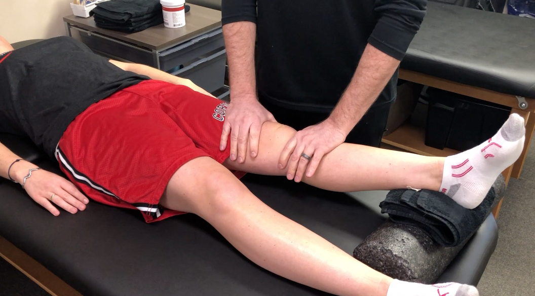
This week, I discussed the progression of someone after a knee surgery. I tried to highlight the key stages and some techniques that I like to use to advance the patient’s mobility and comfort. Take a look at The Week in Research Review, etc 11-26-18 and share with your friends. Hope it helps you improve your patient care tomorrow and beyond!


ACL Reconstruction in a Pediatric and Adolescent Population

1st Day of #PT after an ACL Surgery

Patella Mobilization after Knee Surgery

Knee Flexion PROM after Surgery- Seated or Supine?

Stretching the Quads after Knee Surgery

Assessing for a Cyclops Lesion after an ACL

Assessing for Fat Pad Irritation of the Knee


ACL Reconstruction in a Pediatric and Adolescent Population

17 Year Follow-up After Meniscal Repair With Concomitant ACL Reconstruction in a Pediatric and Adolescent Population. Tagliero et al AJSM 2018

Results: 28% failed meniscal repair and required repeat surgery at the time of final follow-up. They also showed that outcomes and failures rates were comparable across tear complexity.

Guess that means that no matter the tear type, there was no difference in outcomes or retear rates. Although the repair techniques are now outdated and no longer used.

Their study also showed a 30% failure rate for meniscal tear repaired in the medial compartment at index surgery and 7% in the lateral compartment.

Interesting long-term outcomes that may help to guide your rehab and client advancement (and prognosis). Keep these in mind when you treat a future adolescent or pediatric ACL patient.


💥1st Day of #PT after an ACL Surgery 💥

If you have never treated a post-op ACL, then this video should interest you!

This is what the knee looks like that 1st day after surgery and can often set the stage for what’s to come over the next 6-12 months.

Often, the patient is both very curious and ultra-grossed out by the 1st unveiling. It can be stressful for them to see their knee in this condition so you really have to confidently reassure them that it is very normal.

The blood-soaked gauze is mainly saline that was used to irrigate the knee during the reconstruction. Some still leaks out of the incisions the 1st few days and can often be confused with true blood.

Understand that this is quite normal and happens to most every ACL patient’s knee that I’ve seen…nothing to worry about!

From here, I’d work on patella mobility (see the post later today) and then work on flexion ROM at the end of the table.

Again, it’s very important to get the knee moving after surgery. This will help with pain, swelling and gain confidence that the rehab process is moving forward.


Patella Mobilization after Knee Surgery

Get the patella moving early with #patella mobilizations immediately after surgery. One major reason (amongst many others) why we need to get our clients into #PT early.

I am certainly a very loud advocate for early PT and getting the patella moving can help to prevent excessive scarring, which can affect ROM and quadriceps force output.

Glove up and get that patella moving in all directions… medial, lateral, superior, inferior!


🤔Knee Flexion PROM after Surgery- Seated or Supine? 🤔

I’ve treated many patients after an ACL I can honestly say that this may be a huge influence on the early ROM outcomes that you may see.

I’ve tried to bend the knee in both supine or seated, as the video shows, and there’s no doubt that most people tolerate the seated version so much better after a knee surgery. In particular, a big surgery like an ACL, TKA or MPFL reconstruction.

It just seems to be more comfortable and with less stress on the anterior knee because of the position of the tibia (at least I think so!).

My theory, it seems as if the supine position may cause a slight posterior sag which may cause more pain and guarding than when they’re seated at the edge of the table.

I use a similar concept later on in the rehab process when I’m initiating my prone quad stretching. You can see a definitive improvement when I wedge my hand in the popliteal fossa and create a slight anterior translation on the tibia.

Most people say that the anterior knee pain that they were feeling (and not a quad stretch) was replaced by a stretch feeling only and no more anterior knee pain.

Try it out with your ACL patients and see what position they like best…I’ll bet I can covert you over if you still bend your knee patients in supine!


💥Stretching the Quads after Knee Surgery 💥

Continuing my sequence of videos after a knee surgery, I discussed my technique for progressing knee flexion PROM once they hit 120 degrees or so of flexion.

At this point, they’ve probably maxed out how much ROM they can achieve at the edge of the table. They’re ready to get that end range of motion and even some quadriceps flexibility.

In prone, most people will often feel a pain or pressure in the front of their knee when you try to bend it.

To overcome this, I like to wedge my hand into the back of the knee and give an anteriorly directed force through the gastrocnemius (calf) soft tissue and into the tibia.

This seems to create just enough movement of the tibia on the femur to take the pressure off the front of the knee. This may redirect the forces more onto the quadriceps muscle.

You’ll need to play with the amount and direction of force but most often they’ll begin to feel a better quad stretch.

Try this technique out on your next knee surgery client and see if it helps them. I usually initiate this ~4 weeks after an ACL but timeframes will vary person to person.⠀


💥Assessing for a Cyclops Lesion after an ACL 💥

In this video snippet from my YouTube Channel, I discuss how to assess for a Cyclops lesion in a knee. In particular, after knee surgery.

A patient with a potential cyclops lesion, they often present with loss of normal knee extension compared to the other side. They’ll often have anterior knee pain and poor patella mobility. Sometimes a tight feeling in their hamstrings and calves, too.

No matter how they try to regain their extension ROM, the knee just never feels right. Often times, surgical intervention is needed to remove that scar tissue.

Immediate rehab should continue to work on knee extension ROM using low load long duration stretching and aggressive patella mob’s.

No one’s to blame if this occurs. We don’t know exactly why it occurs in some people but we believe a remnant of the ACL stump may be a source of the frustrating issue.


💥Assessing for Fat Pad Irritation of the Knee 💥

Anterior knee pain is very common in the outpatient #PhysicalTherapy setting.

One of my go-to tests to assess for fat pad irritation is simply trying to capture the fatty tissue in the anterior aspect of the knee joint during active and/or passive ROM.

In this snippet from my YouTube channel, you can see that I pinch the fat pads on either side of the patella tendon as @corrine_evelyn is actively extending her knee. I’ll also do it in a relaxed state to assess passive irritability.

I 1st learned this test from @wilk_kevin and it continues to be a mainstay in my knee examination algorithm.

As for a treatment, it usually comes down to a volume issue and/or strength issue or both.

I’ll usually have to address the volume of the activity by relatively easing off of the activity while simultaneously adding in exercises to address an underlying weakness.

Remember the Dye et al study in AJSM 1998 when he talked about the fat pads being super painful during his arthroscopic surgery without anesthesia. Makes sense why they can be so painful if the knee stresses fall upon this tissue.

We talk about this study, fat pad irritability and much much more in our online knee seminar course.


If you want to learn more about how I treat ACL’s or the knee in general, then you can check out our all online knee seminar at www.onlinekneeseminar.com and let me know what you think.

We cover the anatomy, rehab prescription, ACL, meniscal injuries knee replacements and patellofemoral issues. Furthermore, the course covers both the non-operative and post-operative treatment.t

This is an awesome course if you’re interested in learning more about rehabilitating the knee joint. And if you’re a PT, there’s a good chance you can get CEU’s as well.

The Week in Research Review, etc 10-22-18

That was a milestone week as my Instagram account finally hit 10k followers, whatever that means! I’ve really been pushing a daily post to help other rehab professionals better simplify the research. One milestone hit but I still want to keep publishing good quality research reviews. The Week in Research Review, etc 10-22-18 included:

  1. Do baseball Pitchers really have a tight posterior capsule?
  2. ACL strain curve during the squat
  3. Does the pec minor length influence shoulder pain?
  4. What does the literature say about the EMG activity of the rotator cuff, particularly of the supraspinatus, with ROM
  5. Classification of Meniscus Tears and Osteoarthritis

Do baseball Pitchers really have a tight posterior capsule?

My guess is emphatically no based on what I see on a daily basis, the general anatomy of the glenohumeral joint and some research studies.

Anatomy
1️⃣When I stretch a baseball pitcher’s shoulder, it is usually very mobile. I find this in both symptomatic and asymptomatic individuals.

If I try to assess their posterior capsule with a joint play technique, I can often translate the humeral head pretty far over the glenoid rim. Sometimes, I can even sublux the humerus!

2️⃣Anatomically, the posterior capsule is relatively thin compared to the anterior and inferior capsule (see the post).

In general, that capsule is thinner probably because of the glenoid position that is not strictly in the frontal plane.
Because of that, it is theorized that the capsule evolved to have less of a role in stabilizing the humerus.

3️⃣There are a couple of research studies that have specifically looked at baseball pitchers to determine their humeral head translation.

Borsa et al AJSM 2005 reported that posterior translation was actually greater than anterior translation in both the dominant and non-dominant shoulders of professional baseball pitchers.

Crawford et al J Ath Train 2006 found no significant differences in posterior glenohumeral laxity and stiffness between the throwing and non-throwing shoulders.

I understand why the theory exists and think it could be plausible but just don’t think it’s truly responsible for what we think.

We just don’t think we can stretch the posterior capsule with any joint mobilization or contract-relax procedure, including a sleeper stretch. I often giggle at all of those MD prescriptions that say ‘#GIRD, posterior capsule tightness’. I just treat what I find on my examination and ignore the script.


ACL strain curve during the squat

As you can see, the strain curve from the Beynnon et al study is very similar to the strain curve during resisted knee extension in a full ROM (90-0).

We argue all of the time about anterior tibial translation during the open chain exercises but often ignore the other side of the story.

The strain on the ligament is barely 4%, which is in line with many functional activities like walking, descending steps, etc. The argument that we’re going to stretch the ligament out just has not been proven in the literature.

I wanted to show the closed chain strain curve so you could compare it to the open chain strain curve. I know the n=8 argument is present but we really don’t have much more data on the ligament in vivo that shows the true effects of open vs closed chain exercises on the ACL.

Again, as @barbhoogie mentioned, you need to monitor the PF joint, especially after a patella tendon autograft but as long as we’re not aggravating that joint, then I begin early 90-0’s and mini squats as tolerated.

Do you agree with this? Do you prevent squats early on during the ACL rehab process? If you don’t, then why do you hold back on full active knee extension exercises?


Does the pec minor length influence shoulder pain

Does the pectoralis minor length influence acromiohumeral distance, shoulder pain-function, and range of movement? Navarro-Ledesma et al Phys Ther Sport Aug 2018.

Their conclusion: Pectoralis minor length is not a distinguishing factor in shoulder⠀
assessment when a chronic condition exists, and it seems not to play a key role in pain perception and ROM.

54 participants with chronic shoulder pain in their dominant arm were recruited, as well as fifty-four participants with a pain-free shoulder.

The resting muscle length is measured between the caudal edge of the 4th rib to the inferomedial aspect of the coracoid process with a sliding caliper.

The acromiohumeral distance (AHD) was defined as the shortest linear distance between the most inferior aspect of the acromion and the adjacent humeral head, measured by ultrasound.

An interesting study that used an asymptomatic control group along with the contralateral shoulder of the symptomatic subject. A pretty clean study that is very interesting. I’m not going to say that the pec minor doesn’t play a role in shoulder pain but maybe its role is not as prominent as we think.

What do you think? Do you find pec minor length has a substantial role in your patients with shoulder pain?⠀


EMG of the rotator cuff during rehab exercises

What does the literature say about the EMG activity of the rotator cuff, particularly of the supraspinatus, with ROM?

Many PT’s and doctors are uncertain when to safely begin physical therapy after a shoulder surgery, particularly after a rotator cuff repair. In my 15+ years as a PT, I’ve seen docs begin PT post-op day 1 or wait as long as 6 weeks (which drives me bonkers!!)

In this snippet that I’ve taken from an upcoming blog post at LennyMacrina.com. I discuss the research that’s helping to guide best practice, in particular, the research that looks at PROM and AAROM and how much EMG activity is actually going on in the supraspinatus with each movement.

As you can see in the video, there’s minimal supraspinatus activity (<20% is considered minimal) for all motions. Keep in mind, many of these studies are done on healthy individuals but who in their right mind would volunteer their newly repaired RTC repair to have fine-wire EMG done on them?

So, I can only draw my conclusions from a limited body of evidence and my own anecdotal evidence (which consists of 12+ years of immediate PROM POD 1). Many still think it’s safe to get a RTC repair patient’s shoulder moving early for many reasons that I will describe in this blog post.

I just wanted to get this early point out there to get another discussion going. I think our patients can do much better after a RTC repair and this is one of the reasons.

Do you agree? Do you advocate for early PROM after a RTC repair, especially a small-medium repair?


Classification of Meniscus Tears and Osteoarthritis

Great post by @physicaltherapyresearch talking about the various types of meniscus tears. Nice visual & description of each type and the incidence of OA. Take a look! 👇🏼
_______________
Meniscus Tears and Osteoarthritis

💡

Prevalence of meniscal tears is estimated as ~24-31% of some populations, increasing with age and ranging from 19% in women aged 50–59 years to 56% among men between 70 and 90 years and is markedly higher in established OA subjects.
💡

Medial meniscus and/or the posterior horn tears make up 66% of cases, with horizontal and complex tears being the most common.
💡

Most subjects with a meniscal tear are asymptomatic.
💡

Regardless of morphologic type, meniscal tears are strongly associated with OA cross-sectionally and predict OA longitudinally and are considered to be part of the spectrum of early or pre-radiographic disease

📝📝📝

TEAR TYPES INFO:

Often enough, meniscal tear types are categorized into varying groups for comparison rather than separately compared to each other.
📝

There is a striking lack of data on the relevance of different morphologic types of meniscal tears in OA.

📝

Horizontal and complex tears are common findings in knees with OA

📝

Posterior radial tears of the medial meniscus are associated with a high degree of cartilage loss and meniscal extrusion, and appear to be a highly relevant event in the progression of OA in the knee. 📝

Lateral meniscus radial tears affect younger individuals and are considered post-traumatic.

📝
Despite their suggested high relevance, radial tears are more commonly misdiagnosed on MRI than any other type of tear.

📝

While medial meniscus posterior root tears are of “radial” morphology, there is growing interest in regarding them as a separate entity.
📝

Longitudinal and bucket handle tears affect younger individuals and are highly associated with ACL injuries, favoring a traumatic etiology.
📝

MRI is important to detect and locate a possible displaced tear.
📝

Further epidemiologic studies should focus on the morphology of specific meniscal tears to better understand their relevance in the genesis and progression of knee OA.
📚📚📚
SOURCE:
Jarraya et al. 2017 Semin Arthritis Rheum


The Week in Research Review, etc 10-15-18

This week I posted a lot of research and thoughts on shoulder and knee rehab, particularly after an ACL injury. I also shared some others posts that really complimented my posts so there’s some bonus reading to do too. Hope The Physical Therapy Week in Research Review helps your Monday patients and beyond! Take a read and share with your friends!


  1. Co-morbidities in the first 2 years after arthroscopic hip surgery: substantial increases in mental health disorders, chronic pain, substance abuse, and cardiometabolic conditions. Rhon et al BJSM 2018.⠀
  2. Range of Extension Correlates with Posterior Capsule Length after Knee Remobilization Zhou et al Med Sci Sports Exerc 2018⠀
  3. Sidelying External Rotation- The 1 exercise in all upper body programs
  4. @dr.jacob.harden talking Infraspinatus release.
  5. Do you account for Bone Bruises after an ACL
  6.  @cbutlersportspton bone bruises and the specifics
  7. When is it safe to initiate full AROM knee extension after an ACL-PTG autograft
  8. @mickhughes.physio on when it MAY be safe to initiate full knee extension from 90-0 after an ACL reconstruction.


Comorbidities after Hip Arthroscopy

Co-morbidities in the first 2 years after arthroscopic hip surgery: substantial increases in mental health disorders, chronic pain, substance abuse and cardiometabolic conditions. Rhon et al BJSM 2018.

This is an interesting study on 1870 mainly US Military personnel between 2004-13 (~33% were not active duty).

Relative to baseline, cases of:

❇️mental health disorders rose 84%

❇️chronic pain diagnoses increased by 166%

❇️substance abuse disorders rose 57%

❇️cardiovascular disorders rose by 71%

❇️metabolic syndrome cases rose by 85.9%

❇️systemic arthropathy rose 132%

❇️sleep disorders rose 111%

The comorbidity with the greatest increase of new cases was that of mental health disorders (26% of the entire cohort). Age and socioeconomic status had significant associations on outcomes as well.

Just an eye-opening study that followed each subject 2 years after their respective surgeries. One giant variable that jumped out at me was that they used mainly military personnel only as the subjects.

We certainly can’t extrapolate on non-military personnel but need to keep this study in mind for others treating a similar cohort. Did the surgery cause these disorders? Absolutely not! No causation can be associated and that is very important!

What do you think about this study and how mainly military personnel and civilians that were tracked ending up developing many chronic disorders? I say it is very troubling! Let’s chat…and remember, this is not a causation study but just a reminder to educate and monitor your patients’ well-being after a surgery.


Posterior Capsule Limits Knee Extension after an ACL

Range of Extension Correlates with Posterior Capsule Length after Knee Remobilization Zhou et al Med Sci Sports Exerc 2018

This study is a confirmation bias for me because it showed that the knee’s posterior capsule limits extension after immobilization (in rats!) This is why I’m a huge proponent of low load long duration stretching of most joints when they begin to get stiff.

It seems as if the prolonged stretching is needed to regain collagen length and return the ROM. I know it’s in rats so calm down…but we need to get the data from somewhere.

Take it with a grain of salt but know that LLLD is going to be the best mode to return ROM (and not just hamstring stretching).⠀
.⠀
Do you agree? Do you treat rats with stiff knees? Then this study was created for you!


The Best Exercise for the Rotator Cuff

❗️Sidelying External Rotation- The 1 exercise in all upper body programs❗️

I really think this exercise should be in everyone’s program, whether going through rehab for a painful shoulder or a high level, healthy powerlifter. The role of the infraspinatus and other rotator cuff muscles is crucial to maintaining humeral head stability.

Sidelying external rotation has been shown to elicit the highest amount of EMG activity for the infraspinatus so I give this exercise to everyone, once there are no precautions for tissue healing. The infraspinatus and subscapularis (front rotator cuff muscle) are force couples that help to stabilize the humerus within the glenoid. Weakness of the infraspinatus may affect this force couple and create an inefficient movement within the joint.

My goal for all of my clients is to create an efficient movement that allows them to work at their highest level. The infraspinatus is a critical muscle of the shoulder complex so MOST of my programs include this exercise.


Myofascial Release of the Infraspinatus

Great post by @dr.jacob.harden talking Infraspinatus release. Perfect timing for my earlier post today looking at my go to exercise for the shoulder joint. Check his post out below!👉🏻 🔴 𝙃𝙊𝙒 𝙏𝙊 𝙍𝙀𝙇𝙀𝘼𝙎𝙀 𝙄𝙉𝙁𝙍𝘼𝙎𝙋𝙄𝙉𝘼𝙏𝙐𝙎

Coming at ya with a little #throwbackthursday since I’m about to jump on a plane across the pond to London. So we’re looking at how to do a pin and stretch for the rotator cuff, specifically the infraspinatus. The infraspinatus is the main external rotator of your shoulder, so it’s that muscle we see everyone working when they swing there 5 pound plates side to side in their warm-ups. (Side note: if you do that, please use a band or do it sidelying. Standing with plates does nothing but work the bicep.👍)

This can also help with some those little hypersensitive areas in the back of the shoulder. If you’re feeling those spots or having shoulder pain or just want to improve your internal rotation a bit, this release can help.

𝗛𝗲𝗿𝗲’𝘀 𝗵𝗼𝘄 𝘁𝗼 𝗱𝗼 𝗶𝘁:

🔹️Ball placement is below the spine of the scapula.

🔹️Internally rotate, flex, and adduct the shoulder

🔹️Work back and forth for a minute or so


Bone Bruises after an ACL

Do you even consider a bone bruise after an ACL when progressing your patients? I know I certainly do and one of the major reasons why I have gone a bit slower with my latter stage progression, especially to impact activities like plyometrics and running.

There are a few studies that have shown the presence of a bone bruise after an ACL injury but we are not 100% certain this eventually leads to joint degradation.

Hanypsiak et al included 44 patients (82%) who underwent unilateral ACLR without multi-ligament involvement. Thirty-six (82%) patients had a bone bruise on index MRI. Potter et al reported all patients in their cohort sustained chondral damage at the time of injury.

Faber et al examined 23 patients with occult osteochondral lesion (bone bruise) who underwent ACLR. They found that at 6-year follow-up, a significant number of patients had evidence of cartilage thinning adjacent to the site of the initial osteochondral lesion (13/23 patients).

So as you can see, bone bruises are more common than most people think. This may be one reason why osteoarthritis rates are much higher in ACL reconstructed knees.

Additional factors, such as cartilage and meniscus injury, associated with ACL rupture may play an important role in subsequent outcomes following surgical reconstruction independent of a bone bruise.

Do you consider a bone bruise when progressing your patients back from a knee injury like an ACL reconstruction?


Types of Bone Bruises after an ACL Injury

@cbutlersportspton bone bruises, which fits perfectly with my post earlier today. He talks about the 3 different types of common bone bruises…check it out below!

❗️What is a Bone Bruise❗️We often hear that one of our Fantasy Football players has a Bone Bruise and may be out for a few weeks.

It sounds like something that an NFL athlete should be able to tough out, right?

Here’s why you may need to put in a backup for a few games.

A bone bruise occurs when several trabeculae in the bone are broken, whereas a fracture occurs when all the trabeculae in one area have broken. Trabecular bone is also known as spongy bone.

—-Three Types of Bone Bruises—-⠀
1️⃣Subperiosteal hematoma: A bruise that occurs due to an impact on the periosteum that leads to pooling of blood in the region.⠀
2️⃣Intraosseous Bruising: The bruise occurs in the bone marrow and is due to high impact stress on the bone.⠀
3️⃣Subchondral Bruise: This bruise is bleeding between cartilage and bone such as in a joint.

—-Symptoms of Bone Bruises—-

•Pain and tenderness in the region of injury

•Swelling in the region of injury

•Skin discoloration in the region of injury

Bone bruises often occur with joint injuries, such as ankle sprains and ACL tears, therefore a bone bruise can also coincide with stiffness and swelling in the joint.⠀


When is it safe to initiate full AROM knee extension after an ACL-PTG autograft?

I posted this video in my the other day and had a ton of people message me about the exercise.

Most people wanted to know how far out of surgery the patient was and when I felt it was safe to begin full, active knee extension after an ACL.

I’ve always been relatively conservative with my rehab (at least I think so) but I wanted to dig a little deeper. I recently saw a post by @mickhughes.physio and he was talking about the Fukuda et al study from 2013.

The study looked at 90-40 knee extensions and ‘ACLR patients can perform 3×10 at a 70% 1RM load through a restricted 45-90deg ROM between weeks 4-12 post-op, and then the same load full ROM from 12 weeks post-op. ‘

It made me dive a bit deeper and I went to my trusty Beynnon et al AJSM studies from the late 90’s. You can see the strain on the ACL decreases as we approach 40 degrees and stays low out to 90 degrees…but is 3-4% strain on the ligament significant?

If you look at the study (yes, it’s only on 8 subjects) you’ll see a similar strain curve for closed chain exercises as well…but we do mini squats immediately after surgery without 2nd guessing!

In 2011, Beynnon et al AJSM showed that an accelerated program that initiated full resisted knee extension (90-0) at 4 weeks showed similar knee laxity throughout the study. The other group initiated full resisted knee extension at 12 weeks. Also, those who underwent accelerated rehabilitation experienced a significant improvement in thigh muscle strength at the 3-month follow-up.

So, what do we do with this data? I have begun to do full, resisted knee extensions with my patients between 4-6 weeks post-op, as long as it’s a patella tendon autograft. For allografts or HS autografts, I tend to delay it a bit longer because of the soft tissue healing that is delayed.

What do you think? When do you initiate full AROM after an ACL? Do you know of a study that definitively says the strain on the ACL graft is detrimental to the healing ligament?


How much Resistance Should we Recommend Open Chain Exercises After an ACL

This is the post from @mickhughes.physio that made me dive a bit deeper into the research on when it MAY be safe to initiate full knee extension from 90-0 after an ACL reconstruction. Check out his post below! ⠀
____________________

So if we can safely perform OKC exercises (knee extensions) as part of ACLR rehab; how heavy can lift?⠀
*⠀
*

This is a question I often get asked. Based on the work by Fukuda et al (2013), ACLR patients can perform 3×10 at a 70% 1RM load through a restricted 45-90deg ROM between weeks 4-12 post-op, and then the same load full ROM from 12 weeks post-op. *⠀
*⠀

From then you can progressively load as per what can be tolerated. Usually the first sign that the knee is unhappy with the load is that the underneath the kneecap will be sore/painful. That’s a sign you need to back the load off a little so the exercise is felt in the quads only. *

If you’re still unsure about OKC exercises (knee extensions) during ACLR rehab read my blog by clicking on the link in my bio ⠀
#ACL #Physio #Knee #Rehab


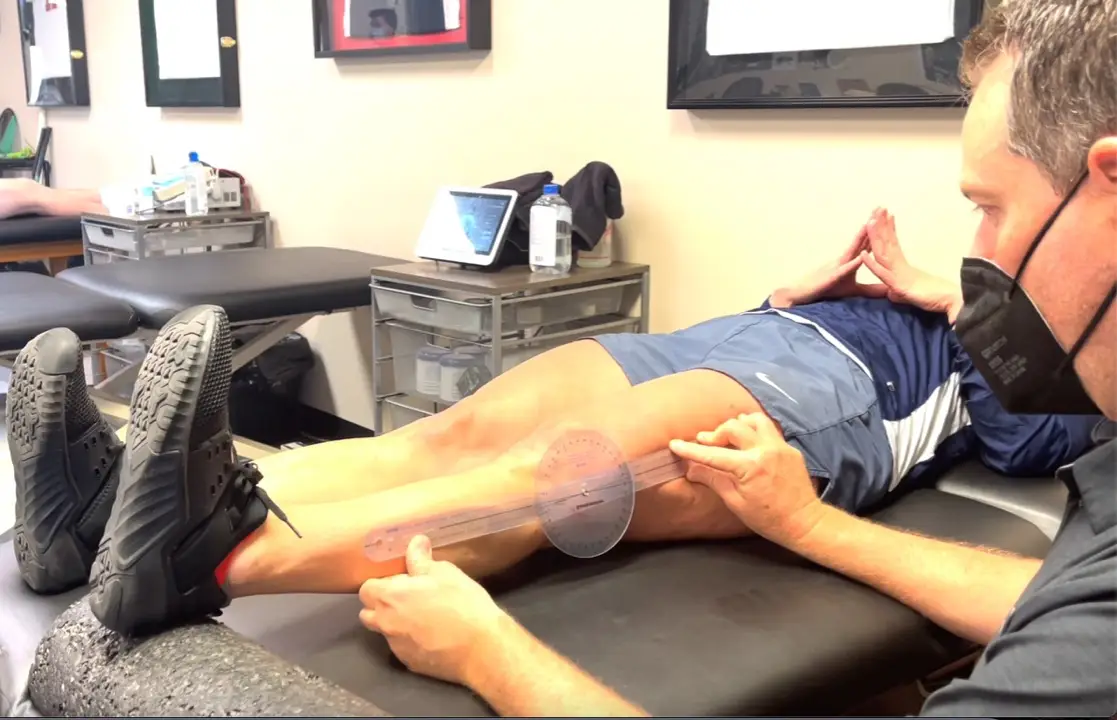
Loss of extension after ACL surgery: How to assess for a cyclops lesion

Loss of extension after an ACL reconstruction can be debilitating for the patient. It’s not as common as you would think but I see it enough in the clinic from people that are months out from surgery. Usually, this loss of knee extension after an ACL reconstruction is caused by a cyclops lesion. Let’s dive deeper into this!

Often times, they’ll present with anterior knee pain, posterior knee soreness and a relatively weakened quadriceps muscle that just won’t return. No matter what they do to get the motion back, the knee just never feels normal.

I’ve written about the loss of extension after an ACL reconstruction in the past. In this post, I discussed how I like to work on knee extension immediately after an ACL surgery. There are a few ways that I think are most effective and with minimal patient efforts.

What is a Cyclops Lesion?

For those not familiar, a cyclops lesion is a wad of scar tissue in the anterior aspect of the knee joint. It is believed to be a remnant of the previous ACL stump that had remained during the reconstruction surgery. At least that’s one theory. Another theory states that it may be fibrocartilage as a result of drilling the tibial tunnels.

Whatever the case, this arthrofibrosis (scar tissue) physically blocks the knee joint from locking out into full extension. Check out this MRI that shows the scar tissue in the anterior knee.

Cyclops lesion in the anterior knee blocking full (hyper) extension

How do I assess for a cyclops lesion after ACL surgery?

In this video, I describe why and how I assess for a cyclops lesion. Check it out.

Is it a Cyclops lesion or just a tight knee?

From the video, you can clearly note that anterior pain, in my experiences, is most often related to a cyclops lesion. Other factors to consider include:

  1. temporary/transient gains in extension
  2. anterior knee pain after increasing activity
  3. poor patella mobility
  4. quads just won’t come back
  5. continued hamstring/calf soreness

These are tell-tale signs that there’s more going on and you should refer back to the doctor so they can order an MRI to rule in/out the anterior scarring. If diagnosed, the best (and only) option is to have a knee scope and remove that scar tissue.

There’s nothing else that can be done. No PT, injections or manual therapy can restore full symmetrical knee extension.

The scar tissue needs to be removed by surgical excision. Aggressive PT should commence immediately after surgery to restore the extension range of motion.

The Best and Easiest Way to Restore Knee Extension after an ACL

Rehab after an ACL is never easy. There are many things that could affect a patient’s outcome. I’ve treated hundreds of patients after an ACL reconstruction and each one is a unique challenge. I wrote about this in a previous post here..check it out and let me know what you think.

I put this video together for Mike Reinold’s website so you could see what I exactly do to gain knee extension back….and why I’m not a fan of prone hangs. Hope it helps with some of your knee patients.

Final Cyclops Thoughts

As a PT or athletic trainer, don’t blame yourself if the patient needs another surgery to remove the scarring. It seems as if it was inevitable and was going to occur no matter the efforts to work on knee extension. I think the lesson here is:

  • work on extension early and often
  • maintain good compliance at home
  • assess/measure each visit to determine gains or losses
  • early patella mobility and knee PROM
  • get the pain and swelling out as quickly as possible
  • refer back to the doctor if the ROM not improving despite your best efforts

Hope this post helps you get better outcomes for your ACL patients!


If you want to learn more about how I treat ACL’s, then you can check out our all online knee seminar. If interested, check it out at www.onlinekneeseminar.com and let me know what you think. We cover the anatomy, rehab prescription, ACL, knee replacements and patellofemoral issues both non-operative treatment and post-operative treatment. This is an awesome course if you’re interested in learning more about rehabilitating the knee joint. And if you’re a PT, there’s a good chance you can get CEU’s as well.