Tag Archive for: rotator cuff exercises

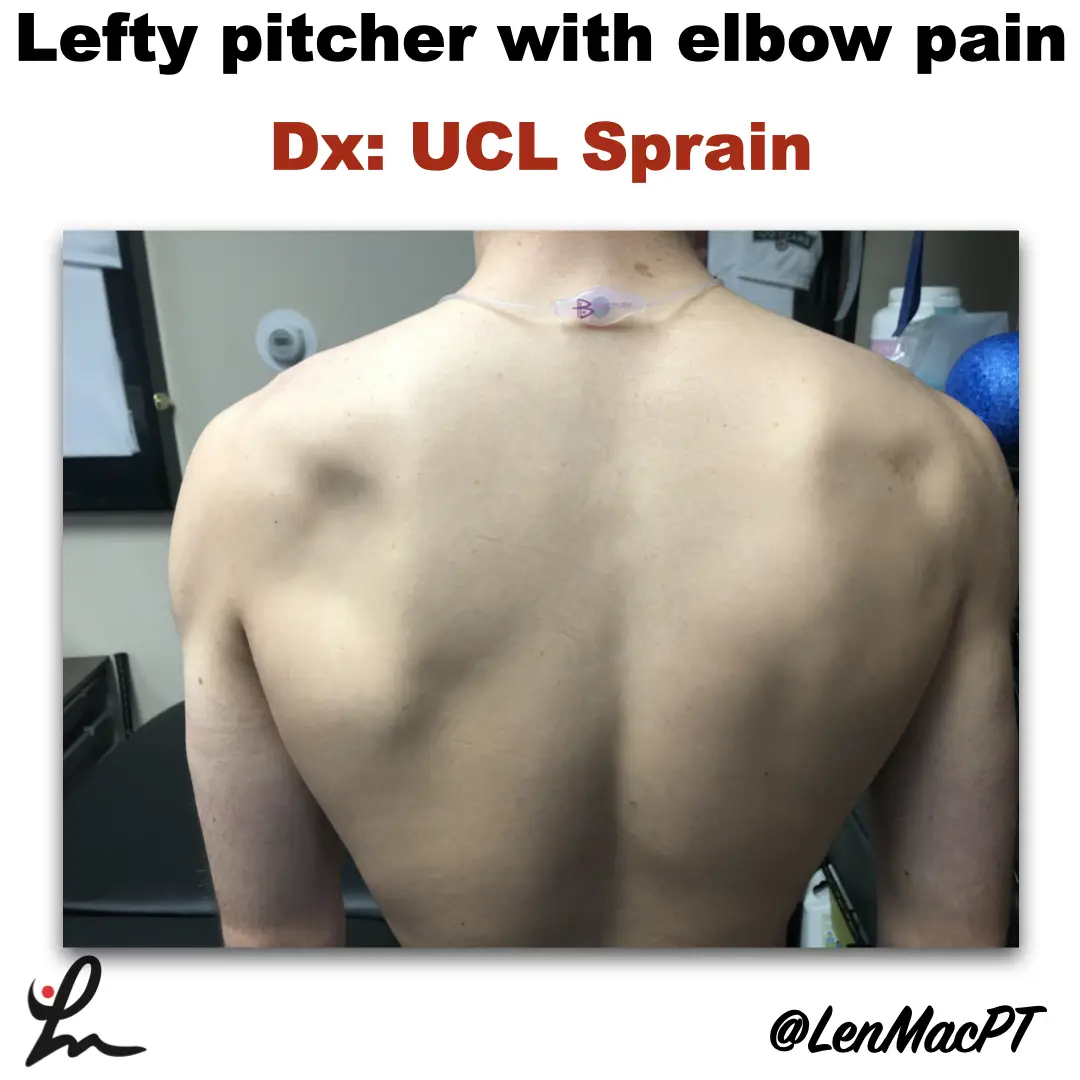
Infraspinatus atrophy in a baseball pitcher with a UCL sprain

Infraspinatus atrophy due to a spinoglenoid notch cyst

This was an incidental finding of mine in a collegiate baseball pitcher. He presented with a UCL sprain during his senior year and was struggling.

He came in looking to salvage his senior year and attempt to pitch at some point. UCL surgery would’ve been warranted if he was anything but a senior.

UCL Evaluation

We evaluated his elbow and he presented with typical findings of painful valgus stress tests. I’ve discussed my UCL exam previously in a post that you can find here.

UCL Tommy John Assessment

His shoulder PROM was slightly limited compared to what we normally see in our overhead throwers, which may have contributed to his elbow issues.

He only had 120 degrees of external rotation compared to 110 degrees on the right side. If you’re wondering how I measure external rotation, then this video below may be a good one to review.

External rotation passive range of motion (PROM)

We use these concepts for our research studies that we have published on weighted ball usage the past few years.

For those that will ask about GIRD, he had 60 degrees of IR on the L side and 50 degrees on the R side. I’m not a big GIRD guy like has been written by others. I still think GIRD is pretty normal and our research has shown that too.

Remember, we showed that differences in total rotational range of motion greater than 5 degrees was significant. This study was published in AJSM back in 2011.

Furthermore, we showed a connection between the loss of passive flexion and elbow injury.

This result was also shown by Camp et al in a separate study, as well.

I had him remove his shirt and noticed a large defect in the infraspinatus area that showed obvious atrophy (see picture below).

Infraspinatus atrophy due to a spinoglenoid notch cyst

The cyst was compressing the suprascapular nerve as it coursed along the spine of the scapula.

His external rotation strength was significantly weaker on his throwing side compared to his right side. This would make complete sense considering the amount of atrophy present.

Often times surgery is warranted to decompress the nerve. There’s often an underlying labral tear that has resulted in a cyst to form. The surgery would remove the cyst and repair the labrum to prevent future issues.

Quick Literature Review on Surgical Outcomes

Reports of improved outcomes are in the literature, here and here and here.

Obviously, this may have contributed to his elbow issues as the posterior cuff muscles are tremendous stabilizers of the arm, especially during the deceleration phase of throwing.

Treatment for infraspinatus muscle atrophy

Our goal was to calm the elbow down through active rest and focus on strengthening of his forearm and shoulder musculature.

We used neuromuscular electrical stimulation (NMES) to the infraspinatus. We felt as if the electrical stimulation would allow for a better muscle contraction than what he could do actively. This is a similar concept to using NMES to the quads after a knee surgery.

NMES to the infraspinatus to help with muscle recruitment

We have previously shown in AJSM that NMES to the infraspinatus was beneficial to patients after a rotator cuff repair. It helped improve their muscle contraction by 22% in the group with the NMES applied during testing.

Outcomes and final thoughts

Believe it or not, we were able to salvage his senior year despite all of the issues that presented.

He made numerous pitching outings and was pretty successful for his team.

The lesson here: Don’t take anything for granted, observe and test.

Make sure you look distal and proximal. It could’ve been very easy to just treat the elbow with some exercises and massage. Don’t get me wrong, we did a bunch of strengthening exercises to his forearm.

His secondary finding was most likely a leading cause for his elbow dysfunction.

This isn’t the 1st time that I’ve run across an issue like this. I’ve found numerous scapula dysfunctions like this involving the infraspinatus. But also a winging issue due to a long thoracic nerve palsy.

You must look at the whole picture… shirts off for a guy and a sports bra for a female to make sure we’re not missing anything obvious!

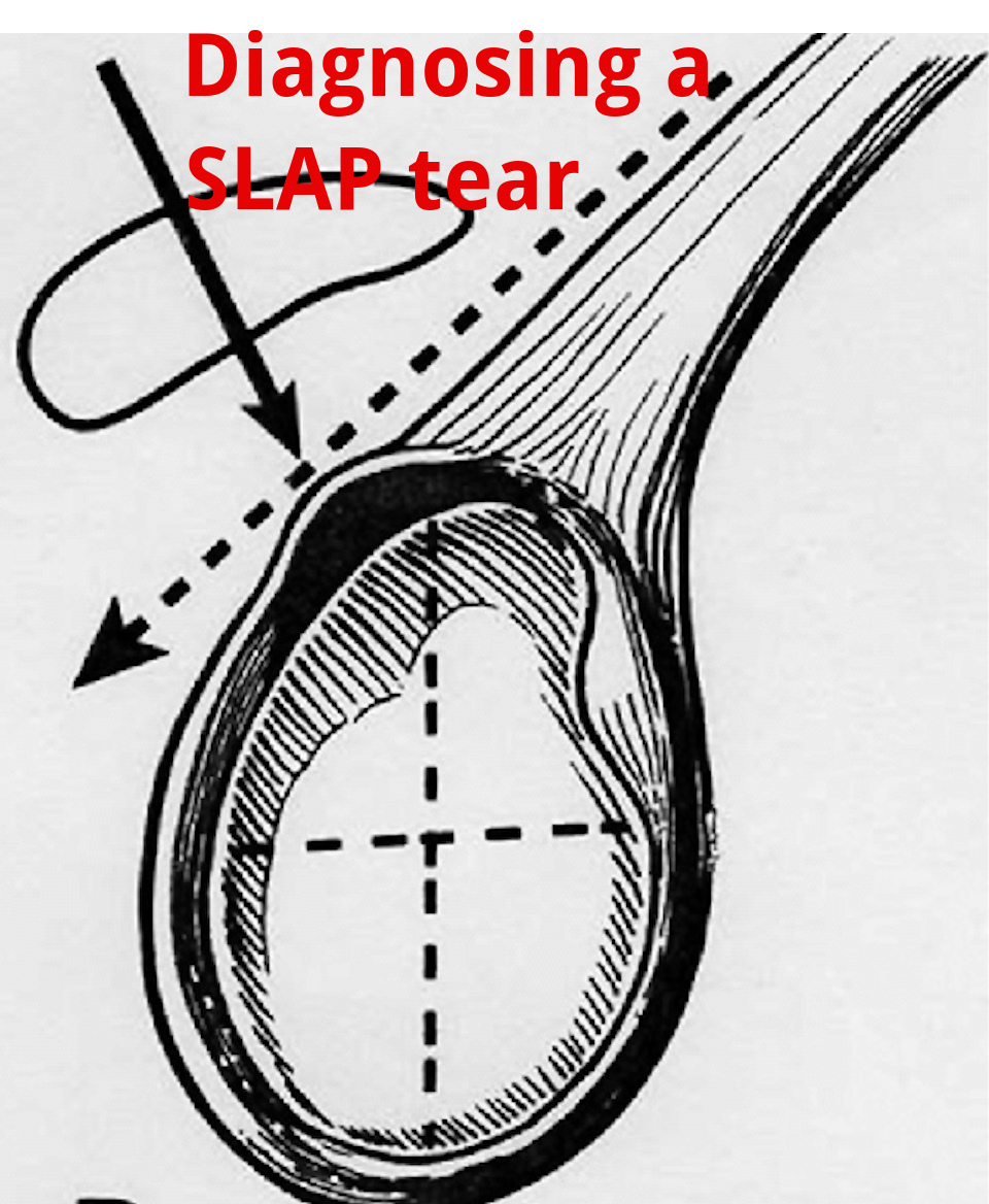
An Update on Diagnosing SLAP tears

Diagnosing a SLAP tear is not easy

We hear a lot about trying to diagnose shoulder pain and to be as specific as possible. It’s often difficult to differentiate SLAP (superior labrum anterior to posterior) tears from other soft tissue injuries of the shoulder.

For a review of the different types of SLAP tears, check out this old blog post that classifies the 10 different types of tears.

from https://commons.wikimedia.org/wiki/File:SLAP-Lesion-front-2.jpg

This paper in IJSPT by Clark et al 2019 attempts to help out the process and recommend a few special tests that MAY aid in diagnosing a SLAP tear.

What do they Recommend to diagnose a slap tear?

They recommend that a combination of at least 3 positive SLAP lesion tests may be clinically useful in diagnosing a shoulder SLAP lesion with greater diagnostic accuracy.

Combo of Tests

The combination of the Biceps Load I/II and O’Brien’s showed the highest sensitivity and specificity.

I have found similar results with this set of special tests so maybe this paper just hits my biases correctly.

In this video at my YouTube channel, I wanted to let you hear my thoughts and small tweaks to the evaluation process.

It’s not easy to diagnose a SLAP tear.

Differential Diagnosis is Critical

Furthermore, does it really matter and will it change the treatment plan much at all? I think it may a little but overall it will remain a pretty similar treatment approach to other similar pathologies like:

  • rotator cuff tendonopathy
  • Biceps strain
  • Latissimus strain
  • Subscapularis strain
  • internal impingement
  • pectoralis major strain

I think one also needs to consider the cervical spine and to make sure the pain is not referred from the neck.

Otherwise, a well thought out program should be implemented that addresses the strain on the shoulder and any strength issues.

I talked about this in an article that I wrote for Medbridge a while back so check out that post here:

You can also check out a snippet of one of my courses at this YouTube video where I discuss rotator cuff and labral issues. Hope it helps too!

Will this Change your Practice?

How ever you look at it, I wanted to use this paper to let you know that there MAY be a cluster of tests that better diagnose a suspected SLAP tear in your next patient’s shoulder.

Check out the paper and comment so we can talk it through. Are these tests similar to what you use in your clinical practice? Will this paper change what you do in your clinical practice?

The Week in Research Review, etc 10-15-18

This week I posted a lot of research and thoughts on shoulder and knee rehab, particularly after an ACL injury. I also shared some others posts that really complimented my posts so there’s some bonus reading to do too. Hope The Physical Therapy Week in Research Review helps your Monday patients and beyond! Take a read and share with your friends!


  1. Co-morbidities in the first 2 years after arthroscopic hip surgery: substantial increases in mental health disorders, chronic pain, substance abuse, and cardiometabolic conditions. Rhon et al BJSM 2018.⠀
  2. Range of Extension Correlates with Posterior Capsule Length after Knee Remobilization Zhou et al Med Sci Sports Exerc 2018⠀
  3. Sidelying External Rotation- The 1 exercise in all upper body programs
  4. @dr.jacob.harden talking Infraspinatus release.
  5. Do you account for Bone Bruises after an ACL
  6.  @cbutlersportspton bone bruises and the specifics
  7. When is it safe to initiate full AROM knee extension after an ACL-PTG autograft
  8. @mickhughes.physio on when it MAY be safe to initiate full knee extension from 90-0 after an ACL reconstruction.


Comorbidities after Hip Arthroscopy

Co-morbidities in the first 2 years after arthroscopic hip surgery: substantial increases in mental health disorders, chronic pain, substance abuse and cardiometabolic conditions. Rhon et al BJSM 2018.

This is an interesting study on 1870 mainly US Military personnel between 2004-13 (~33% were not active duty).

Relative to baseline, cases of:

❇️mental health disorders rose 84%

❇️chronic pain diagnoses increased by 166%

❇️substance abuse disorders rose 57%

❇️cardiovascular disorders rose by 71%

❇️metabolic syndrome cases rose by 85.9%

❇️systemic arthropathy rose 132%

❇️sleep disorders rose 111%

The comorbidity with the greatest increase of new cases was that of mental health disorders (26% of the entire cohort). Age and socioeconomic status had significant associations on outcomes as well.

Just an eye-opening study that followed each subject 2 years after their respective surgeries. One giant variable that jumped out at me was that they used mainly military personnel only as the subjects.

We certainly can’t extrapolate on non-military personnel but need to keep this study in mind for others treating a similar cohort. Did the surgery cause these disorders? Absolutely not! No causation can be associated and that is very important!

What do you think about this study and how mainly military personnel and civilians that were tracked ending up developing many chronic disorders? I say it is very troubling! Let’s chat…and remember, this is not a causation study but just a reminder to educate and monitor your patients’ well-being after a surgery.


Posterior Capsule Limits Knee Extension after an ACL

Range of Extension Correlates with Posterior Capsule Length after Knee Remobilization Zhou et al Med Sci Sports Exerc 2018

This study is a confirmation bias for me because it showed that the knee’s posterior capsule limits extension after immobilization (in rats!) This is why I’m a huge proponent of low load long duration stretching of most joints when they begin to get stiff.

It seems as if the prolonged stretching is needed to regain collagen length and return the ROM. I know it’s in rats so calm down…but we need to get the data from somewhere.

Take it with a grain of salt but know that LLLD is going to be the best mode to return ROM (and not just hamstring stretching).⠀
.⠀
Do you agree? Do you treat rats with stiff knees? Then this study was created for you!


The Best Exercise for the Rotator Cuff

❗️Sidelying External Rotation- The 1 exercise in all upper body programs❗️

I really think this exercise should be in everyone’s program, whether going through rehab for a painful shoulder or a high level, healthy powerlifter. The role of the infraspinatus and other rotator cuff muscles is crucial to maintaining humeral head stability.

Sidelying external rotation has been shown to elicit the highest amount of EMG activity for the infraspinatus so I give this exercise to everyone, once there are no precautions for tissue healing. The infraspinatus and subscapularis (front rotator cuff muscle) are force couples that help to stabilize the humerus within the glenoid. Weakness of the infraspinatus may affect this force couple and create an inefficient movement within the joint.

My goal for all of my clients is to create an efficient movement that allows them to work at their highest level. The infraspinatus is a critical muscle of the shoulder complex so MOST of my programs include this exercise.


Myofascial Release of the Infraspinatus

Great post by @dr.jacob.harden talking Infraspinatus release. Perfect timing for my earlier post today looking at my go to exercise for the shoulder joint. Check his post out below!👉🏻 🔴 𝙃𝙊𝙒 𝙏𝙊 𝙍𝙀𝙇𝙀𝘼𝙎𝙀 𝙄𝙉𝙁𝙍𝘼𝙎𝙋𝙄𝙉𝘼𝙏𝙐𝙎

Coming at ya with a little #throwbackthursday since I’m about to jump on a plane across the pond to London. So we’re looking at how to do a pin and stretch for the rotator cuff, specifically the infraspinatus. The infraspinatus is the main external rotator of your shoulder, so it’s that muscle we see everyone working when they swing there 5 pound plates side to side in their warm-ups. (Side note: if you do that, please use a band or do it sidelying. Standing with plates does nothing but work the bicep.👍)

This can also help with some those little hypersensitive areas in the back of the shoulder. If you’re feeling those spots or having shoulder pain or just want to improve your internal rotation a bit, this release can help.

𝗛𝗲𝗿𝗲’𝘀 𝗵𝗼𝘄 𝘁𝗼 𝗱𝗼 𝗶𝘁:

🔹️Ball placement is below the spine of the scapula.

🔹️Internally rotate, flex, and adduct the shoulder

🔹️Work back and forth for a minute or so


Bone Bruises after an ACL

Do you even consider a bone bruise after an ACL when progressing your patients? I know I certainly do and one of the major reasons why I have gone a bit slower with my latter stage progression, especially to impact activities like plyometrics and running.

There are a few studies that have shown the presence of a bone bruise after an ACL injury but we are not 100% certain this eventually leads to joint degradation.

Hanypsiak et al included 44 patients (82%) who underwent unilateral ACLR without multi-ligament involvement. Thirty-six (82%) patients had a bone bruise on index MRI. Potter et al reported all patients in their cohort sustained chondral damage at the time of injury.

Faber et al examined 23 patients with occult osteochondral lesion (bone bruise) who underwent ACLR. They found that at 6-year follow-up, a significant number of patients had evidence of cartilage thinning adjacent to the site of the initial osteochondral lesion (13/23 patients).

So as you can see, bone bruises are more common than most people think. This may be one reason why osteoarthritis rates are much higher in ACL reconstructed knees.

Additional factors, such as cartilage and meniscus injury, associated with ACL rupture may play an important role in subsequent outcomes following surgical reconstruction independent of a bone bruise.

Do you consider a bone bruise when progressing your patients back from a knee injury like an ACL reconstruction?


Types of Bone Bruises after an ACL Injury

@cbutlersportspton bone bruises, which fits perfectly with my post earlier today. He talks about the 3 different types of common bone bruises…check it out below!

❗️What is a Bone Bruise❗️We often hear that one of our Fantasy Football players has a Bone Bruise and may be out for a few weeks.

It sounds like something that an NFL athlete should be able to tough out, right?

Here’s why you may need to put in a backup for a few games.

A bone bruise occurs when several trabeculae in the bone are broken, whereas a fracture occurs when all the trabeculae in one area have broken. Trabecular bone is also known as spongy bone.

—-Three Types of Bone Bruises—-⠀
1️⃣Subperiosteal hematoma: A bruise that occurs due to an impact on the periosteum that leads to pooling of blood in the region.⠀
2️⃣Intraosseous Bruising: The bruise occurs in the bone marrow and is due to high impact stress on the bone.⠀
3️⃣Subchondral Bruise: This bruise is bleeding between cartilage and bone such as in a joint.

—-Symptoms of Bone Bruises—-

•Pain and tenderness in the region of injury

•Swelling in the region of injury

•Skin discoloration in the region of injury

Bone bruises often occur with joint injuries, such as ankle sprains and ACL tears, therefore a bone bruise can also coincide with stiffness and swelling in the joint.⠀


When is it safe to initiate full AROM knee extension after an ACL-PTG autograft?

I posted this video in my the other day and had a ton of people message me about the exercise.

Most people wanted to know how far out of surgery the patient was and when I felt it was safe to begin full, active knee extension after an ACL.

I’ve always been relatively conservative with my rehab (at least I think so) but I wanted to dig a little deeper. I recently saw a post by @mickhughes.physio and he was talking about the Fukuda et al study from 2013.

The study looked at 90-40 knee extensions and ‘ACLR patients can perform 3×10 at a 70% 1RM load through a restricted 45-90deg ROM between weeks 4-12 post-op, and then the same load full ROM from 12 weeks post-op. ‘

It made me dive a bit deeper and I went to my trusty Beynnon et al AJSM studies from the late 90’s. You can see the strain on the ACL decreases as we approach 40 degrees and stays low out to 90 degrees…but is 3-4% strain on the ligament significant?

If you look at the study (yes, it’s only on 8 subjects) you’ll see a similar strain curve for closed chain exercises as well…but we do mini squats immediately after surgery without 2nd guessing!

In 2011, Beynnon et al AJSM showed that an accelerated program that initiated full resisted knee extension (90-0) at 4 weeks showed similar knee laxity throughout the study. The other group initiated full resisted knee extension at 12 weeks. Also, those who underwent accelerated rehabilitation experienced a significant improvement in thigh muscle strength at the 3-month follow-up.

So, what do we do with this data? I have begun to do full, resisted knee extensions with my patients between 4-6 weeks post-op, as long as it’s a patella tendon autograft. For allografts or HS autografts, I tend to delay it a bit longer because of the soft tissue healing that is delayed.

What do you think? When do you initiate full AROM after an ACL? Do you know of a study that definitively says the strain on the ACL graft is detrimental to the healing ligament?


How much Resistance Should we Recommend Open Chain Exercises After an ACL

This is the post from @mickhughes.physio that made me dive a bit deeper into the research on when it MAY be safe to initiate full knee extension from 90-0 after an ACL reconstruction. Check out his post below! ⠀
____________________

So if we can safely perform OKC exercises (knee extensions) as part of ACLR rehab; how heavy can lift?⠀
*⠀
*

This is a question I often get asked. Based on the work by Fukuda et al (2013), ACLR patients can perform 3×10 at a 70% 1RM load through a restricted 45-90deg ROM between weeks 4-12 post-op, and then the same load full ROM from 12 weeks post-op. *⠀
*⠀

From then you can progressively load as per what can be tolerated. Usually the first sign that the knee is unhappy with the load is that the underneath the kneecap will be sore/painful. That’s a sign you need to back the load off a little so the exercise is felt in the quads only. *

If you’re still unsure about OKC exercises (knee extensions) during ACLR rehab read my blog by clicking on the link in my bio ⠀
#ACL #Physio #Knee #Rehab


The Week in Research Review, etc 10-1-18

Another week of some great discussions and learning opportunities. The Week in Research Review included:

  • Risk Factors for Patellofemoral pain
  • Shoulder ROM and elbow injuries
  • Rotator Cuff Exercises
  • Eccentric or Concentric exercise for Tendinopathy
  • Hamstrings Protect the ACL
  • Stretching the Shoulder in the Overhead Athlete

Share with your friends and have them subscribe to the weekly newsletter!


Risk factors for patellofemoral pain: a systematic review and meta-analysis Neal et al BJSM 2018.

This systematic review and meta-analysis of 18 studies involved 4818 participants, of whom 483 developed patellofemoral pain syndrome (PFPS).

First off, PFPS is a wastebasket term that basically tells the client that they have knee pain…that’s it.

My 1st job is to educate the client about this fancy term because they often come in confused and wanting more information.

I use a good subjective exam to have the patient help me narrow in on a potential cause so I can answer the question ‘why’.

My clinical exam will attempt to diagnose the particular culprit…whether it’s mechanical, overuse or something else.

Back to the study…it showed that in patients with PFPS, quadriceps weakness in military recruits and higher hip strength in adolescents were risk factors for PFP.

Not surprised by the quadriceps weakness but kinda surprised by the hip weakness!

The same authors showed this in JOSPT 2012 Lankhorst et al that weaker knee extension strength, expressed by peak torque, appears to be a risk factor for PFPS.

Not sure what to do with the hip strength as a risk factor in adolescents but maybe it becomes a biomechanical issue if the hips are stronger than the quadriceps, relatively.

Do you guys see this out there as well? The key, as usual, is to strengthen the quadriceps!

I would also say activity modification that is causing the quad weakness (overuse) and a progressive return to their activity.

Chime in and let’s talk this out…thanks!⠀


Deficits in glenohumeral passive range of motion increase risk of elbow injury in professional baseball pitchers: a prospective study. @wilk_Kevin, Macrina et al AJSM 2014.

In this paper, we looked to determine whether decreased ROM of the throwing shoulder is correlated with the onset of elbow injuries in professional baseball pitchers.

This one took years to get all of the data collected through multiple spring training trips to the @raysbaseball facilities.

In the end, we were able to show that: ⚾️pitchers with deficits of >5° in total rotation in their throwing shoulders had a 2.6x greater risk for injury.

⚾️Pitchers with deficit of ≥ 5° in flexion of the throwing shoulder had a 2.8x greater risk for injury.

These findings have guided our evaluation and treatment strategies at @championptp.

We hypothesize that loss of flexion may be a result of some soft tissue limitation of the lats, teres, pecs and other muscles.

We focus much of our attention on these muscle groups during our arm care to help regain the flexion and may even help gain back some of the ER in those that are tighter than normal…whatever that means.

After soft tissue work, we look to work on dynamic stability and strength in the newly gained ROM.
Do you use these similar concepts with your baseball pitchers too? Tag a friend who may be interested in this study…thanks!⠀


Rotator Cuff Exercises

In this post, I wanted to discuss my go-to exercises for the shoulder when someone presents with an injury or pain.

Of course, my exam TRIES to determine the tissue involved but most of our clinical exam tests cannot pinpoint the exact pain generator and pathological tissue.

With that, I have certain exercises that I think, through the available EMG data, are the best to help regain strength and confidence prior to beginning their return to sport (or life) activities.

Numerous studies have looked at the EMG during these specific motions and have determined that the supraspinatus and infraspinatus have higher relative levels compared to other positions, say the full can vs empty can debate, for example.

Take a look at these classic studies to help guide your programs:⠀

❇️Blackburn et al JAT 1990

❇️Townsend et al AJSM 1991

❇️Reinold et al JOSPT 2002

❇️Reinold et al JAT 2007

❇️Kelly et al AJSM 1996

❇️Worrell et al Med Sci Sports Exerc 1992

❇️Jobe et al 1982

❇️Decker et al AJSM 2003

These papers have provided the foundation for today’s shoulder programs and are some that I discuss during my Biomechanics lectures that I give when teaching my course.

Are you familiar with these papers and do you keep them in mind when building your shoulder programs for your clients?

Tag a colleague or friend that may want to see this post…thanks!⠀


Eccentric or Concentric Exercises for the Treatment of Tendinopathies? Couppe et al JOSPT Nov 2015

Interesting clinical commentary from a few years ago talking about tendinopathy treatments.

Most PT’s and ATC’s generally talk about eccentric loading of tendons to help treat suspected tendon pain.

In this review, they discuss the potential mechanisms that may aid in helping people suffering from tendon pain.

I found this statement very interesting:

👉🏼”There is little evidence for isolating the eccentric component of a loading-based regime.

👉🏼The basic mechanisms that are likely to influence tendon adaptations appear to be related mainly to tendon load/strain magnitude and duration, and there is no theoretical basis for greater tendon loads in eccentric exercises at a given force (body weight or external load).” 🤯

As always, it makes me think that as specific as we think we are with some of our exercises, maybe just putting any strain through the muscle-tendon unit is good enough.

Have you guys read this review? What do you think? is this similar to what you see in your practice?

Tag a friend who may want to read or comment on this post…thanks!⠀


𝐇𝐚𝐦𝐬𝐭𝐫𝐢𝐧𝐠𝐬 & 𝐓𝐡𝐞 𝐀𝐂𝐋

Great post by @rehabscience talking about the influence of the hamstrings on the #ACL. A big focus of my rehab for my patients that have had an ACL reconstruction involves building hamstring strength.

Check out his original post below!

💥𝐇𝐚𝐦𝐬𝐭𝐫𝐢𝐧𝐠𝐬 & 𝐓𝐡𝐞 𝐀𝐂𝐋💥
———–
📌The anterior cruciate ligament (ACL) is an extremely important ligament in terms of overall knee integrity and stability. Specifically, the ACL connects the femur (thigh bone) to the tibia (shin bone) and runs at an oblique angle from the posterior aspect of the femur to the anterior aspect of the tibia. Due to this arrangement, the ACL is responsible for preventing anterior translation of the tibia or posterior translation of the femur.

🔎Now, many of us are aware of the importance of the quadriceps to knee health, but, often times, the hamstrings get neglected. The hamstrings run along the posterior (backside) of the thigh and insert onto the posterior surfaces of the tibia and fibula (shin bones).

When contracting, the hamstrings work to bend the knee, but also pull the tibia posteriorly. In this way, the hamstrings can serve as a dynamic protector of the ACL by limiting excessive anterior displacement of the tibia and strain on the ligament.

✅If you are looking to reduce your risk of ACL injury or recovering from an ACL reconstruction, don’t forget to include hamstring work in your strength training program as this group has an instrumental role in protecting the ACL.

⬅️Swipe left to see several exercises from myself, @jasonbombard@zerenpt and @strengthcoachtherapy that can be incorporated to increase hamstring strength.


⚾️Stretching the Overhead Athlete ⚾️

In this post, I wanted to give a glimpse into the stretching routine I use on some of my OH athletes before and after a workout, bullpen or a game.

I like to stretch the shoulder into external rotation to make sure the athlete can maintain that important ROM, especially to keep that layback or late cocking position.

I also like to work on horizontal adduction with the lateral border of the scapula stabilized. It’s important that the athlete feels the stretch in the back of the shoulder and nowhere close to the front of the shoulder.

This is the lone reason why I have gone away from the sleeper stretch and focus on horizontal adduction.

I also stretch out the forearm flexors by extending the elbow/wrist and all of the fingers, including the thumb (don’t forget about the thumb!)

I also like to stretch the shoulder joint into flexion by pinning down the scapula and hope I’m somewhere on the lats and/or subscapularis to be able to stretch these muscles out and improve that overhead position.

Remember, in 2014 we showed a loss of flexion increased the risk of medial elbow injuries by almost 3x.
I like to repeat the process a few times until I feel like we maximized the amount of new ROM.⠀
.⠀
At the same time, we’re chatting about the session, how it went, what’s to come, how their fantasy football team is doing, etc.

It’s my way to connect with each client before and after they have a session with me. I feel this is very important and often overlooked by other PT’s.

Do you have any other stretches you like to do? Tag a friend who may want to check out this video…thanks!⠀