Tag Archive for: exercise

The Week in Research Review, etc 12-24-18

The Week in Research Review, etc 12-24-18 only had two posts to social media this week but hopefully two very helpful posts for your practice.

The back pain post was a repost from a previous time but I thought it was very important to share it again. I also put a new post from my YouTube channel where I discussed patellar mobility assessment for instability. Check the post out at the link here or below to see the full version.

Physical Therapy First to Treat Low Back Pain

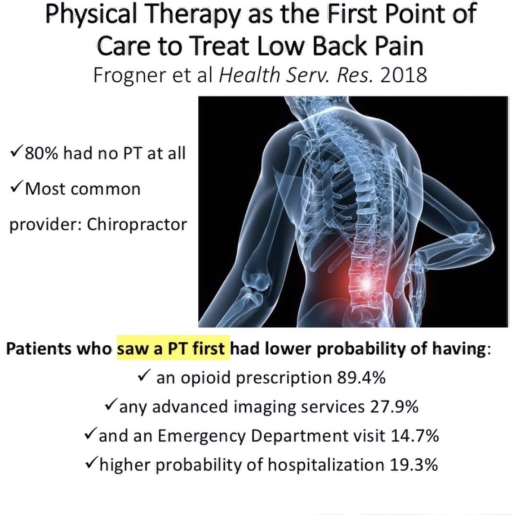
[ICYMI} Physical Therapy as the First Point of Care to Treat Low Back Pain: An Instrumental Variables Approach to Estimate Impact on Opioid Prescription, Health Care Utilization, and Costs. Frogner et al Health Serv. Res. 2018

The Week in Research Review, etc 12-24-18This study compared the differences in opioid prescription, health care utilization, and costs among patients with low back pain (LBP) who saw a physical therapist as the first point of care, at any time during the episode, or not at all.

Patients aged 18-64 years with a new primary diagnosis of LBP, living in the northwest United States, were observed over a 1-year period.

Patients who saw a PT first had:

  • a lower probability of having an opioid prescription (89.4 percent),
  • any advanced imaging services (27.9 percent),
  • and an Emergency Department visit (14.7 percent), yet 19.3 percent higher probability of hospitalization.

Interestingly enough, 80% of the patients in the sample had no PT at all. Furthermore, 8.7% saw a PT first and 11.5% saw a PT later (avg 38 days). The most common provider seen 1st was a chiropractor.


Assessing for Patella Instability

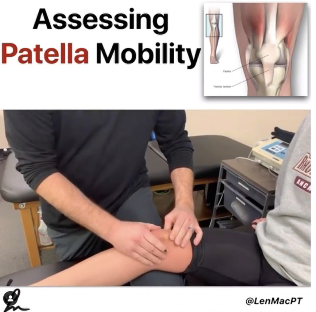
Assessing Patella Mobility

💥Assessing for Patella Hypermobility💥

This Instagram snippet shows how I assess a patient with suspected patella hypermobility who may have sustained a subluxation, dislocation or instability episode.

To see the full video at my YouTube Channel, click the link here!

Basically, we’re looking at how mobile the patella is when the knee is locked at full extension compared to when the knee is flexed to about 25 degrees.

Normally, the patella should become relatively stable when the knee is flexed to 25 degrees because it engages the trochlea groove.

In patients with underlying patella hyper-mobility, the amount of mobility with the knee slightly flexed will be similar to when the knee is in full extension (and not locked into the trochlea groove).

This is often the case when the patient’s trochlea groove is too shallow to offer bony stability.

The test should help the clinician gain a better understanding of the patient’s anatomical make-up and prognosis for the long term.

Check out the full video at my YouTube Channel.

The Week in Research Review, etc 11-5-18

The Week in Research Review, etc 11-5-18 was filled with more informative and eye-opening posts! Lots of visually stimulating posts to help clarify what exactly is going on in the hip joint with PROM. Another post that shows the suction effect from an intact hip labrum… amongst other great posts.  Just some great stuff..hope you enjoy!


  1. Manual Forearm Resistance Drills
  2. ACL Graft Healing Times to Maturation
  3. Hip Capsule Stress with PROM External Rotation
  4. Muscle Activation Affected by Hip Thrust Variation
  5. Hip Thrust Form by Bret Contreras
  6. Hip Joint Suction Affected by labral Status


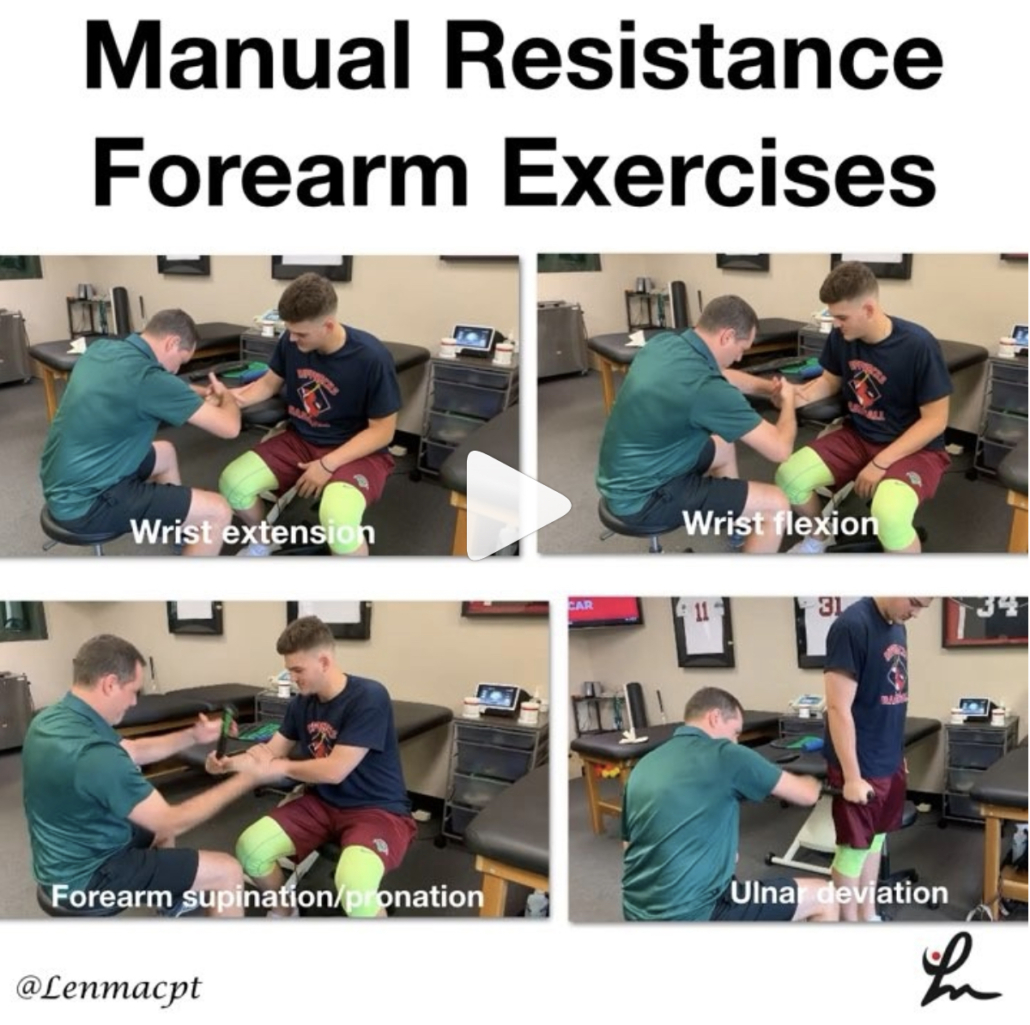
Manual Resistance Forearm Exercises

In this post, I wanted to show you guys some of the manual resistance drills we use @championptp on our shoulder and elbow clients, especially our baseball players. We love to use these drills because we can control so many variables with each athlete and tailor it for their specific needs.

We can control the speed and tempo, the direction of forces (eccentric, concentric), and the magnitude of the forces. Plus it’s a great way to interact with our clients. It’s also a great way to feel how well they’re progressing in their programs instead of just giving them dumbbells.

I have found these manual resistance drills to be very helpful with my overhead athletes and hope you give them a try on your clients soon! Let me know what you think or tag a friend below who may like to use these drills too.

In my course that I teach around the US, I try to include these concepts so you can practice and be able to utilize these drills for your clients…thanks!


ACL Graft Harvesting and Healing times

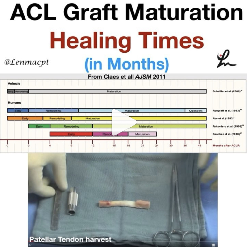
In this post, I wanted to show some research studies on graft healing times and why we need to respect tissue biology.

The systematic review from AJSM 2011 looked at ‘The ‘‘Ligamentization’’ Process in Anterior Cruciate Ligament Reconstruction.’

They essentially looked at 4 different biopsy studies on BPTB and Hamstring autograft reconstructions. They concluded that maturation of the graft, as determined by mainly vascularity and cellularity, was not complete until 12 months at the earliest. The healing time even extended to 24+ months as well.

The ligamentization endpoint is defined as the time point from which no further changes are witnessed in the remodeled grafts. The surgical procedure is quite involved, as you can see in the video that I took from @drlylecain on #YouTube.

As I’m rehabbing my clients, my decision making and post-op progressions often take into account:

✔️Healing biology

✔️Graft harvesting

✔️Graft Type

✔️Bone bruise presence (often!)

✔️Other concomitant issues (meniscus, articular cartilage).

So, respect the tissue and allow natural healing to occur before you add more exercises or are concerned that they’re not making the gains you’d expect.⠀


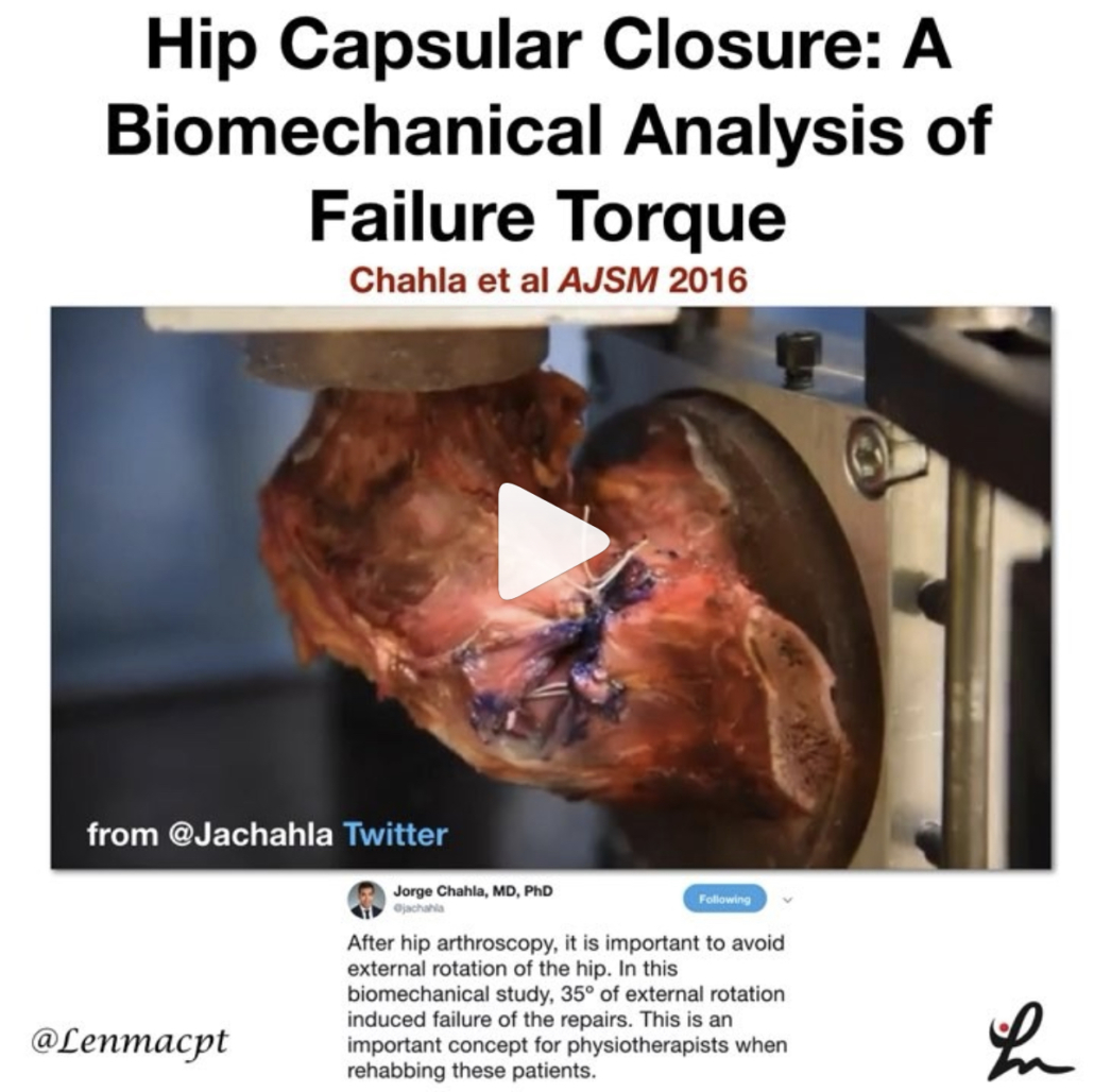
Hip Capsular Closure: A Biomechanical Analysis of Failure Torque

Chahla et al AJSM 2016

Interesting look at tissue failure, albeit in a cadaver graft, that should help to guide the physical therapist or ATC early in the rehab process after a hip scope.

The purpose of this study was to determine the failure torques of 1-, 2-, and 3-suture constructs for hip capsular closure to resist external rotation and extension.

The 3-suture construct withstood a significantly higher torque (91.7 Nm) than the 1-suture construct (67.4 Nm) but no significant difference was found between the 2- and 3- suture construct.

The hip external rotation degree in which the capsule failed was:

✅1-suture construct: 34 degrees

✅2-suture construct: 44.3 degrees

✅3-sutures: 30.3 degrees (yes, smaller than 2-suture construct)

I think as a #PT, we need to keep this study in mind and respect the healing tissues after a hip scope.

Love when we can get this information and put it into practice, similar to RTC repairs, ACL, etc.

Obviously, this was on a cadaver where there’s no guarding, pain or muscle contraction. We still need to know that there MAY be enough tension on the capsule to create potential issues (like tissue failure).

If you treat patients after hip scopes, then I recommend you read this cadaveric study.


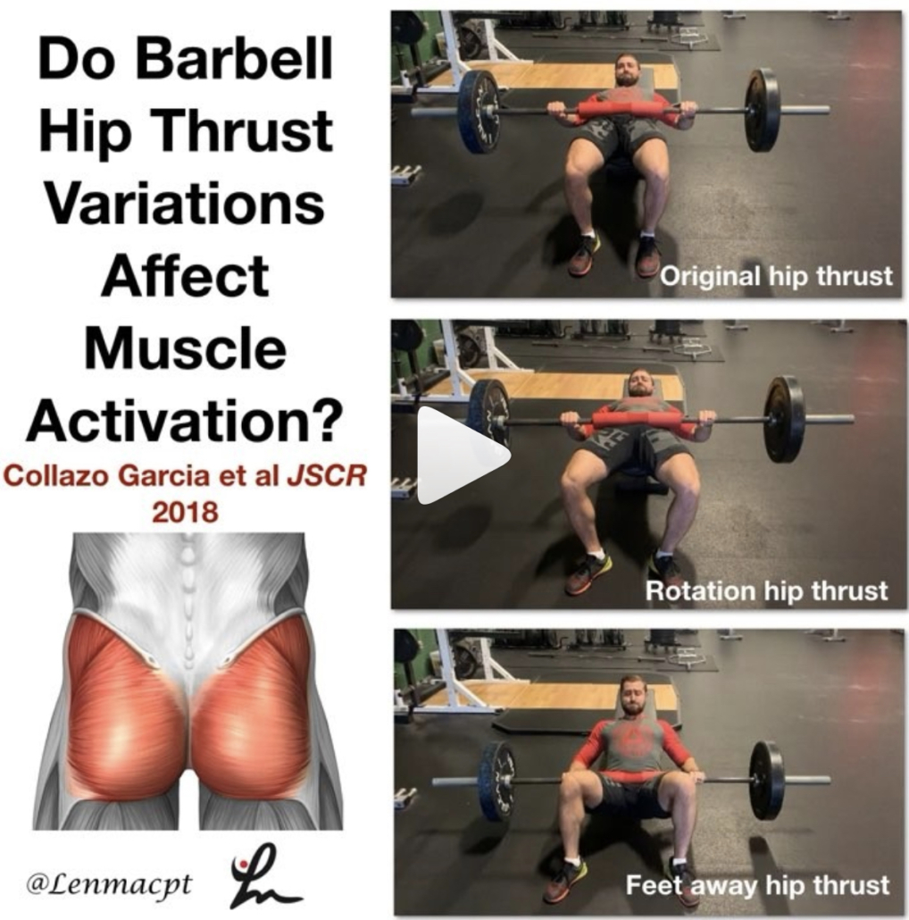
Barbell Hip Thrust Variations Affect Muscle Activation

COLLAZO GARCIA et al JSCR 2018

This study looked at the EMG activity of various lower body muscles while performing the hip thrust in various positions.

Their results showed that by varying the foot position into more external rotation, you can recruit the glute max and medius more than by the traditional hip thrust.⠀ …”the activity of the gluteus maximus increases significantly reaching up to 90% MVIC with only 40% of 1RM” with this hip ER variation.

Also, ‘when the distance between the feet is increased, the activity of knee flexors increases. Therefore, this is a very recommendable option to increase hamstring: quadriceps co-activation ratio.’

I like this study because it helps guide our rehab if we’re targeting a specific muscle group a bit more because of an injury or surgery.

It’s one of my go exercises for anyone with a lower body injury, especially after an ACL reconstruction. But I do use this exercise for most of my clients rehabbing from any injury, including the upper body.

It’s a great way to recruit the gluteus maximus and medius, which we know are hugely? (is that a word?) important to help produce and dissipate forces during athletic movements.

The exercise was widely researched by @bretcontreras1 and should be a staple in your rehab programs.

Check it out and add this to your go-to exercise list…thanks!


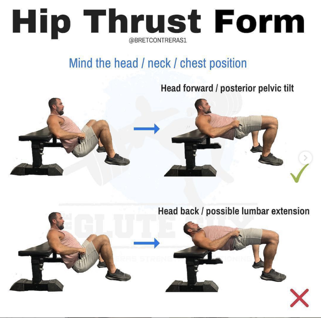
Hip Thrust Form

[REPOST] and a great one from @bretcontreras1 talking hip thrust form, which is perfectly coinciding with my post earlier today on variations to the hip thrust and how they affect muscle activation. Check out his original post below…highly recommended!

Teaching optimal hip thrust form is complicated. While the occasional lifter prefers and functions better staying fairly neutral in the head, neck, and spine, the vast majority of lifters do best maintaining a forward head position, which leads to ribs down and a posterior pelvic tilt.

It’s not just the forward eye gaze; the whole head has to maintain its forward position. You’re not hinging around the bench; the body mass above the bench stays relatively put, while the body mass below the bench is where the movement occurs.

The astute science geeks out there will rightfully point out that posterior pelvic tilt is associated with some lumbar flexion, and that lumbar flexion under load can be problematic. However, lumbar flexion is only dangerous when the discs are simultaneously subjected to compressive forces. With this style of hip thrust, the glutes are driving hip extension and posterior pelvic tilt, and erector spinae activation is greatly diminished. Core activation is what creates the bulk of the compressive forces, so with the erectors more “silenced,” the discs aren’t as compressed. This makes the exercise very safe. In fact, it’s safer than the “neutral” technique because as you rep to failure or go a bit too heavy, you will inevitably arch the chest and hyperextend the spine, which can lead to lower back pain. ⁣

We have 200 members at Glute Lab hip thrusting day in and day out, and there have been zero injuries to date. Considering how heavy we go, this is astounding.⠀
⁣⠀
#gluteguy #glutelab #thethrustisamust


Hip Joint Suction and Stability

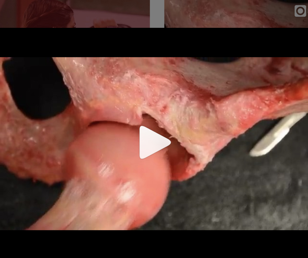
[REPOST] From @chicagosportsdoc and a very cool look at the suction within the hip joint that contributes to its stability. As the video progresses, they have simulated a labral tear that shows how easily the joint can dislocate. Once the labrum is repaired, the suction effect is recreated, and joint stability is re-established.

That’s 2 posts this week on the hip…if you want to see some awesome posts, then follow him. He just got on Instagram but his visual posts really aid in learning the mechanics of the various joints…see below!

An impressive demonstration of the powerful hip suction seal. When the hip labrum is injured, the seal is disrupted which can potentially produce microinstability. A labral reconstruction can restore the suction seal #labrum #sportsmedicine #hip #anatomy#orthopedicsurgery #medicine


The Week in Research Review, etc 10-15-18

This week I posted a lot of research and thoughts on shoulder and knee rehab, particularly after an ACL injury. I also shared some others posts that really complimented my posts so there’s some bonus reading to do too. Hope The Physical Therapy Week in Research Review helps your Monday patients and beyond! Take a read and share with your friends!


  1. Co-morbidities in the first 2 years after arthroscopic hip surgery: substantial increases in mental health disorders, chronic pain, substance abuse, and cardiometabolic conditions. Rhon et al BJSM 2018.⠀
  2. Range of Extension Correlates with Posterior Capsule Length after Knee Remobilization Zhou et al Med Sci Sports Exerc 2018⠀
  3. Sidelying External Rotation- The 1 exercise in all upper body programs
  4. @dr.jacob.harden talking Infraspinatus release.
  5. Do you account for Bone Bruises after an ACL
  6.  @cbutlersportspton bone bruises and the specifics
  7. When is it safe to initiate full AROM knee extension after an ACL-PTG autograft
  8. @mickhughes.physio on when it MAY be safe to initiate full knee extension from 90-0 after an ACL reconstruction.


Comorbidities after Hip Arthroscopy

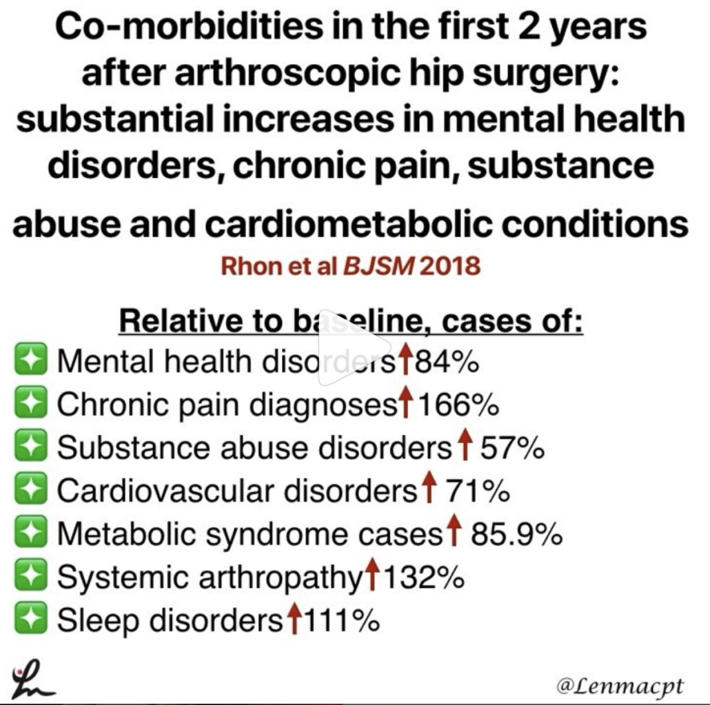
Co-morbidities in the first 2 years after arthroscopic hip surgery: substantial increases in mental health disorders, chronic pain, substance abuse and cardiometabolic conditions. Rhon et al BJSM 2018.

This is an interesting study on 1870 mainly US Military personnel between 2004-13 (~33% were not active duty).

Relative to baseline, cases of:

❇️mental health disorders rose 84%

❇️chronic pain diagnoses increased by 166%

❇️substance abuse disorders rose 57%

❇️cardiovascular disorders rose by 71%

❇️metabolic syndrome cases rose by 85.9%

❇️systemic arthropathy rose 132%

❇️sleep disorders rose 111%

The comorbidity with the greatest increase of new cases was that of mental health disorders (26% of the entire cohort). Age and socioeconomic status had significant associations on outcomes as well.

Just an eye-opening study that followed each subject 2 years after their respective surgeries. One giant variable that jumped out at me was that they used mainly military personnel only as the subjects.

We certainly can’t extrapolate on non-military personnel but need to keep this study in mind for others treating a similar cohort. Did the surgery cause these disorders? Absolutely not! No causation can be associated and that is very important!

What do you think about this study and how mainly military personnel and civilians that were tracked ending up developing many chronic disorders? I say it is very troubling! Let’s chat…and remember, this is not a causation study but just a reminder to educate and monitor your patients’ well-being after a surgery.
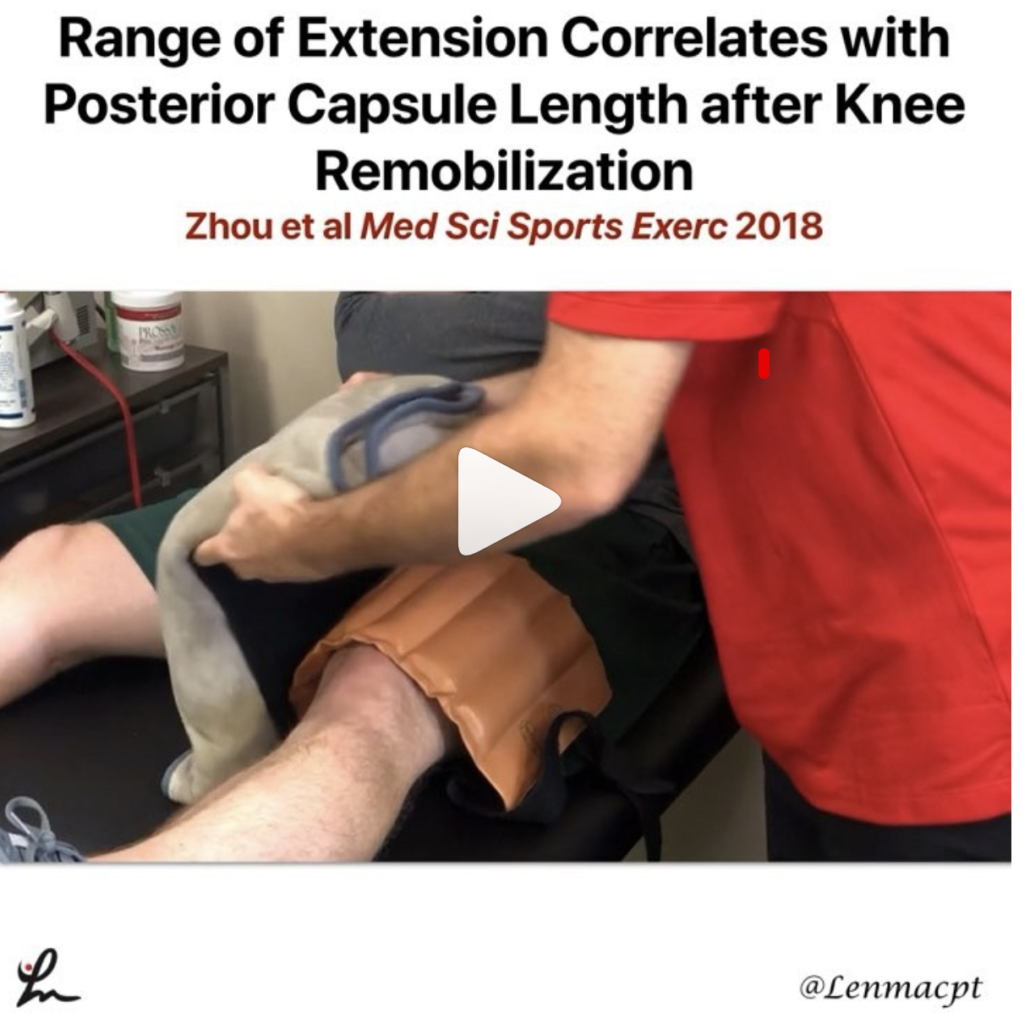


Posterior Capsule Limits Knee Extension after an ACL

Range of Extension Correlates with Posterior Capsule Length after Knee Remobilization Zhou et al Med Sci Sports Exerc 2018

This study is a confirmation bias for me because it showed that the knee’s posterior capsule limits extension after immobilization (in rats!) This is why I’m a huge proponent of low load long duration stretching of most joints when they begin to get stiff.

It seems as if the prolonged stretching is needed to regain collagen length and return the ROM. I know it’s in rats so calm down…but we need to get the data from somewhere.

Take it with a grain of salt but know that LLLD is going to be the best mode to return ROM (and not just hamstring stretching).⠀
.⠀
Do you agree? Do you treat rats with stiff knees? Then this study was created for you!
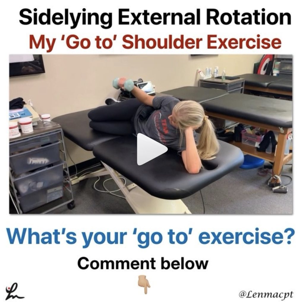


The Best Exercise for the Rotator Cuff

❗️Sidelying External Rotation- The 1 exercise in all upper body programs❗️

I really think this exercise should be in everyone’s program, whether going through rehab for a painful shoulder or a high level, healthy powerlifter. The role of the infraspinatus and other rotator cuff muscles is crucial to maintaining humeral head stability.

Sidelying external rotation has been shown to elicit the highest amount of EMG activity for the infraspinatus so I give this exercise to everyone, once there are no precautions for tissue healing. The infraspinatus and subscapularis (front rotator cuff muscle) are force couples that help to stabilize the humerus within the glenoid. Weakness of the infraspinatus may affect this force couple and create an inefficient movement within the joint.

My goal for all of my clients is to create an efficient movement that allows them to work at their highest level. The infraspinatus is a critical muscle of the shoulder complex so MOST of my programs include this exercise.
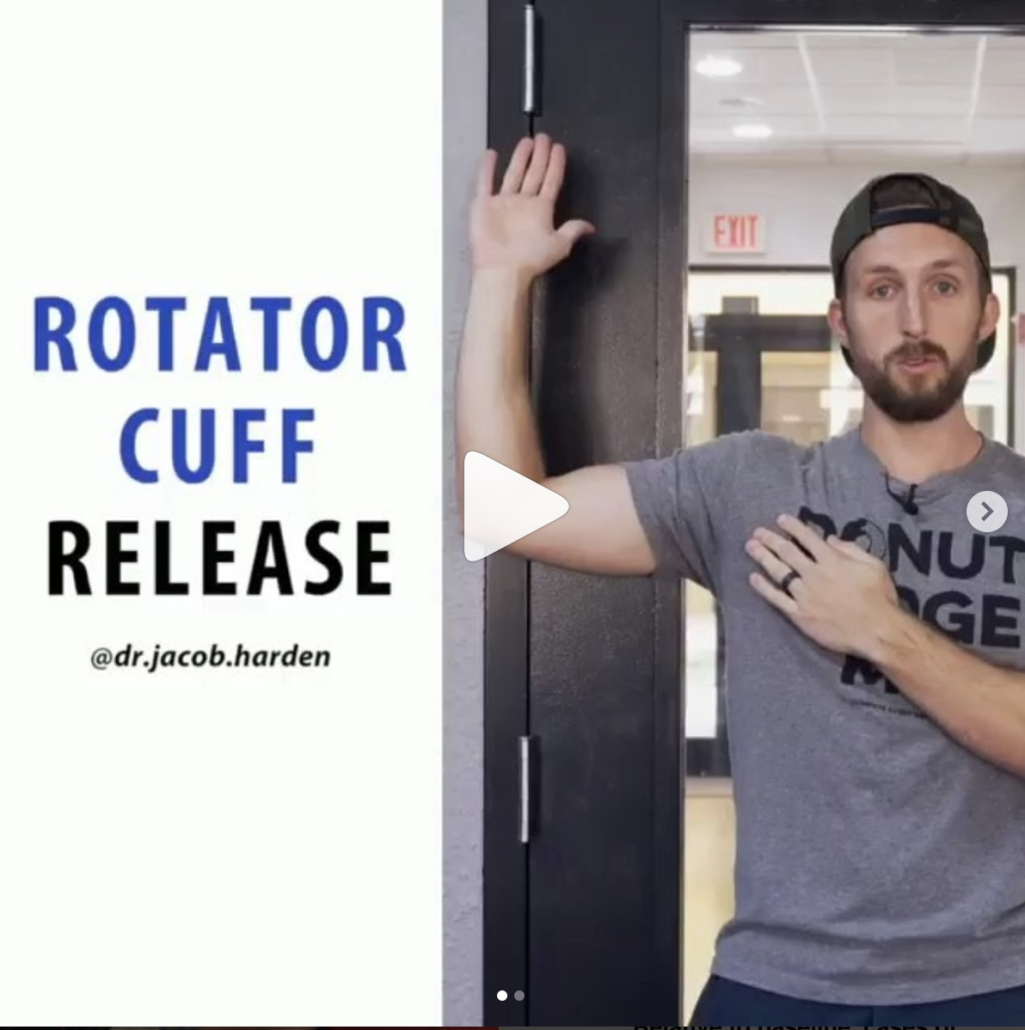


Myofascial Release of the Infraspinatus

Great post by @dr.jacob.harden talking Infraspinatus release. Perfect timing for my earlier post today looking at my go to exercise for the shoulder joint. Check his post out below!👉🏻 🔴 𝙃𝙊𝙒 𝙏𝙊 𝙍𝙀𝙇𝙀𝘼𝙎𝙀 𝙄𝙉𝙁𝙍𝘼𝙎𝙋𝙄𝙉𝘼𝙏𝙐𝙎

Coming at ya with a little #throwbackthursday since I’m about to jump on a plane across the pond to London. So we’re looking at how to do a pin and stretch for the rotator cuff, specifically the infraspinatus. The infraspinatus is the main external rotator of your shoulder, so it’s that muscle we see everyone working when they swing there 5 pound plates side to side in their warm-ups. (Side note: if you do that, please use a band or do it sidelying. Standing with plates does nothing but work the bicep.👍)

This can also help with some those little hypersensitive areas in the back of the shoulder. If you’re feeling those spots or having shoulder pain or just want to improve your internal rotation a bit, this release can help.

𝗛𝗲𝗿𝗲’𝘀 𝗵𝗼𝘄 𝘁𝗼 𝗱𝗼 𝗶𝘁:

🔹️Ball placement is below the spine of the scapula.

🔹️Internally rotate, flex, and adduct the shoulder

🔹️Work back and forth for a minute or so
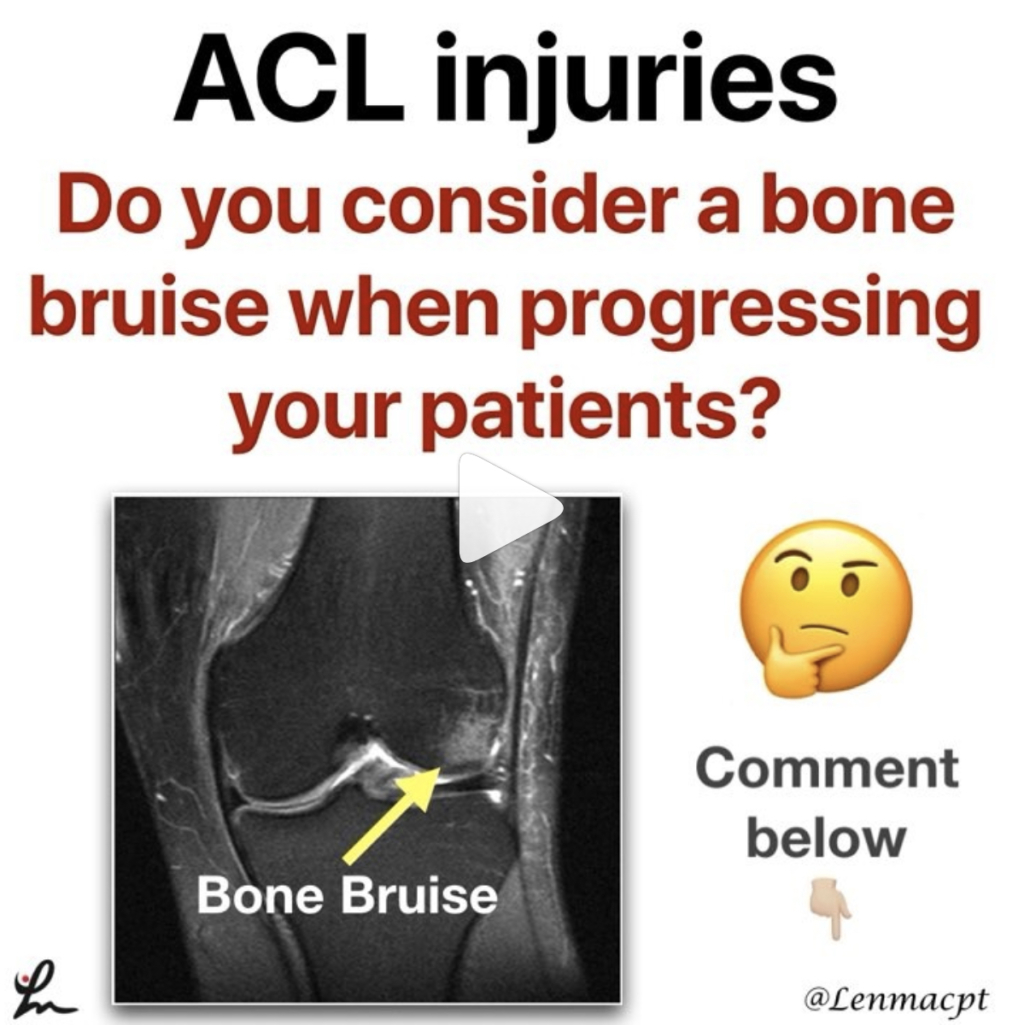


Bone Bruises after an ACL

Do you even consider a bone bruise after an ACL when progressing your patients? I know I certainly do and one of the major reasons why I have gone a bit slower with my latter stage progression, especially to impact activities like plyometrics and running.

There are a few studies that have shown the presence of a bone bruise after an ACL injury but we are not 100% certain this eventually leads to joint degradation.

Hanypsiak et al included 44 patients (82%) who underwent unilateral ACLR without multi-ligament involvement. Thirty-six (82%) patients had a bone bruise on index MRI. Potter et al reported all patients in their cohort sustained chondral damage at the time of injury.

Faber et al examined 23 patients with occult osteochondral lesion (bone bruise) who underwent ACLR. They found that at 6-year follow-up, a significant number of patients had evidence of cartilage thinning adjacent to the site of the initial osteochondral lesion (13/23 patients).

So as you can see, bone bruises are more common than most people think. This may be one reason why osteoarthritis rates are much higher in ACL reconstructed knees.

Additional factors, such as cartilage and meniscus injury, associated with ACL rupture may play an important role in subsequent outcomes following surgical reconstruction independent of a bone bruise.

Do you consider a bone bruise when progressing your patients back from a knee injury like an ACL reconstruction?
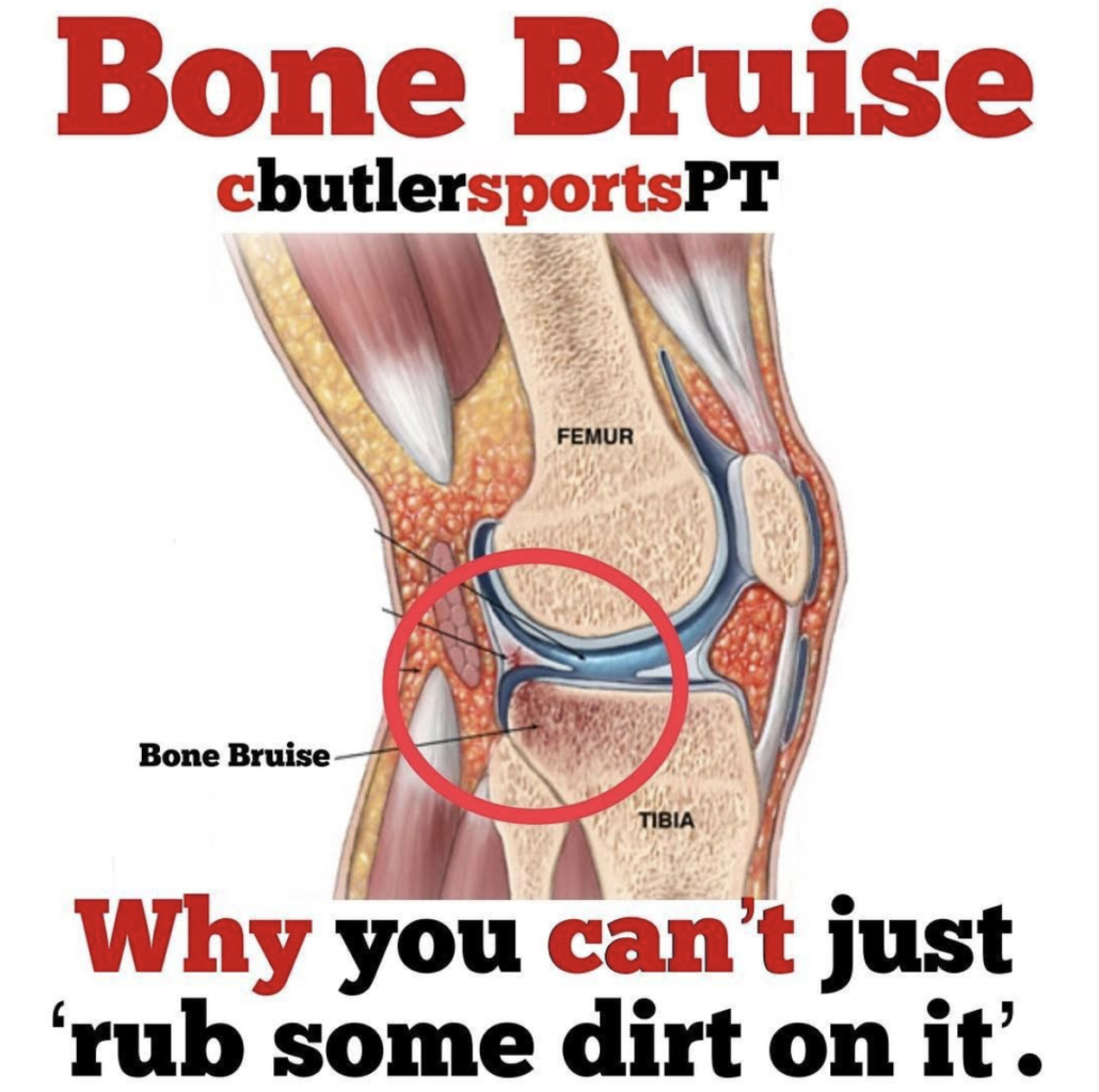


Types of Bone Bruises after an ACL Injury

@cbutlersportspton bone bruises, which fits perfectly with my post earlier today. He talks about the 3 different types of common bone bruises…check it out below!

❗️What is a Bone Bruise❗️We often hear that one of our Fantasy Football players has a Bone Bruise and may be out for a few weeks.

It sounds like something that an NFL athlete should be able to tough out, right?

Here’s why you may need to put in a backup for a few games.

A bone bruise occurs when several trabeculae in the bone are broken, whereas a fracture occurs when all the trabeculae in one area have broken. Trabecular bone is also known as spongy bone.

—-Three Types of Bone Bruises—-⠀
1️⃣Subperiosteal hematoma: A bruise that occurs due to an impact on the periosteum that leads to pooling of blood in the region.⠀
2️⃣Intraosseous Bruising: The bruise occurs in the bone marrow and is due to high impact stress on the bone.⠀
3️⃣Subchondral Bruise: This bruise is bleeding between cartilage and bone such as in a joint.

—-Symptoms of Bone Bruises—-

•Pain and tenderness in the region of injury

•Swelling in the region of injury

•Skin discoloration in the region of injury

Bone bruises often occur with joint injuries, such as ankle sprains and ACL tears, therefore a bone bruise can also coincide with stiffness and swelling in the joint.⠀
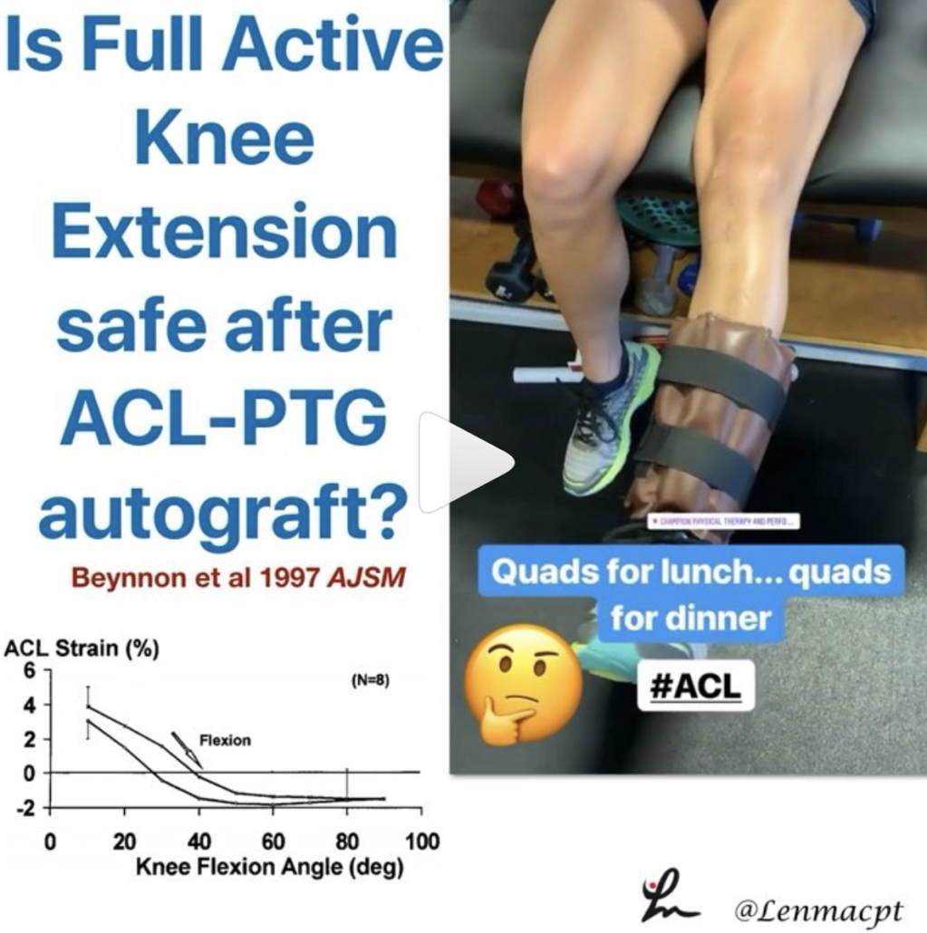


When is it safe to initiate full AROM knee extension after an ACL-PTG autograft?

I posted this video in my the other day and had a ton of people message me about the exercise.

Most people wanted to know how far out of surgery the patient was and when I felt it was safe to begin full, active knee extension after an ACL.

I’ve always been relatively conservative with my rehab (at least I think so) but I wanted to dig a little deeper. I recently saw a post by @mickhughes.physio and he was talking about the Fukuda et al study from 2013.

The study looked at 90-40 knee extensions and ‘ACLR patients can perform 3×10 at a 70% 1RM load through a restricted 45-90deg ROM between weeks 4-12 post-op, and then the same load full ROM from 12 weeks post-op. ‘

It made me dive a bit deeper and I went to my trusty Beynnon et al AJSM studies from the late 90’s. You can see the strain on the ACL decreases as we approach 40 degrees and stays low out to 90 degrees…but is 3-4% strain on the ligament significant?

If you look at the study (yes, it’s only on 8 subjects) you’ll see a similar strain curve for closed chain exercises as well…but we do mini squats immediately after surgery without 2nd guessing!

In 2011, Beynnon et al AJSM showed that an accelerated program that initiated full resisted knee extension (90-0) at 4 weeks showed similar knee laxity throughout the study. The other group initiated full resisted knee extension at 12 weeks. Also, those who underwent accelerated rehabilitation experienced a significant improvement in thigh muscle strength at the 3-month follow-up.

So, what do we do with this data? I have begun to do full, resisted knee extensions with my patients between 4-6 weeks post-op, as long as it’s a patella tendon autograft. For allografts or HS autografts, I tend to delay it a bit longer because of the soft tissue healing that is delayed.

What do you think? When do you initiate full AROM after an ACL? Do you know of a study that definitively says the strain on the ACL graft is detrimental to the healing ligament?
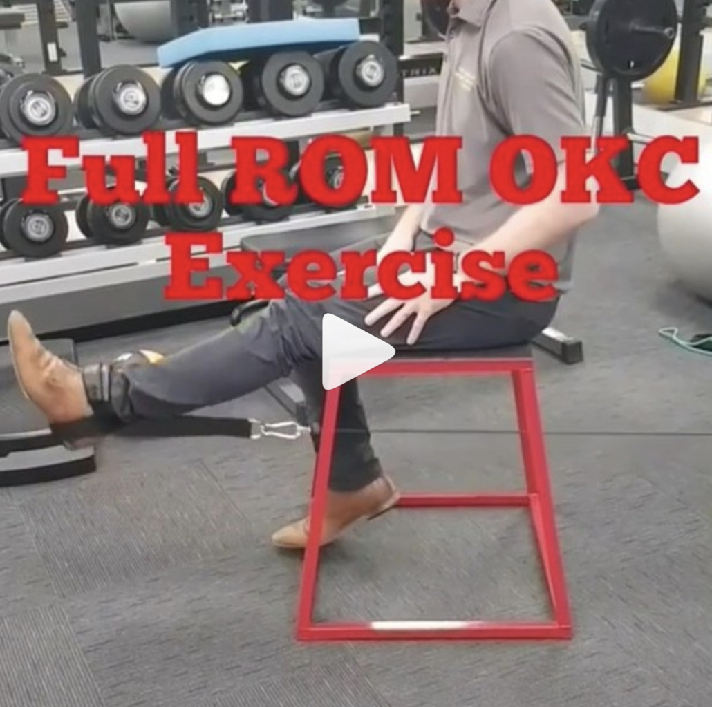


How much Resistance Should we Recommend Open Chain Exercises After an ACL

This is the post from @mickhughes.physio that made me dive a bit deeper into the research on when it MAY be safe to initiate full knee extension from 90-0 after an ACL reconstruction. Check out his post below! ⠀
____________________

So if we can safely perform OKC exercises (knee extensions) as part of ACLR rehab; how heavy can lift?⠀
*⠀
*

This is a question I often get asked. Based on the work by Fukuda et al (2013), ACLR patients can perform 3×10 at a 70% 1RM load through a restricted 45-90deg ROM between weeks 4-12 post-op, and then the same load full ROM from 12 weeks post-op. *⠀
*⠀

From then you can progressively load as per what can be tolerated. Usually the first sign that the knee is unhappy with the load is that the underneath the kneecap will be sore/painful. That’s a sign you need to back the load off a little so the exercise is felt in the quads only. *

If you’re still unsure about OKC exercises (knee extensions) during ACLR rehab read my blog by clicking on the link in my bio ⠀
#ACL #Physio #Knee #Rehab


The Week in Research Review, etc 8-5-18

The Week in Research Review, etc 8-5-18 we discuss a wide variety of topics including:

  • Long-term disability if weak during adolescence
  • Using heat during rehabilitation
  • OKC vs CKC exercises after an ACL
  • Live look at an Achilles rupture (with sound too!)
  • A fun look at the different types of PT’s
  • Congrats to all of the newly licensed PT’s!

Muscular weakness in adolescence is associated with disability 30 years later: a population-based cohort study of 1.2 million Swedish men. Henriksson et al BJSM June 2018.

Conclusion: There was a strong association between muscular weakness and disability. A combination of muscular weakness and low aerobic fitness was an especially important risk factor for disability. This adds weight to call for muscular strength and fitness-enhancing exercise for adolescents in all BMI categories.

This study out of Sweden looked at the associations of muscular strength in adolescence with later disability pension.

A total of 1 212 503 adolescent males aged 16–19 years, recruited from the Swedish military conscription register between 1969 and 1994.

Moral of the story: exercise as an adolescent may help to reduce issues later in life, including the potential for disability.

Taking it 1 step further… why is physical education being cut out of school requirements when studies like this show the potential negative effects of inactivity?!


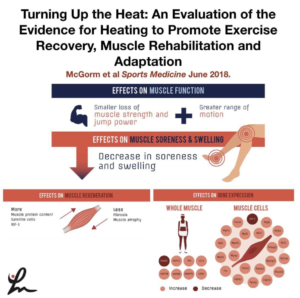
Turning Up the Heat: An Evaluation of the Evidence for Heating to Promote Exercise Recovery, Muscle Rehabilitation, and Adaptation McGorm et al Sports Medicine June 2018.

Key Points: Animal and human trials have shown that various forms of heating can be used in conjunction with exercise or stress to enhance recovery, adaptation and limit muscle atrophy.

Heating muscle activates protective mechanisms, reduces oxidative stress and inflammation, and stimulates genes and proteins involved in muscle hypertrophy.

Further studies highlighting the differences between various heating modalities will help inform athletes and coaches on the best heating practices for specific situations.

This article has a ton of great information that I highly recommend any PT, strength coach, athletic trainer or massage therapist.

It is a review of the literature and there are still many questions to be answered so, as always, take with a grain of salt.

I am a fan of heating before treatment…I do it daily with 99% of the clients that I see and they love it…so that says something.

What do you think? Do you like to heat your clients up before treatment or before a workout? Tag a friend that may benefit from this post! Thanks, guys!


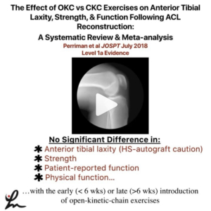
The Effect of Open- Versus Closed-Kinetic-Chain Exercises on Anterior Tibial Laxity, Strength, and Function Following Anterior Cruciate Ligament Reconstruction: A Systematic Review and Meta-analysis Perriman et al JOSPT July 2018 Level 1a

FINDINGS: There was no significant difference in anterior tibial laxity, strength, patient-reported function, or physical function with the early or late introduction of open-kinetic-chain exercises in those who have had anterior cruciate ligament reconstruction, when compared to closed-kinetic-chain exercises, at all follow-up time points.

They wanted to determine whether OKC quadriceps exercises result in differences in anterior laxity, when compared to CKC exercises, at any time point following ACLr.

Also, they wanted to determine whether there are differences in strength, function, quality of life, and adverse events with OKC quadriceps exercises when compared to CKC exercises at any time point.

Overall, calculated effect sizes showed a slight increased laxity in the OKC groups, particularly for the hamstrings graft. The⠀
pooled difference was not statistically significant (P>.05)

When considering all graft types, there was low- to moderate-quality evidence from 3 studies suggesting that there were no between-group differences in laxity at any time point when OKC exercises were introduced earlier than 6 weeks post ACLR, compared to CKC exercises.

There seemed to be a trend that showed early OKC knee extension was safer after a PTG than a hamstring autograft but protocols varied so data was inconsistent.

Of interest to me, they said “The early introduction of OKC quadriceps exercises did not appear to offer additional significant benefits in function and strength for the average patient post ACLR; therefore, this early introduction is questionable, especially in patients with a hamstring graft.


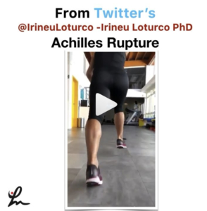
From Twitter’s @IrineuLoturco showing the moment this athlete ruptured their achilles tendon. Pretty impressive and you can see the eccentric loading of the tendon that caused the rupture. See his original post below…

A very impressive recording of the exact moment when an elite sprinter had an acute and complete rupture of the Achilles tendon. Pay attention to the “boom”.


[REPOST] If there was an ESPY for a post by a PT then @theperformancedoc would definitely get it for these videos! Great job and keep pumping out great content. Give him a follow if you haven’t already!

👇🏻
💥Different Types of Physical Therapists In the Real World💥 SWIPE 👉🏽 (Turn on 🔊) Sometimes we have trouble “turning it off” when we are outside of the clinic. Which one are you?! Tag, Comment, & Share with a Physical Therapist‼️

▪️
#ThePerformanceDoc #RehabWithTheDoc
#TeamMovement


Congrats to all of the newly licensed PT’s out there…Welcome to the profession!

My advice to you:

Stay humble and put the patient first, always

Keep learning and try to avoid complacency

The road will seem rough but it does get a little easier. Get experience…as much as you can. Each interaction with a patient is a snapshot to help guide your future interactions.

Put yourself in their shoes… give them the best experience as you would expect to receive if you were sitting on the plinth being asked questions.

Follow people on social media that help you to learn and keep an open mind. Don’t get pulled into 1 system. Take a little from each and package it nicely.

The research is often biased. Opinions come and go. Stay somewhere in the middle…remember the bell curve, always!

Each “system” has huge overlap despite their ‘trademarked’ proprietary information…they all involve motion and strengthening. That’s the key to PT- keep people moving and keep them stronger…or at least keep them positive and hopeful.

I recently wrote a blog post discussing the evolution of a PT. Take a look…the link is in my bio on Instagram.

These words are the basis of my practice. Take what you think is important and apply it to your practice. Good luck, now work on your dives!


A great week of content that I hope you found valuable and willing to share with your friends and colleagues! Thanks for reading!