Tag Archive for: anterior knee pain

The Week in Research Review, etc 12-10-18


This week we’re still playing with formats and learning these Instagram changes. With that, in the week in research review 12-10-18, we discussed many topics that I wanted to share!


Surgery vs Physical Therapy for Carpal Tunnel Syndrome

Cost-Effectiveness Evaluation of Manual Physical Therapy Versus Surgery for Carpal Tunnel Syndrome: Evidence From a Randomized Clinical Trial Fernández-de-las-Peñas et al JOSPT 2018.

This Level 1b study looked to evaluate cost-effectiveness differences of manual physical therapy versus surgery in women with carpal tunnel syndrome (CTS).

Performed in Spain, 120 women with a clinical and electromyographic diagnosis of CTS were randomized through concealed allocation to either manual physical therapy or surgery.

They concluded that manual PT including desensitization maneuvers of the central nervous system has found to be equally effective but less costly, i.e., more cost-effective than surgery for women with CTS.

From a cost-benefit perspective, the proposed manual PT intervention of CTS can be considered.

Interesting results but 2 obvious limitations to this study:
1️⃣No control group. What if the symptoms could spontaneously improve over time
2️⃣ They only looked at 1-year improvement and not short-term improvements. I would’ve liked to have seen 3 months and 6 months results as well to see the acute effects.

Not sure what to make of this study but it does seem as if a population of Spanish women may respond to Rx of CTS without surgical intervention.

This could be a huge cost/time saver for society!


Return to Sport Criteria and Reinjury Rates

The Association Between Passing Return-to-Sport Criteria and Second ACL Injury Risk: A Systematic Review With Meta-Analysis  Losciale et al JOSPT 2018.

Not going to lie, this study caught my attention because the results match my confirmation bias.⠀

I’ve been saying for years that hop tests, even combined with other tests, just don’t cut it.⠀

I wrote a blog post about this too for @mikereinold. This study, although with its limitations, did show that passing RTS criteria did not show a statistically significant association with risk of a second ACL injury. 

This review also determined that 12% of those who failed RTS testing suffered a graft injury, compared to 5.9% of patients who passed. 

It seems as if quadriceps strength measured via isokinetic testing or isometric testing may be an important factor to consider for RTS decision making.


Also, hamstring-quadriceps strength ratio symmetry should also be considered.

So with this review demonstrating that current objective criteria-based RTS decisions did not show an association with the risk of a second ACLI, how does this affect your practice?


Physical Therapy vs Knee Scope for Meniscus Tears

💥PT vs Scope for Meniscus Tear 💥
.
Effect of Early Surgery vs Physical Therapy on Knee Function Among Patients With Non-obstructive Meniscal Tears: The ESCAPE Randomized Clinical Trial. van de Graaf VA et al JAMA Oct. 2018

Among patients with non-obstructive meniscal tears, PT was equal to arthroscopy for improving patient-reported knee function over a 24-month follow-up period. 

They went on to say that “Based on these results, PT may be considered an alternative to surgery for patients with non-obstructive meniscal tears.”

So basically, if there’s no bucket handle tear present that may be blocking joint range of motion, then it is highly encouraged that the patient #GetPT1st and not do surgery.

Without going out on a limb, I’d say this is a much more cheaper treatment option as well and would save society many costs associated with the surgery and lost time from work.

I do note a couple limitations: the surgical group did not get PT after surgery if they did ‘as expected’ but they could get PT to help improve their symptoms.

The PT group did pretty basic exercises although leg press, lunges, and balance type exercises were included.

Have you read this paper? It was a multi-center, randomized controlled trial performed in 9 hospitals in the Netherlands.

So, are we encouraged or surprised? Let me know by commenting below…thanks!


Shoulder Health Accessory Exercises

by @kieferlammi

Want strong and healthy shoulders!?

Shoulder strength is about more than pushing big lifts like strict pressing, push pressing, etc.

If you want a robust, healthy, well moving shoulder you should be including lower level drills that more specifically address scapular and RTC strength and control. –

I will always be a fan of traditional exercises like side like ERs, Prone Ys, Ts, etc.

Lately, I’ve thrown in more band work because it’s easy for me to do for higher volumes on a frequent basis and I enjoy the constant tension that the band provides. 

Give these two exercises a try:

✅ Band Front Raise Pull-Apart

✅ Band Overhead Y Raise

I find that these two do a great job of targeting my mid back and posterior shoulder without much compensation through a big range of motion.

Give them a try either in a warm-up for 1-3 sets of 10-15 reps or at the end of a training session for 2-4 sets of 10-25 reps depending on the difficulty of your band and your capacity. 


Should we Brace after an ACL Surgery?

View this post on Instagram

[NEW BLOG POST]⠀ 💥Knee Bracing Immediately After an ACL Reconstruction 💥⠀ In this post, I review some recent Twitter, Facebook and Instagram discussions about the usage of a hinged knee brace after an ACL surgery.⠀ .⠀ You'd be surprised what I found in the literature and the differences that exist throughout the US and the world.⠀ .⠀ Go to my website <LINK in my BIO> and read/share with your friends/colleagues.⠀ .⠀ It was a bit eye-opening and I'm curious to hear what others have to say.⠀ .⠀ I still like to recommend a brace for my clients because it seems to give them an added security after a pretty painful surgery.⠀ .⠀ I typically keep them braced 4-6 weeks, depending on their quadriceps activity and if they can do an active straight leg raise without a lag.⠀ .⠀ Many others don't even bother bracing at anytime post-op, which was surprising.⠀ .⠀ What do you think? Read the blog post and let me know. Let's try to educate and come to a better consensus...thanks!⠀ .⠀ #kneerehab #knee #kneepain #kneesurgery #acl #aclsurgery #ROM #physio #physiotherapist #crossfit #exercise #deadlift #physiotherapy #physicaltherapy #physicaltherapist #athletictraining #athletictrainer #ATC #PT #teamchampion #dptstudent #lenmacpt #instagram

A post shared by Lenny Macrina MSPT, SCS, CSCS (@lenmacpt) on

You can also get to the blog post by clicking this link

Let me know what you think about this new blog post or any of my social media posts…thanks!

The Week in Research Review, etc 12-3-18

Hey everyone,  The Week in Research Review, etc for this week has a new look, compliments of Instagram’s new algorithm. Hope the new format doesn’t throw you too big of a curveball (maybe you’ll like it better), so here goes…


ACL Injury Rates Higher on Synthetic Turf than Natural Grass in the NFL

Preventing low back pain by @joegambinodpt

Female Soccer Players have a 5x Increased Risk of a Second ACL injury

Anatomy of the Proximal Humerus


ACL Injury Rates Higher on Synthetic Turf than Natural Grass in the NFL


View this post on Instagram


A post shared by Lenny Macrina MSPT, SCS, CSCS (@lenmacpt) on

Preventing low back pain by @joegambinodpt


View this post on Instagram


A post shared by Lenny Macrina MSPT, SCS, CSCS (@lenmacpt) on

Female Soccer Players have a 5x Increased Risk of a Second ACL injury


View this post on Instagram


A post shared by Lenny Macrina MSPT, SCS, CSCS (@lenmacpt) on

Anatomy of the Proximal Humerus


View this post on Instagram


A post shared by Lenny Macrina MSPT, SCS, CSCS (@lenmacpt) on

The Week in Research Review, etc 11-26-18

This week, I discussed the progression of someone after a knee surgery. I tried to highlight the key stages and some techniques that I like to use to advance the patient’s mobility and comfort. Take a look at The Week in Research Review, etc 11-26-18 and share with your friends. Hope it helps you improve your patient care tomorrow and beyond!


ACL Reconstruction in a Pediatric and Adolescent Population

1st Day of #PT after an ACL Surgery

Patella Mobilization after Knee Surgery

Knee Flexion PROM after Surgery- Seated or Supine?

Stretching the Quads after Knee Surgery

Assessing for a Cyclops Lesion after an ACL

Assessing for Fat Pad Irritation of the Knee


ACL Reconstruction in a Pediatric and Adolescent Population

17 Year Follow-up After Meniscal Repair With Concomitant ACL Reconstruction in a Pediatric and Adolescent Population. Tagliero et al AJSM 2018

Results: 28% failed meniscal repair and required repeat surgery at the time of final follow-up. They also showed that outcomes and failures rates were comparable across tear complexity.

Guess that means that no matter the tear type, there was no difference in outcomes or retear rates. Although the repair techniques are now outdated and no longer used.

Their study also showed a 30% failure rate for meniscal tear repaired in the medial compartment at index surgery and 7% in the lateral compartment.

Interesting long-term outcomes that may help to guide your rehab and client advancement (and prognosis). Keep these in mind when you treat a future adolescent or pediatric ACL patient.


💥1st Day of #PT after an ACL Surgery 💥

If you have never treated a post-op ACL, then this video should interest you!

This is what the knee looks like that 1st day after surgery and can often set the stage for what’s to come over the next 6-12 months.

Often, the patient is both very curious and ultra-grossed out by the 1st unveiling. It can be stressful for them to see their knee in this condition so you really have to confidently reassure them that it is very normal.

The blood-soaked gauze is mainly saline that was used to irrigate the knee during the reconstruction. Some still leaks out of the incisions the 1st few days and can often be confused with true blood.

Understand that this is quite normal and happens to most every ACL patient’s knee that I’ve seen…nothing to worry about!

From here, I’d work on patella mobility (see the post later today) and then work on flexion ROM at the end of the table.

Again, it’s very important to get the knee moving after surgery. This will help with pain, swelling and gain confidence that the rehab process is moving forward.


Patella Mobilization after Knee Surgery

Get the patella moving early with #patella mobilizations immediately after surgery. One major reason (amongst many others) why we need to get our clients into #PT early.

I am certainly a very loud advocate for early PT and getting the patella moving can help to prevent excessive scarring, which can affect ROM and quadriceps force output.

Glove up and get that patella moving in all directions… medial, lateral, superior, inferior!


🤔Knee Flexion PROM after Surgery- Seated or Supine? 🤔

I’ve treated many patients after an ACL I can honestly say that this may be a huge influence on the early ROM outcomes that you may see.

I’ve tried to bend the knee in both supine or seated, as the video shows, and there’s no doubt that most people tolerate the seated version so much better after a knee surgery. In particular, a big surgery like an ACL, TKA or MPFL reconstruction.

It just seems to be more comfortable and with less stress on the anterior knee because of the position of the tibia (at least I think so!).

My theory, it seems as if the supine position may cause a slight posterior sag which may cause more pain and guarding than when they’re seated at the edge of the table.

I use a similar concept later on in the rehab process when I’m initiating my prone quad stretching. You can see a definitive improvement when I wedge my hand in the popliteal fossa and create a slight anterior translation on the tibia.

Most people say that the anterior knee pain that they were feeling (and not a quad stretch) was replaced by a stretch feeling only and no more anterior knee pain.

Try it out with your ACL patients and see what position they like best…I’ll bet I can covert you over if you still bend your knee patients in supine!


💥Stretching the Quads after Knee Surgery 💥

Continuing my sequence of videos after a knee surgery, I discussed my technique for progressing knee flexion PROM once they hit 120 degrees or so of flexion.

At this point, they’ve probably maxed out how much ROM they can achieve at the edge of the table. They’re ready to get that end range of motion and even some quadriceps flexibility.

In prone, most people will often feel a pain or pressure in the front of their knee when you try to bend it.

To overcome this, I like to wedge my hand into the back of the knee and give an anteriorly directed force through the gastrocnemius (calf) soft tissue and into the tibia.

This seems to create just enough movement of the tibia on the femur to take the pressure off the front of the knee. This may redirect the forces more onto the quadriceps muscle.

You’ll need to play with the amount and direction of force but most often they’ll begin to feel a better quad stretch.

Try this technique out on your next knee surgery client and see if it helps them. I usually initiate this ~4 weeks after an ACL but timeframes will vary person to person.⠀


💥Assessing for a Cyclops Lesion after an ACL 💥

In this video snippet from my YouTube Channel, I discuss how to assess for a Cyclops lesion in a knee. In particular, after knee surgery.

A patient with a potential cyclops lesion, they often present with loss of normal knee extension compared to the other side. They’ll often have anterior knee pain and poor patella mobility. Sometimes a tight feeling in their hamstrings and calves, too.

No matter how they try to regain their extension ROM, the knee just never feels right. Often times, surgical intervention is needed to remove that scar tissue.

Immediate rehab should continue to work on knee extension ROM using low load long duration stretching and aggressive patella mob’s.

No one’s to blame if this occurs. We don’t know exactly why it occurs in some people but we believe a remnant of the ACL stump may be a source of the frustrating issue.


💥Assessing for Fat Pad Irritation of the Knee 💥

Anterior knee pain is very common in the outpatient #PhysicalTherapy setting.

One of my go-to tests to assess for fat pad irritation is simply trying to capture the fatty tissue in the anterior aspect of the knee joint during active and/or passive ROM.

In this snippet from my YouTube channel, you can see that I pinch the fat pads on either side of the patella tendon as @corrine_evelyn is actively extending her knee. I’ll also do it in a relaxed state to assess passive irritability.

I 1st learned this test from @wilk_kevin and it continues to be a mainstay in my knee examination algorithm.

As for a treatment, it usually comes down to a volume issue and/or strength issue or both.

I’ll usually have to address the volume of the activity by relatively easing off of the activity while simultaneously adding in exercises to address an underlying weakness.

Remember the Dye et al study in AJSM 1998 when he talked about the fat pads being super painful during his arthroscopic surgery without anesthesia. Makes sense why they can be so painful if the knee stresses fall upon this tissue.

We talk about this study, fat pad irritability and much much more in our online knee seminar course.


If you want to learn more about how I treat ACL’s or the knee in general, then you can check out our all online knee seminar at www.onlinekneeseminar.com and let me know what you think.

We cover the anatomy, rehab prescription, ACL, meniscal injuries knee replacements and patellofemoral issues. Furthermore, the course covers both the non-operative and post-operative treatment.t

This is an awesome course if you’re interested in learning more about rehabilitating the knee joint. And if you’re a PT, there’s a good chance you can get CEU’s as well.

The Week in Research Review, etc 11-19-18

Great ‘Week in Research Review, etc 11-19-18’ that I hope you find helpful to your practice.

I’ve always touted the importance of the subjective portion of the exam so I wanted to share a slide from a recent talk I gave to a group in Canandaigua, NY. Obviously, the squat is a fundamental movement and I wanted to give some basic positions that I use to help assess. So excited that I’ve launched a brand new Medbridge course that helps the rehab specialist better eval and treat the baseball pitcher. On my YouTube channel, I discussed my thoughts on setting the scapula with various upper and lower body exercises. And finally, my co-worker Kiefer Lammi discusses the landmine with exercise.


Importance of the Subjective Exam

Assessing the Squat

My New Baseball Medbridge Course

Set the Scapula with Shoulder Exercises?

6 Ways to use the Landmine by @kieferlammi


💥Subjective the most important aspect of the Evaluation💥

This slide, taken from this past weekend’s course in Canandaigua, NY is always a favorite of mine.

I try to keep a slide like this in all of my lectures because I have found that this portion of the examination can give the rehab specialist a huge look into what is going on with the person in front of them.

Don’t get me wrong, I still consider the biomechanical aspect of what may be causing their symptoms.

It often comes down to a tissue capacity issue but it’s up to me to determine the appropriate course of treatment.

These questions will help build confidence in your client and guide the early stages of rehab.

Do you have any specific questions that you like to ask your clients during their 1st few sessions? Remember, these questions are just not for the evaluation. You should be asking these questions periodically to gauge progress and help guide the next phases of rehab, too!


🔅Assessing the Squat 🔅

Squatting is a fundamental movement that all of us have to do on a daily basis.

Utilizing several different positions can help the rehab specialist better assess the squat and develop a treatment plan that enables their client the ability to improve their squat pattern.

In the above videos, I have utilized 3 different squat patterns and will outline them by the degree of difficulty.

✅The Overhead Squat- by far the most challenging version which challenges the shoulders, thoracic spine, lumbar spine, pelvis, knee and ankles.

A movement limitation at any of these joints will most likely cause the squat pattern to break down. Using overhead resistance would further challenge the system and potentially cause the squat to further breakdown.

✅Arms Crossed Chest Squat- alters the challenge by taking most of the shoulder and thoracic spine out of the equation and isolates the motions to the lumbar spine, hips, knees and ankles.

I often use this position as my fundamental motion because most people don’t have to squat with any weights over their head. This position, in my opinion, should be the most informational and utilized.

✅Counter-weight Squat

This position changes the center of mass by moving some of the weight distribution more anteriorly (front) and making the squat motion slightly easier. I use this position as a regression, for some, which allows them to squat with less stress and potential difficulty.

There are many other variations to the squat that you can make but I wanted to highlight a few of the major changes that you cause successfully. Assessing the squat is essential and can give the rehab specialist a nice picture of the function of multiple joints during a common movement.


My BRAND NEW course on Medbridge’s platform

…that helps the sports and ortho rehab specialist (PT, OT, ATC) better understand the anatomy and biomechanics involved in the baseball pitching motion.

Advanced Rehab for the Baseball Pitcher to Improve ROM & Strength@medbridge_education

The goal of this course was to allow the clinician to be able to evaluate and treat the baseball pitcher using evidence-based guidelines that I use on a daily basis.

Numerous research studies discuss the adaptive changes that occur with the pitching motion followed by numerous videos to help guide the treatment process.

If you’re already a Medbridge subscriber, then you have immediate access today.

If you’re not a Medbridge member, then you can use my promo code “Lenny2018” to save up to 40% off a yearly membership.

This gets you unlimited CEU’s for 1 year and potential access to their online HEP and a lot more!

Students can also get 1 year of unlimited courses (no CEU’s) by using promo code LennySTUDENT2018 and pay only $100.

Check out my other shoulder courses as well by using the Medbridge platform…along with many other great speakers!

Hope you enjoy and good luck!


💥Should you Set the Scapula with your Shoulder Exercise?💥

In this video excerpt from my YouTube channel, I wanted to discuss my opinion on setting the scapula during common exercises.

I think there’s an obvious role for setting the scapula during a heavier lower body lift like a deadlift.

But for a classic upper body exercise like the Full Can (Scaption Raises) or prone T (horizontal abduction), prone Y (Prone full can), etc then I definitely want the scapula to freely move along the rib cage.

I did a quick literature search and didn’t see anything obvious that helped to guide my thoughts so most of this is anecdotal. Check out the video and comment below.

Do you coach your clients to set their scapulae before a rotator cuff workout? If so, why? If not, do you think we should reconsider?


6 WAYS TO USE THE LANDMINE!⁣

Great post from our own @kieferlammi at @championptp on various ways to use the landmine in your client’s workout routine.

If you don’t have one, then I’d highly recommend you try to obtain one because they are highly versatile and can be used in many stages of rehab. See Kiefer’s original post below 👏🏼

_____________

6 WAYS TO USE THE LANDMINE!⁣

The landmine attachment is a super versatile tool for loading that is traditionally known for being used for angled pressing variations. While that’s probably my most programmed use for it, it also provides benefit to a ton of other movements by placing the load and direction of force at a bit of an angle, which can help to promote a particular path of movement, like sitting back more in a squat or lunge. Here are 6 of my favorite ways to use the landmine:⁣

1️⃣1-Leg RDL⁣

2️⃣Split Stance Row⁣

3️⃣Reverse Lunge⁣

4️⃣Deadlift⁣

5️⃣Squat⁣

6️⃣Russian Twist⁣⠀


Save 25% off our OnLine Knee Seminar Course…all this week!

Expires Sunday, November 25th at midnight ET

If you want to learn more about how I treat ACL’s or the knee in general, then you can check out our all online knee seminar at www.onlinekneeseminar.com and let me know what you think.

We cover the anatomy, rehab prescription, ACL, meniscal injuries knee replacements and patellofemoral issues. Furthermore, the course covers both the non-operative and post-operative treatment.t

This is an awesome course if you’re interested in learning more about rehabilitating the knee joint. And if you’re a PT, there’s a good chance you can get CEU’s as well.

The Week in Research Review, etc 11-12-18

This week in research review for 11-12-18 we focused a bit more on assessment and also dabbled in some basic treatment strategies for the back and shoulder. Check out the topics below and like them or comment on Instagram to keep the conversation going…thanks all!


  • A quick fix for a sore low back?
  • Knee Fat Pad Testing and Diagnosis
  • How to Assess the Elbow for a Tommy John (UCL) Sprain
  • Lumbopelvic control on shoulder and elbow kinetics in elite baseball pitchers
  • Full Can or Empty Can? – by @mikereinold


Looking for a quick fix for a sore low back?

I’m speaking from personal experiences when I post a few of the common exercises that have helped me tremendously in the past.

I’m not saying that this is all you have to do but I do think that new onset of low back soreness, you know that tightness that you feel on either side of your spine, can be somewhat alleviated with some foam rolling and active range of motion.

I would definitely include more focal strengthening of the core like deadbugs and bird dogs, squats, deadlifts (when they’re ready), etc.

But for the purpose of this post, I think some foam rolling and motion to the area can take the edge off of someone’s soreness and get them feeling a little better. That’s my goal for many and hopefully those small gins can add up to big gains in the long run!

Do you utilize these techniques as well? If you don’t, then I suggest that you try! They’ve helped me numerous times and continue to help me when my soreness gets a bit out of control.

Tag a friend who may want to check out this post…thanks!

Thanks @corrine_evelyn for the demos!


Knee Fat Pad Testing and Diagnosis

Here’s an excerpt from a previous blog post where I talked about anterior knee pain fat pad irritation. Link in bio!

Keep in mind, my differential diagnosis is all over the place at times. With knee pain you need to consider:

Meniscus (see my previous blog post)⠀

ITB

Osteochondral lesion

Patella tendonitis

Pes anserine bursitis

MPFL sprain

Hamstring strain

Plica syndrome

MCL/LCL

Tumor

Infrapatellar fat pad irritation can be functionally debilitating. I believe it presents itself pretty often in the clinic, more than most PT’s realize.

Use this test to see if it truly is a fat pad issue.


How to Assess the Elbow for a Tommy John (UCL) Sprain

In this excerpt from my YouTube channel, I discuss the tests that I use to help identify an elbow sprain, typically seen in the baseball players that I treat.

In the full video, I discuss:

✅Joint Palpation

✅Seated Milking Sign

✅Prone Valgus Test (maybe a new one for you!)

✅Supine end range External Rotation with Valgus Extension Overload (VEO)

I also wrote a blog post about this topic so hopefully you’ll go to my site and read a bit more about this.

If you treat baseball players of all ages, then you should know how to diagnose a UCL sprain.


The influence of lumbopelvic control on shoulder and elbow kinetics in elite baseball pitchers

Laudner et al JSES 2018.

This study looked at 43 asymptomatic, #NCAA Division I and professional minor league baseball pitchers. They measured the bilateral amount of anterior-posterior lumbopelvic tilt during a single-leg stance trunk stability test.

The Level Belt Pro (Perfect Practice, Columbus, OH, USA) was used to assess anterior-posterior lumbopelvic control. The LevelBelt Pro consists of an iPod–based digital level secured to a belt using hook-and-loop fasteners.

This test has been used and studied previously by Chaudhari et al (JSCR 2011) and he showed that pitchers with less lumbopelvic control produced more walks and hits per inning than those with more control.

Also, pitchers with less lumbopelvic control have been shown to have an increased likelihood of spending more days on the disabled list than those with more control (Chaudhari et al AJSM 2014).

“The results of our study show that as lumbopelvic control of the drive leg decreases, shoulder horizontal abduction torque and elbow valgus torque increase.”

Have you tried this simple test? I will say that having the ability to detect millimeters of motion is clinically difficult.

It is good to see such a simple test utilized clinically can help aid in determining the need for more core/hip exercises for our pitchers. In all, I think it’s a safe bet to incorporate these exercises in all pitchers’ programs.


Full Can or Empty Can?

– by @mikereinold 

Great Post by @mikereinold on which motion is BEST to isolate the supraspinatus during arm elevation. I know you can’t isolate the supraspinatus but numerous studies have (Kelly et al 1996, Reinold et al 2004) shown that the full can (or thumb up position) is better than the empty can position.

Check it out below! 👇🏼

Full Can or Empty Can? – by @mikereinold⠀⠀
-⠀⠀
🧠 WANT TO LEARN MORE FROM ME? Head to my website MikeReinold.com, link in bio.⠀⠀
-⠀⠀
I’m still surprised after all these years that I still see the empty can exercise kicking around. I analyzed these two movements many years ago in an article in JOSPT and showed that the full can exercise (thumbs up 👍) had similar EMG of the supraspinatus with lower levels of deltoid EMG, while the empty can (thumbs down 👎) had higher levels of deltoid EMG.

Why does this matter?

Well, think about it. If you are performing this exercise you probably are trying to strengthen the rotator cuff. And if you are weak and performing an exercise with more deltoid, the ratio of cuff to deltoid will be lower and you’ll have more potential for superior humeral head migration.

Plus, let’s be honest, the empty can just hurts… It’s also a provocative test, and I don’t like to use provocative tests as exercises. 😂😂😂⠀


The Week in Research Review, etc 11-5-18

The Week in Research Review, etc 11-5-18 was filled with more informative and eye-opening posts! Lots of visually stimulating posts to help clarify what exactly is going on in the hip joint with PROM. Another post that shows the suction effect from an intact hip labrum… amongst other great posts.  Just some great stuff..hope you enjoy!


  1. Manual Forearm Resistance Drills
  2. ACL Graft Healing Times to Maturation
  3. Hip Capsule Stress with PROM External Rotation
  4. Muscle Activation Affected by Hip Thrust Variation
  5. Hip Thrust Form by Bret Contreras
  6. Hip Joint Suction Affected by labral Status


Manual Resistance Forearm Exercises

In this post, I wanted to show you guys some of the manual resistance drills we use @championptp on our shoulder and elbow clients, especially our baseball players. We love to use these drills because we can control so many variables with each athlete and tailor it for their specific needs.

We can control the speed and tempo, the direction of forces (eccentric, concentric), and the magnitude of the forces. Plus it’s a great way to interact with our clients. It’s also a great way to feel how well they’re progressing in their programs instead of just giving them dumbbells.

I have found these manual resistance drills to be very helpful with my overhead athletes and hope you give them a try on your clients soon! Let me know what you think or tag a friend below who may like to use these drills too.

In my course that I teach around the US, I try to include these concepts so you can practice and be able to utilize these drills for your clients…thanks!


ACL Graft Harvesting and Healing times

In this post, I wanted to show some research studies on graft healing times and why we need to respect tissue biology.

The systematic review from AJSM 2011 looked at ‘The ‘‘Ligamentization’’ Process in Anterior Cruciate Ligament Reconstruction.’

They essentially looked at 4 different biopsy studies on BPTB and Hamstring autograft reconstructions. They concluded that maturation of the graft, as determined by mainly vascularity and cellularity, was not complete until 12 months at the earliest. The healing time even extended to 24+ months as well.

The ligamentization endpoint is defined as the time point from which no further changes are witnessed in the remodeled grafts. The surgical procedure is quite involved, as you can see in the video that I took from @drlylecain on #YouTube.

As I’m rehabbing my clients, my decision making and post-op progressions often take into account:

✔️Healing biology

✔️Graft harvesting

✔️Graft Type

✔️Bone bruise presence (often!)

✔️Other concomitant issues (meniscus, articular cartilage).

So, respect the tissue and allow natural healing to occur before you add more exercises or are concerned that they’re not making the gains you’d expect.⠀


Hip Capsular Closure: A Biomechanical Analysis of Failure Torque

Chahla et al AJSM 2016

Interesting look at tissue failure, albeit in a cadaver graft, that should help to guide the physical therapist or ATC early in the rehab process after a hip scope.

The purpose of this study was to determine the failure torques of 1-, 2-, and 3-suture constructs for hip capsular closure to resist external rotation and extension.

The 3-suture construct withstood a significantly higher torque (91.7 Nm) than the 1-suture construct (67.4 Nm) but no significant difference was found between the 2- and 3- suture construct.

The hip external rotation degree in which the capsule failed was:

✅1-suture construct: 34 degrees

✅2-suture construct: 44.3 degrees

✅3-sutures: 30.3 degrees (yes, smaller than 2-suture construct)

I think as a #PT, we need to keep this study in mind and respect the healing tissues after a hip scope.

Love when we can get this information and put it into practice, similar to RTC repairs, ACL, etc.

Obviously, this was on a cadaver where there’s no guarding, pain or muscle contraction. We still need to know that there MAY be enough tension on the capsule to create potential issues (like tissue failure).

If you treat patients after hip scopes, then I recommend you read this cadaveric study.


Barbell Hip Thrust Variations Affect Muscle Activation

COLLAZO GARCIA et al JSCR 2018

This study looked at the EMG activity of various lower body muscles while performing the hip thrust in various positions.

Their results showed that by varying the foot position into more external rotation, you can recruit the glute max and medius more than by the traditional hip thrust.⠀ …”the activity of the gluteus maximus increases significantly reaching up to 90% MVIC with only 40% of 1RM” with this hip ER variation.

Also, ‘when the distance between the feet is increased, the activity of knee flexors increases. Therefore, this is a very recommendable option to increase hamstring: quadriceps co-activation ratio.’

I like this study because it helps guide our rehab if we’re targeting a specific muscle group a bit more because of an injury or surgery.

It’s one of my go exercises for anyone with a lower body injury, especially after an ACL reconstruction. But I do use this exercise for most of my clients rehabbing from any injury, including the upper body.

It’s a great way to recruit the gluteus maximus and medius, which we know are hugely? (is that a word?) important to help produce and dissipate forces during athletic movements.

The exercise was widely researched by @bretcontreras1 and should be a staple in your rehab programs.

Check it out and add this to your go-to exercise list…thanks!


Hip Thrust Form

[REPOST] and a great one from @bretcontreras1 talking hip thrust form, which is perfectly coinciding with my post earlier today on variations to the hip thrust and how they affect muscle activation. Check out his original post below…highly recommended!

Teaching optimal hip thrust form is complicated. While the occasional lifter prefers and functions better staying fairly neutral in the head, neck, and spine, the vast majority of lifters do best maintaining a forward head position, which leads to ribs down and a posterior pelvic tilt.

It’s not just the forward eye gaze; the whole head has to maintain its forward position. You’re not hinging around the bench; the body mass above the bench stays relatively put, while the body mass below the bench is where the movement occurs.

The astute science geeks out there will rightfully point out that posterior pelvic tilt is associated with some lumbar flexion, and that lumbar flexion under load can be problematic. However, lumbar flexion is only dangerous when the discs are simultaneously subjected to compressive forces. With this style of hip thrust, the glutes are driving hip extension and posterior pelvic tilt, and erector spinae activation is greatly diminished. Core activation is what creates the bulk of the compressive forces, so with the erectors more “silenced,” the discs aren’t as compressed. This makes the exercise very safe. In fact, it’s safer than the “neutral” technique because as you rep to failure or go a bit too heavy, you will inevitably arch the chest and hyperextend the spine, which can lead to lower back pain. ⁣

We have 200 members at Glute Lab hip thrusting day in and day out, and there have been zero injuries to date. Considering how heavy we go, this is astounding.⠀
⁣⠀
#gluteguy #glutelab #thethrustisamust


Hip Joint Suction and Stability

[REPOST] From @chicagosportsdoc and a very cool look at the suction within the hip joint that contributes to its stability. As the video progresses, they have simulated a labral tear that shows how easily the joint can dislocate. Once the labrum is repaired, the suction effect is recreated, and joint stability is re-established.

That’s 2 posts this week on the hip…if you want to see some awesome posts, then follow him. He just got on Instagram but his visual posts really aid in learning the mechanics of the various joints…see below!

An impressive demonstration of the powerful hip suction seal. When the hip labrum is injured, the seal is disrupted which can potentially produce microinstability. A labral reconstruction can restore the suction seal #labrum #sportsmedicine #hip #anatomy#orthopedicsurgery #medicine


The Week in Research Review, etc 10-29-18

This week we started the week off with a couple shoulder posts, specifically the rotator cuff and SLAP tears. As usual, I can’t resist a good ACL paper so included that NM control program that should be in all knee patients’ programs. We ended the week with a recorded knee scope as the surgeon was mobilizing the patella. It was a very informative and fun way to see the patella. We closed the week off with an old school video of myself performing a proprioception drill for the shoulder. I recommend you read these posts and like them on Instagram. Take a look at The Week in Research Review, etc 10-29-18


  1. Topics on the Rotator Cuff including post-op
  2. Classifying SLAP tears
  3. Essential Components of a neuromuscular control program
  4. Live Patellar scope during mobilization
  5. Shoulder Proprioception Drill


Topics on the Rotator Cuff including post-op

A Systematic Summary of Systematic Reviews on the Topic of the Rotator Cuff- Jancuska et al OJSM 2018

Nice summary of systematic reviews for you guys if you treat patients after a rotator cuff surgery. I’ve been doing a pretty good literature on the topic and wanted to share some of the articles that I have found helpful.

Their conclusions:

❇️There is substantial evidence indicating that the most accurate physical examinations for diagnosing RC tears are a positive painful arc and positive ER lag test

❇️Considerable evidence showing that rehabilitation is better than no rehabilitation for non-op management of RC tears, although RC repair was shown to be superior to rehabilitation alone.⠀

❇️No evidence to support the use of injections for nonoperative management of RC tears.

❇️Double Row repair results in better outcomes and fewer re-tears than Single Row repairs, especially for tears >3 cm.

❇️Predictors of re-tears and poor postoperative outcomes:⠀

✔️older age⠀

✔️female sex⠀

✔️smoking⠀

✔️increased tear size⠀

✔️preoperative fatty infiltration⠀

✔️preoperative shoulder stiffness⠀

✔️diabetes⠀

✔️workers’ compensation claim⠀

✔️decreased preoperative muscle strength⠀

✔️concomitant procedures.

Overall, a good review of the literature on rotator cuffs and anything associated.⠀


Classification of SLAP Tears

If you treat patients with shoulder pain, then you may run into different labral tears of the shoulder.

This post hopes to summarize the 10 different types of #SLAP tears that are currently known.

Type 1️⃣: Fraying but intact biceps

Type 2️⃣: Superior Labrum and biceps detached from the glenoid rim

Type 3️⃣: Bucket handle tear of the superior labrum but biceps anchor attached

Type 4️⃣: Bucket handle tear of the superior labrum that extends up into the biceps tendon

Type 5️⃣: BankartTear and also a detached biceps anchor

Type 6️⃣: an unstable flap of the superior labrum with a detached biceps anchor

Type 7️⃣: Anterior superior labral tear that extends to the middle Glenohumeral ligament; Biceps anchor detached

Type 8️⃣: Superior and posterior labral tear along with detached biceps anchor

Type 9️⃣: 360° labral tear

Type 🔟: Superior labral tear along with reverse Bankart tear and a detached biceps anchor.

That’s a lot and some are pretty rare but it helps to be able to communicate effectively with the medical team or to read an operative report.⠀


Neuromuscular training to reduce ACL injuries in female athletes

Critical components of neuromuscular training to reduce ACL injury risk in female athletes: meta-regression analysis. Sugimoto et al BJSM 2016.

This meta-regression analysis looked at the effects of combining key components in neuromuscular training (NMT) that optimize ACL injury reduction in female athletes.

They looked at a total of 14 studies that met the inclusion criteria of the current analyses. A total of 23 544 athletes were included.

They showed that there are 4 Key components

✅14-18 years old better than other age groups

✅2x/week for 30 minutes/session

✅Balance, planks, ‘posterior chain’ and plyometrics

✅Verbal cues like ‘Land softly’ or ‘Don’t let knees cave in’

Furthermore, inclusion of 1 of the 4 components in NMT could reduce ACL injury risk by 17.2–17.7% in female athletes. A great look that really specifics what age groups would best benefit from a NMT program. Do you incorporate any of these key concepts into your programs, even 1-2 of them?

I know I try to with most of my clients, whether or not they’re returning from an ACL or not.


Patella mobility during a knee scope

Great video by @physionetwork looking at the patella during a knee scope. This stuff is just exciting to see (in my opinion) because it gives us a little bit of insight into what is exactly going on during a patella mobilization.

In my opinion, the PF joint is often overlooked when it comes to knee surgery and it can affect joint mechanics, quadriceps activation and patient function. You need to mobilize the patella and normalize the motion…can’t stress this enough!

Check out the post below…good stuff!

Patellar mobilization is important to avoid stiffness after surgery. In this video, you can see from an arthroscopic view that little motion outside the knee, translates into a significant motion inside the knee. Mobilization may help prevent the formation of scar tissue and allow for better biomechanics of the knee joint.

We review the latest and most clinically relevant research in physiotherapy. Click link in bio to learn more and boost your knowledge 🔗

Video by Jorge Chahla, MD, PhD – Orthopaedic Surgeon -Sports Medicine Specialist


Active Reposition Drill after a Passive Motion

Loss of proprioception after a shoulder injury has been documented numerous times in the literature and can affect long-term function.

This drill may help the rehab specialist to test proprioception by measuring the exact active position difference that the patient attains.

You can also use this drill as a treatment reproduce the exact position that you passively brought them into.

Give it a shot and see what you think…you can use this drill for any joint in which you have assessed proprioception loss.


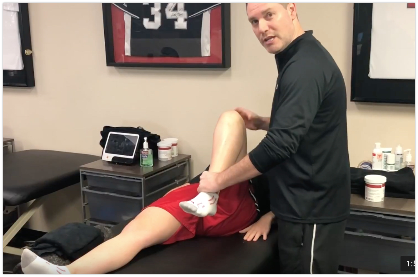
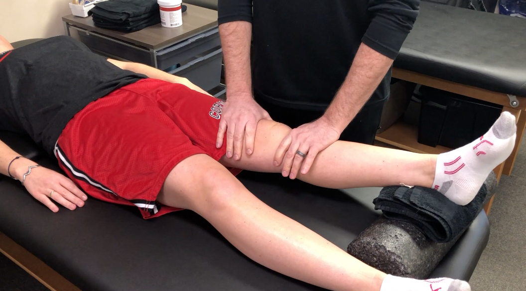
Quadriceps Stretching after Knee Surgery: A tweak to the technique

Obtaining full knee flexion after a knee surgery or knee injury can be difficult for some. The transition from passive knee flexion in seated (my preferred) or supine (not preferred!) can be a challenge for the physical therapist, once they are starting to work on quadriceps stretching. This blog post serves to help modify the prone quadriceps stretching technique after a knee surgery. The goal is to better localize the stretch to the muscle and not cause further pain and discomfort to the patient.

In the past, I’ve talked about restoring knee extension after surgery, particularly after an ACL reconstruction. If you missed that blog post, you can read it here here and here. You might say I have a slight obsession with talking about ROM after surgeries.

For a common surgery like an ACL reconstruction, I often see people present to me without full ROM. That’s why I continue to discuss techniques that can help other clinicians and patients restore their ROM before it’s too late.

Why is full ROM important after knee surgery?

Well, we think there’s a pretty close link between long-term issues and not restoring knee ROM.

In Shelbourne’s article in AJSM 2012, he states that “abnormal knee flexion at early follow-up, abnormal knee extension at final follow-up, abnormal knee flexion at final follow-up, partial medial meniscectomy, and articular cartilage damage were significant factors related to the presence of osteoarthritis on radiographs.”

He also went on to say that you had a 2x increased risk of knee arthritis if you don’t get full ROM. This was similar to having had a meniscectomy surgery or articular cartilage loss.

For such a simple concept, we PT’s can really affect the long-term outcomes for our patients if we don’t get full knee ROM. So why are people still struggling years after their knee surgery? I don’t know… but it’s troubling and definitely avoidable in most patients.

Check out this study from the Journal of Athletic Training in 2015. They discuss how a patient’s flexion ROM can help significantly improve their IKDC scores (functional score) during the 1st 2 months post-op.

Simple Modification to Prone Quadriceps Stretching

I began to think about this topic when I was reading a Facebook post in one of the PT groups and it made me think. We always talk about knee extension and there is some research that discusses the importance of safely obtaining knee extension ROM. I put this study in a recent Instagram post and got some pretty good response.

It seems that whenever I talk about knee ROM after a surgery, people’s ears perk up. Let’s go to the video and talk about a simple technique I use to improve knee flexion ROM after a surgery or injury.

As you can see, a small tweak to your technique can really make a big difference. Again, I’m not sure what exactly is going on. It seems like I’m adjusting the position of the tibia just slightly and that is influencing the location of the stretch toward the quads. If I take my hand away, then they feel the pain and pressure in the front of the knee and it does not feel good.

Slow and Steady Knee Flexion

For the record, I’m not one of those PT’s that likes to be super aggressive and push my patients to tears. I’ve inherited those patients from other PT’s and that technique often fails.

When I talk about improving prone knee flexion, I’m talking about improving quadriceps flexibility and length. Remember, if you have someone lie on their stomach and you try to flex their knee, you’re either limited by pain, joint capsule or the rectus femoris (2-joint muscle.)

It’s not until I think the knee has reached a period of calm that I begin pushing into prone flexion. I’m not using this to crank and get 5 more degrees for my documentation. I’m using this at an appropriate time to improve muscle flexibility and maybe get that end range of motion that is so important.

Remember, obtaining full knee ROM is a process, but a very important process. it’s not going to happen quickly, especially if you’re wicked aggressive (my Boston comes out when I get fired up!!)

Try it for yourself

So this week, try this technique out on one of your clients who may be struggling with regaining their knee flexion ROM. I’m curious to know what you think and if it works for you. As we’ve seen, it’s very important to get that full ROM back after a surgery and this is one modification that I think can help you immediately.


If you want to learn more about how I treat ACL’s and other knee injuries, then you can check out our all online knee seminar. If interested, check it out at www.onlinekneeseminar.com and let me know what you think. We cover the anatomy, rehab prescription, ACL, knee replacements and patellofemoral issues both non-operative treatment and post-operative treatment. This is an awesome course if you’re interested in learning more about rehabilitating the knee joint. And if you’re a PT, there’s a good chance you can get CEU’s as well.

Loss of extension after ACL surgery: How to assess for a cyclops lesion

Loss of extension after an ACL reconstruction can be debilitating for the patient. It’s not as common as you would think but I see it enough in the clinic from people that are months out from surgery. Usually, this loss of knee extension after an ACL reconstruction is caused by a cyclops lesion. Let’s dive deeper into this!

Often times, they’ll present with anterior knee pain, posterior knee soreness and a relatively weakened quadriceps muscle that just won’t return. No matter what they do to get the motion back, the knee just never feels normal.

I’ve written about the loss of extension after an ACL reconstruction in the past. In this post, I discussed how I like to work on knee extension immediately after an ACL surgery. There are a few ways that I think are most effective and with minimal patient efforts.

What is a Cyclops Lesion?

For those not familiar, a cyclops lesion is a wad of scar tissue in the anterior aspect of the knee joint. It is believed to be a remnant of the previous ACL stump that had remained during the reconstruction surgery. At least that’s one theory. Another theory states that it may be fibrocartilage as a result of drilling the tibial tunnels.

Whatever the case, this arthrofibrosis (scar tissue) physically blocks the knee joint from locking out into full extension. Check out this MRI that shows the scar tissue in the anterior knee.

Cyclops lesion in the anterior knee blocking full (hyper) extension

How do I assess for a cyclops lesion after ACL surgery?

In this video, I describe why and how I assess for a cyclops lesion. Check it out.

Is it a Cyclops lesion or just a tight knee?

From the video, you can clearly note that anterior pain, in my experiences, is most often related to a cyclops lesion. Other factors to consider include:

  1. temporary/transient gains in extension
  2. anterior knee pain after increasing activity
  3. poor patella mobility
  4. quads just won’t come back
  5. continued hamstring/calf soreness

These are tell-tale signs that there’s more going on and you should refer back to the doctor so they can order an MRI to rule in/out the anterior scarring. If diagnosed, the best (and only) option is to have a knee scope and remove that scar tissue.

There’s nothing else that can be done. No PT, injections or manual therapy can restore full symmetrical knee extension.

The scar tissue needs to be removed by surgical excision. Aggressive PT should commence immediately after surgery to restore the extension range of motion.

The Best and Easiest Way to Restore Knee Extension after an ACL

Rehab after an ACL is never easy. There are many things that could affect a patient’s outcome. I’ve treated hundreds of patients after an ACL reconstruction and each one is a unique challenge. I wrote about this in a previous post here..check it out and let me know what you think.

I put this video together for Mike Reinold’s website so you could see what I exactly do to gain knee extension back….and why I’m not a fan of prone hangs. Hope it helps with some of your knee patients.

Final Cyclops Thoughts

As a PT or athletic trainer, don’t blame yourself if the patient needs another surgery to remove the scarring. It seems as if it was inevitable and was going to occur no matter the efforts to work on knee extension. I think the lesson here is:

  • work on extension early and often
  • maintain good compliance at home
  • assess/measure each visit to determine gains or losses
  • early patella mobility and knee PROM
  • get the pain and swelling out as quickly as possible
  • refer back to the doctor if the ROM not improving despite your best efforts

Hope this post helps you get better outcomes for your ACL patients!


If you want to learn more about how I treat ACL’s, then you can check out our all online knee seminar. If interested, check it out at www.onlinekneeseminar.com and let me know what you think. We cover the anatomy, rehab prescription, ACL, knee replacements and patellofemoral issues both non-operative treatment and post-operative treatment. This is an awesome course if you’re interested in learning more about rehabilitating the knee joint. And if you’re a PT, there’s a good chance you can get CEU’s as well.

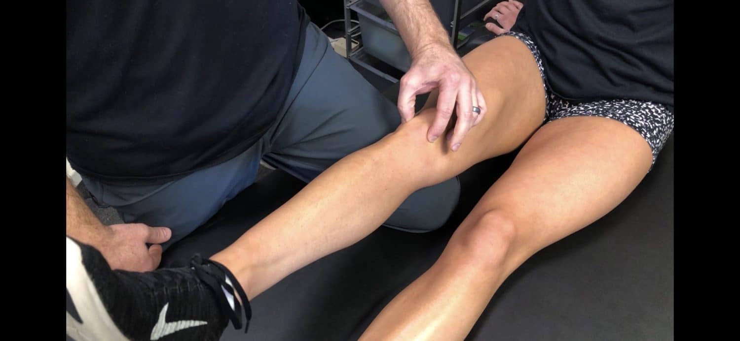
Anterior Knee Pain- A Test for Fat Pad Irritation

We as physical therapists are constantly seeing patients with anterior knee pain with a very vague history. Often times, there’s not a specific onset or mechanism of injury. With that, it seems as if the retro patellar fat pad is a common source of pain in many people and is commonly overlooked.

What actually hurts in the knee?

Whenever I am evaluating someone with knee pain, I always keep in mind the Dye et al AJSM article from 1998. In my opinion, this is a keeper for all PT’s and future PT’s to have in their arsenal of top literature papers.

For those that are not familiar, let me explain it a bit.

Basically, San Francisco orthopaedic surgeon Dr. Scott Dye had his knee scoped without any anesthesia. That’s right, classic beast mode!

He did have local anesthesia (for the record), so they could make the incisions…otherwise, he was awake and alert for the whole procedure.

This allowed him to report back to his colleagues (one being his brother) an actual pain response as they were poking away at the different structures within the knee. He ranked the pain on a 0-4 scale with 0 being pain-free (patella cartilage) and 4 being a severe pain (fat pad, anterior synovium, joint capsule.)

You can read the article to get the full gist (and I HIGHLY RECOMMEND this!) but my point is to let you realize that there are only certain structures capable of causing severe pain in the anterior knee.

Their Findings

Cartilage is avascular and aneural. It has no blood supply or nerves that innervate it. None! So the whole chondromalacia patella diagnosis attempting to implicate a maltracking patella is often hogwash.

But the fat pad, anterior synovium, and anterior capsule are extremely painful and are often involved, I believe, in patients that we see on a daily basis.

In my opinion, this is a diagnosis that sees us more than we see it. Like thoracic outlet syndrome… we can talk about that a different day though.

Back to the point of the blog.

1 Test for Fat Pad Assessment

Assessing the knee and asking the right questions is critical. Besides a tremendously great subjective, there’s one test that I use to rule in or out a fat pad irritation. Check out the video below:

I promise you this test is a great way to establish a pretty clear diagnosis and reassure the patient that a specific structure may be the issue.

When it’s painful, its pretty obvious and the patient can immediately report back to you their symptoms. I test for this a bunch in a given week and it’s not always present. But when someone presents with a positive finding, it’s pretty relieving to them (believe it or not.)

Differential Diagnosis

Keep in mind, my differential diagnosis is all over the place at times.  With knee pain you need to consider:

  1. Meniscus (see my previous blog post)
  2. ITB
  3. Osteochondral lesion
  4. Patella tendonitis
  5. Pes anserine bursitis
  6. MPFL sprain
  7. Hamstring strain
  8. Plica syndrome
  9. MCL/LCL
  10. Tumor

That’s a bunch to consider for someone who can’t really explain why or when their injury occurred. That’s why I still believe this overuse injury sees us more than we see it.

Treatment of Fat Pad Irritation

This one can get a bit tricky. Again, it’s often due to an overuse injury. In others, it’s attributed to kneeling on it for too long or banging it against something. In those more acute cases, the answer is pretty straightforward. Ice, motion and progressive return to their function.

In the overuse group, which is more prevalent in my opinion, we need to figure out which stimulus is causing the issue. It’s often due to starting a new program or ramping up too quickly during some training event.

In these people, I need to modify their volume of training or destress the area by giving them new activities that they can do pain-free…but only for a short period of time.

Along with activity modification, a course of some form of modality (yup, the M-word was used) can help expedite the pain control. I still have a special place in my heart for iontophoresis with dexamethasone. I’ve had tremendous results in patients with fat pad irritation.

I also like to use a low-level laser to help with the healing process (future blog post alert!) I’ve used it on my self for various ailments and even my dog after her ACL surgery. I’m definitely a believer in the healing power of the laser when applied in the correct situation.

Besides all of that, I would also want to assess the person’s movements and attempt to adjust any motions or movements that I thought could contribute to the knee pain. Often times, there’s an underlying weakness or misconception of a weakness that needs to be explained to the patient.

These things run their course for a few weeks but should improve pretty quickly if handled correctly. Patient gratification is pretty obvious because their pain diminishes after a session or 2 and they buy into the program pretty quickly.

Fat Pad Conclusions

Infrapatellar fat pad irritation can be functionally debilitating. I believe it presents itself pretty often in the clinic, more than most PT’s realize. Use my test above to see if it truly is a fat pad issue. Here’s a nice open-access article that you can read to learn more about the fat pad.

Remember, the test should be pretty obvious and locally oriented. If pain-free, then move on. If not, then try some of my above recommendations. It’s usually an overuse issue so you need to adjust their volumes and maybe some form of mechanics.

Mike Reinold and I discuss this and much more about how we treat the knee conservatively. Check it out in our acclaimed all online knee course www.onlinekneeseminar.com.