Tag Archive for: physiotherapy

The Week in Research Review, etc 7-29-18

Last week was the 1st of my research review that summarized my social media posts from the previous week. It seemed to be well received so I figured I’d continue it. My goal is to help summarize some of the research that I found interesting and package it nicely for my readers.

Each photo contains a link back to a social media feed where you can see the conversation that ensued and maybe chime in…or just be a passive reader and see where the conversation went. You never know where the conversation may go on social media…so be ready! haha!


Socioeconomic Factors for Sports Specialization and Injury in Youth Athletes Jayanthi et al Sports Health Journal 2018.

This study looked at the effect of socioeconomic status (SES) on rates of sports specialization and injury among youth athletes.

They looked at injured athletes between the ages of 7 to 18 years that were recruited from 2 hospital-based sports medicine clinics. They compared these with uninjured athletes presenting for sports physicals at primary care clinics between 2010 and 2013.

They concluded that:
✅High-SES athletes reported more serious overuse injuries than low-SES athletes
✅More hours/wk playing organized sports
✅Higher ratio of weekly hours in organized sports to free play
✅Greater participation in individual sports

I applaud the authors for attempting to bring this very difficult collection of data into a formal research paper. I will say some of the statistics and standard deviations may not make the conclusions as powerful.

I do think this is a good paper to help educate our athletes on injury rates, especially in those that specialize in 1 sport.

What do you think? Tag a friend that may benefit from this article!


From #Twitter’s @retlouping that perfectly sums up what I’ve observed recently on social media with many PT’s.

For some reason, pain science has overtaken most diagnosis and treatment conversations.

It’s as if you get bullied into talking pain science and ignoring our clinical judgment and diagnosis skills. I understand there’s a constant tug-of-war between the biomechanical PT’s and the pain science PTs.

But as usual, the answer usually lies somewhere in between and both groups are correct. The biomechanics of an injury are often important as well as the language we use to explain these tissue biomechanics.

To my fellow clinicians, especially the newer grads and #dptstudent, remember this little cartoon for every future encounter.

Yeah, speak to people in non-threatening tones (in my world it’s just being respectful) but trust me, they WANT to hear what could be going wrong or what may be causing their pain.

Don’t blow off their symptoms and don’t go into depth about pain science because they won’t understand.

Trust me, the clinicians that try to do that often end up losing their patients in the long run.

I hear these stories day after day of people coming to me because the last PT either only talks to them or made them ONLY do strength exercises and it didn’t help their pain.

The PT didn’t listen to them and was so blinded by their pain science background that they ignored the person sitting right in front of them. Remember, the person sitting there will tell you what is going on and what treatment will most help them feel/move better.


Influence of Body Position on Shoulder and Trunk Muscle Activation During Resisted Isometric Shoulder External Rotation Krause et al Sports Health 2018.

The purpose of this study was to examine ER torque and electromyographic (EMG) activation of shoulder and trunk muscles while performing resisted isometric shoulder ER in 3 positions:
✔️Standing
✔️Side-lying
✔️Side plank

Using surface EMG and a hand-held dynamometer, the researchers tried to determine EMG activity of the:
✔️infraspinatus
✔️Posterior Deltoids
✔️Mid traps
✔️Multifidi
✔️External/internal obliques (dominant side)
✔️External/internal obliques (non-dominant side)

EMG values for the infraspinatus were greatest in the side plank position. In general, EMG values for the trunk muscles were also greatest in the side plank position.

✅Their Conclusions: If the purpose of a rehabilitation program is to strengthen the rotator cuff, in particular, the infraspinatus, the side plank is preferred over standing or side lying. If the goal is to simultaneously strengthen both the rotator cuff and trunk muscles, the side plank position again is preferred.

Makes sense but good to see the research and have concrete evidence to back up what we think actually goes on.

Tag a friend who may be interested in this research paper!


Reliability of heel-height measurement for documenting knee extension deficits. Schlegel et al AJSM 2002

Prone heel-height difference of 1cm equates to 1.2 degree difference in knee extension ROM.

Do you use this method to assess knee ROM? I still measure knee extension ROM is supine but find this method helpful as well.

I know my friend and colleague @wilk_kevin has measured this way for many years. i originally saw his use this technique at @ChampionSportsM

I don’t want people to confuse this with prone hangs for knee extension ROM. I am not a fan of that method as I’ve stated in the past.

This is a method to assess knee extension differences, particularly after an ACL reconstruction. I have gone back to using this method for some people that have subtle ROM differences side-to-side.

The patella position (on the plinth or off) did not matter in the study and thigh girth did not appear to make a difference.

I would recommend stabilizing the pelvis to prevent excess ROM from occurring at that region and to better isolate the knee joint.

Have you tried this method? Tag a friend who may benefit from using this ROM method…thanks!


Evidence-Based Best-Practice Guidelines for Preventing #ACL Injuries in Young Female Athletes: A Systematic Review and Meta-analysis Petushek et al AJSM 2018.

Injury prevention neuromuscular training (NMT) programs reduce the risk for anterior cruciate ligament (ACL) injury.

Eighteen studies were included in the meta-analyses, with a total of 27,231 participants, 347 sustaining an ACL injury.

The overall mean training amount was 57 sessions totaling 18.17 hours (roughly 24 minutes per session, 2.5 times per week).

They concluded:

✔️Interventions targeting middle school or high school–aged athletes reduced injury risk to a greater degree than did interventions for college or professional-aged athletes.

✔️Continued exposure to neuromuscular training throughout the sport season seems to enhance prophylactic effects of NMT.

✔️NMT interventions were effective for female basketball, and handball athletes and interventions including various athletes were potentially effective (eg, soccer, basketball, and volleyball).

✔️ Interventions included some form of implementer training (eg, instructional workshop, video, or brochure) on proper program implementation.

✔️Programs including more landing stabilization and lower body strength exercises during each session were most effective.

🤔Programs including balance, core-strengthening, stretching, or agility exercises were no more effective than programs that did not incorporate these components.

✔️ Specifically, programs that included more landing stabilization exercises (eg, drop landings, jump/hop and holds), hamstring strength (eg, Nordic hamstring), lunges, and heel-calf raises reduced the risk for ACL injury to a greater degree than did programs without these exercises.

✅ Wow, lots of great information here. Please share this with a friend or colleague who may benefit from knowing this information.


Hope that helped to catch you up on my posts from this week.

Do you like these weekly updates? Let me know if I should continue…love your feedback!

Thanks for reading!

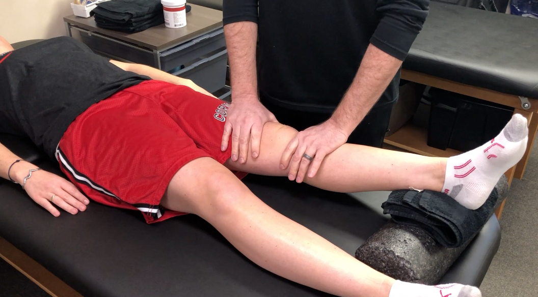
Loss of extension after ACL surgery: How to assess for a cyclops lesion

Loss of extension after an ACL reconstruction can be debilitating for the patient. It’s not as common as you would think but I see it enough in the clinic from people that are months out from surgery. Usually, this loss of knee extension after an ACL reconstruction is caused by a cyclops lesion. Let’s dive deeper into this!

Often times, they’ll present with anterior knee pain, posterior knee soreness and a relatively weakened quadriceps muscle that just won’t return. No matter what they do to get the motion back, the knee just never feels normal.

I’ve written about the loss of extension after an ACL reconstruction in the past. In this post, I discussed how I like to work on knee extension immediately after an ACL surgery. There are a few ways that I think are most effective and with minimal patient efforts.

What is a Cyclops Lesion?

For those not familiar, a cyclops lesion is a wad of scar tissue in the anterior aspect of the knee joint. It is believed to be a remnant of the previous ACL stump that had remained during the reconstruction surgery. At least that’s one theory. Another theory states that it may be fibrocartilage as a result of drilling the tibial tunnels.

Whatever the case, this arthrofibrosis (scar tissue) physically blocks the knee joint from locking out into full extension. Check out this MRI that shows the scar tissue in the anterior knee.

Cyclops lesion in the anterior knee blocking full (hyper) extension

How do I assess for a cyclops lesion after ACL surgery?

In this video, I describe why and how I assess for a cyclops lesion. Check it out.

Is it a Cyclops lesion or just a tight knee?

From the video, you can clearly note that anterior pain, in my experiences, is most often related to a cyclops lesion. Other factors to consider include:

  1. temporary/transient gains in extension
  2. anterior knee pain after increasing activity
  3. poor patella mobility
  4. quads just won’t come back
  5. continued hamstring/calf soreness

These are tell-tale signs that there’s more going on and you should refer back to the doctor so they can order an MRI to rule in/out the anterior scarring. If diagnosed, the best (and only) option is to have a knee scope and remove that scar tissue.

There’s nothing else that can be done. No PT, injections or manual therapy can restore full symmetrical knee extension.

The scar tissue needs to be removed by surgical excision. Aggressive PT should commence immediately after surgery to restore the extension range of motion.

The Best and Easiest Way to Restore Knee Extension after an ACL

Rehab after an ACL is never easy. There are many things that could affect a patient’s outcome. I’ve treated hundreds of patients after an ACL reconstruction and each one is a unique challenge. I wrote about this in a previous post here..check it out and let me know what you think.

I put this video together for Mike Reinold’s website so you could see what I exactly do to gain knee extension back….and why I’m not a fan of prone hangs. Hope it helps with some of your knee patients.

Final Cyclops Thoughts

As a PT or athletic trainer, don’t blame yourself if the patient needs another surgery to remove the scarring. It seems as if it was inevitable and was going to occur no matter the efforts to work on knee extension. I think the lesson here is:

  • work on extension early and often
  • maintain good compliance at home
  • assess/measure each visit to determine gains or losses
  • early patella mobility and knee PROM
  • get the pain and swelling out as quickly as possible
  • refer back to the doctor if the ROM not improving despite your best efforts

Hope this post helps you get better outcomes for your ACL patients!


If you want to learn more about how I treat ACL’s, then you can check out our all online knee seminar. If interested, check it out at www.onlinekneeseminar.com and let me know what you think. We cover the anatomy, rehab prescription, ACL, knee replacements and patellofemoral issues both non-operative treatment and post-operative treatment. This is an awesome course if you’re interested in learning more about rehabilitating the knee joint. And if you’re a PT, there’s a good chance you can get CEU’s as well.